. 2022 Nov 30;6(2):e202201372.
doi: 10.26508/lsa.202201372. Print 2023 Feb.

Revisiting degron motifs in human AURKA required for its targeting by APC/CFZR1

Affiliations

Revisiting degron motifs in human AURKA required for its targeting by APC/CFZR1

Ahmed Abdelbaki et al. Life Sci Alliance. .

Abstract

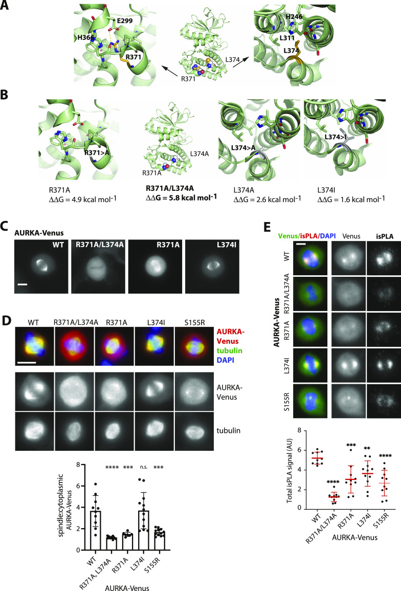
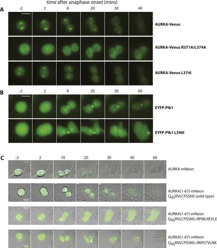
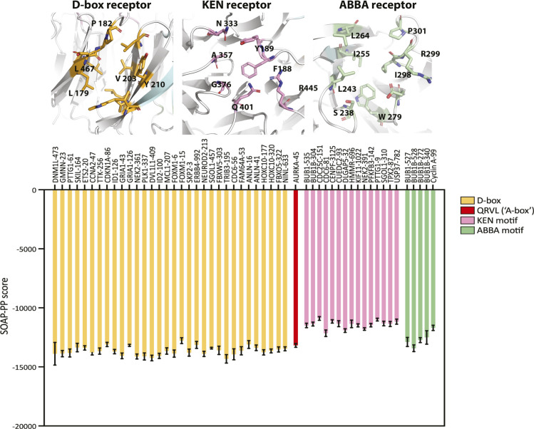
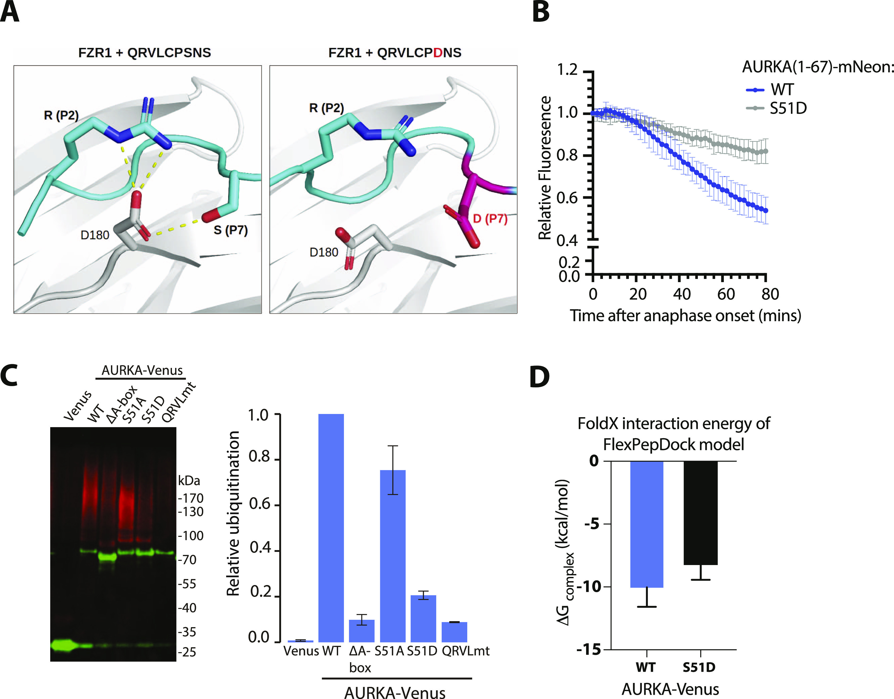
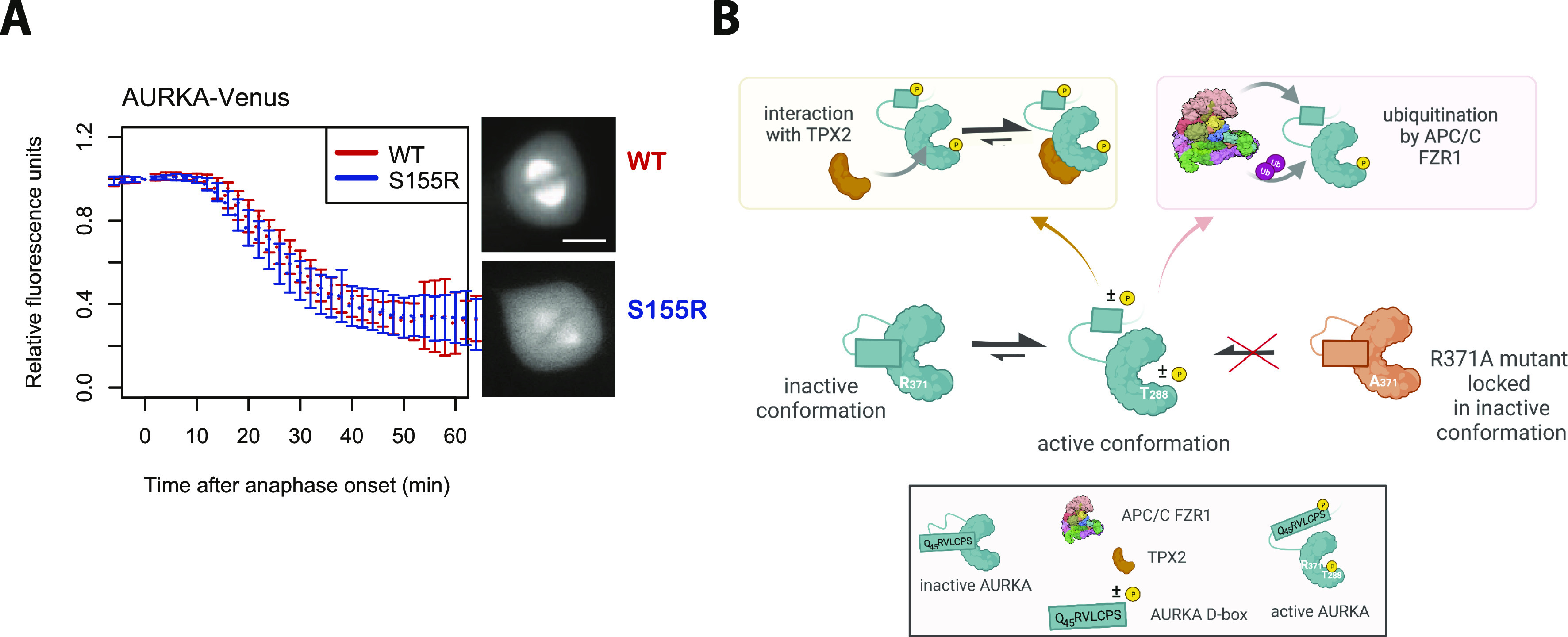
Mitotic kinase Aurora A (AURKA) diverges from other kinases in its multiple active conformations that may explain its interphase roles and the limited efficacy of drugs targeting the kinase pocket. Regulation of AURKA activity by the cell is critically dependent on destruction mediated by the anaphase-promoting complex (APC/CFZR1) during mitotic exit and G1 phase and requires an atypical N-terminal degron in AURKA called the "A-box" in addition to a reported canonical D-box degron in the C-terminus. Here, we find that the reported C-terminal D-box of AURKA does not act as a degron and instead mediates essential structural features of the protein. In living cells, the N-terminal intrinsically disordered region of AURKA containing the A-box is sufficient to confer FZR1-dependent mitotic degradation. Both in silico and in cellulo assays predict the QRVL short linear interacting motif of the A-box to be a phospho-regulated D-box. We propose that degradation of full-length AURKA also depends on an intact C-terminal domain because of critical conformational parameters permissive for both activity and mitotic degradation of AURKA.


Conflict of interest statement

The authors declare that they have no conflict of interest.

Figures

Figure 1.
Figure 1.. C-terminal R371xxL motif of AURKA plays a critical role in folding and function.
(A, B) In silico testing of the C-terminal D-box–like motif. (A) Published structures of the AURKA C-terminal region (here, PDB ID:1MQ4) show the R371xxL motif buried within the kinase domain. Arginine residue R371 (orange) establishes salt bridges with conserved glutamic acid residue E299 (green). Leucine L374 fits into the hydrophobic aliphatic pocket on the kinase domain. (A, B) Interactions shown in (A) are lost in predicted structures modelled for R371A and L374A substitutions. Gibbs free energy variations (ΔΔG = ΔGmut-ΔGwt) for the protein folding state were predicted using the FoldX3 software and show that R371A and L374A substitutions are more strongly destabilizing to the structure than the conserved substitution L374I. (C, D, E) Characterization of different versions of Venus-tagged AURKA in human U2OS cells. (C) Panels showing localization of AURKA-Venus in live cells during interphase and at mitosis. (D) U2OS cells transfected with different variants of AURKA-Venus were MeOH-fixed and processed for immunofluorescence using antibodies against GFP (red), and β-tubulin (green) and DAPI (blue). Representative images of mitotic cells are shown (upper panels), with quantified data on spindle localization of AURKA variants shown in the accompanying graph (lower panel): average pixel values within an ROI of fixed size were measured at spindle (next to but not overlapping centrosome) and in the cytoplasm (midway between spindle and cortex) and, after subtraction of background values (neighbouring the cell), used to calculate the spindle:cytoplasmic ratio. Data from individual mitotic cells (n ≥ 10) are plotted, with a bar chart indicating the mean and SDs. (E) Interaction with TPX2 assayed by isPLA (see also Fig S1). isPLA signal revealed AURKA-Venus–TPX2 interaction on mitotic spindles (examples in upper panels) with quantified data shown in the accompanying graph (lower panel): total isPLA signal was measured per mitotic cell, corrected for background, and normalized to the mean of the WT values in each experiment. Data for each condition (n ≥ 10) were plotted in a scatter plot with bar and whiskers to indicate the mean and SDs. (D, E) Each mutant was compared with WT by ordinary one-way ANOVA. **P ≤ 0.01; ***P ≤ 0.001; ****P ≤ 0.0001; and n.s., non-significant. Results are representative of two identical repeats of experiments.
Figure S1.
Figure S1.. Controls for isPLA experiment to measure AURKA–TPX2 interaction.
U2OS cells were co-transfected with WT AURKA-Venus and processed for isPLA detection of AURKA-Venus and endogenous TPX2 using mouse GFP and rabbit TPX2 antibodies alone and in combination. Figure shows example images of mitotic cells (left-hand panels) from which isPLA signals were measured and plotted (right-hand graph). The scatter plot shows whole-cell measurements from single cells, with mean values ± SDs indicated in red. ****P ≤ 0.0001 by a t test.
Figure S2.
Figure S2.. C-terminal mutants of AURKA-Venus unable to interact with TPX2 are inactive according to the pT288 marker.
U2OS cells were transiently transfected with WT or different versions of AURKA-Venus mutated in the putative C-terminal D-box or the non–TPX-binding S155R control. Extracts from cells arrested in mitosis were probed for pT288-AURKA by immunoblot. The blot is representative of two identical experiments.
Figure 2.
Figure 2.. R371xxL motif is not a D-box.
(A, B) In cellulo mitotic degradation assays of AURKA-Venus. Graphs show quantified Venus levels from fluorescence time-lapse imaging of single cells undergoing mitotic exit. Venus levels from individual cells are normalized against the Venus level at anaphase onset and in silico–synchronized so that the mean and SDs can be plotted for each version of AURKA-Venus. (A) Mutations predicted to cause disruption of the C-terminus block AURKA degradation during mitotic exit: R371A/L374A (top graph, n ≥ 10 cells, pooled from two experiments) blocks degradation of AURKA-Venus in a similar way to deletion of the N-terminal A-box (bottom graph, n ≥ 6 cells, from a single experiment) (Floyd et al, 2008). (B) Conservative substitution L374I has no effect on kinetics of degradation of AURKA-Venus (n ≥ 10 cells, pooled from two experiments). (C) Correlation plot for percentage degradation of each version of AURKA-Venus versus predicted ΔΔG of substituted residues in R371xxL. (D) Schematic of known and proposed degrons in AURKA and Plk1. (E) In cellulo mitotic degradation assays of YFP-Plk1 WT and L341I version (n ≥ 10 cells, pooled from two experiments). The L > I substitution at P4 of the R337xxL motif abrogates degradation at mitotic exit whilst having no effect on localization of the protein (right-hand panels).
Figure S3.
Figure S3.. Time-lapse from degradation movies.
Frames from time-lapse movies analysed in Figs 2 and 3. (A, B, C) Fluorescence images are shown for AURKA-Venus variants (A) and EYFP-Plk1 variants (B), whereas combined DIC and fluorescence images are shown for AURKA (1-67)-mNeon constructs (C). Times are indicated in minutes relative to anaphase onset timing. Scale bar, 10 μm.
Figure S4.
Figure S4.. AURKA (1-133)-GFP is degraded at mitotic exit in an FZR1-sensitive manner.
U2OS parental or FZR1KO cells were transfected with full-length or 1-133 AURKA tagged with GFP and live cells filmed through mitosis. GFP fluorescence was quantified in individual cells and plotted as a fraction of protein remaining relative to the level at anaphase onset. Curves show mean values and standard deviations from n ≥ 5 cells. The result was representative of two experiments and consistent with Fig 3 data.
Figure 3.
Figure 3.. Q45RVL motif within the AURKA A-box displays properties of a D-box degron.
(A, B) In cellulo mitotic degradation assays for full-length AURKA-mNeon and AURKA (1-67)-mNeon expressed in U2OS or FZR1KO U2OS cells. mNeon levels from individual cells are normalized against the anaphase onset level and in silico–synchronized so that the mean and SDs can be plotted for each protein (n ≥ 20 cells pooled from ≥ 3 experiments). Degradation curves show that (A) residues 1-67 are sufficient for mitotic exit–specific FZR1-dependent degradation and (B) mitotic degradation of AURKA (1-67)-mNeon depends on SLiMs at K5 and Q45RVL. (C) Energetics of in silico docking of proposed AURKA degrons into known binding pockets on FZR1, scored by statistically optimized atomic potential for protein–protein docking, using the FlexPepDock server. (D) A-box (QRVLCPSNS) peptide docked to the H.s. FZR1 DBR (top panel), modelled upon structure PDB ID:4BH6 that shows S.c. Cdh1 bound to the D-box peptide (RIALKD). PDB ID:4BH6 is shown for comparison in the bottom panel (He et al, 2013). (E) In cellulo mitotic degradation assays of AURKA-Venus WT and L48I (L > I at P4 of A-box motif, n = 26 cells pooled from two experiments). (F) In cellulo mitotic degradation assays of AURKA (1-67)-mNeon in which the Q45RVLCPSNS peptide (the so-called “A-box”) is substituted with the bona fide D-box from Plk1 (R337KPLTVLNK) or with the C-terminal R371PMLREVLE motif of AURKA.
Figure S5.
Figure S5.. In silico docking of the A-box motif to the DBR site compared with a panel of other degrons.
Peptides corresponding to a range of validated D-box and KEN degrons, the AURKA A-box (Q45RVLxxx), and ABBA motifs that also bind to co-activators (all taken from Davey & Morgan [2016]) were in silico–docked into the DBR cleft of FZR1. Energies of binding are expressed as statistically optimized atomic potential scores and shown that the A-box docks to the DBR and many D-boxes.
Figure 4.
Figure 4.. Modelling of the Q45RVL motif at the DBR explains the role of AURKA S51 phosphorylation.
(A) Docking of Q45RVLPSNSS peptide on FZR1. Predicted pose for QRVL in the DBR by in silico docking, showing orientation of the P4 leucine and novel contacts afforded at P2 and P7. (B) In cellulo mitotic degradation assays of AURKA-Venus WT and S51D. (C) Ubiquitination of WT and different versions of AURKA carrying mutations in the A-box; transiently expressed Venus-tagged proteins were purified from U2OS cells synchronized in mitotic exit and blotted for GFP (in green) and ubiquitin conjugates (FK1 antibody, in red). Relative ubiquitination was plotted as the ratio of ubiquitin-conjugated:unmodified protein, normalized against the WT protein; error bars show SDs from three repeats of the experiment. (D) Free energy values for WT and S51D peptides computed using FoldX3. In silico docking models were rebuilt using the mutant peptide QRVLCPDNS, models were scored with FoldX3, and the average binding free energies of 10 models for each were plotted. The higher binding energy of the mutant is significant according to a Mann–Whitney test (P = 0.0147).
Figure 5.
Figure 5.. R371xxL motif plays a critical role in conformational regulation of AURKA.
(A) In cellulo mitotic degradation assays of AURKA-Venus WT and S155R. Venus levels from individual cells are normalized against the anaphase onset level and in silico–synchronized so that the mean and SDs can be plotted for each protein (n ≥ 10 cells, representative of two experiments). (B) Schematic proposing that the link between R371 and degradability of AURKA is mediated by a conformational step that simultaneously activates AURKA (leading to phosphorylation on T288) and makes it degradable by the APC/C. The Q45RVL motif is “buried” in the autoinhibited state of the WT protein (green) and once released is autoregulated by phosphorylation on S51. The R371A mutant (orange) is unable to undergo the critical conformational step required for both activation and degradation. Schematic created in BioRender.com.

References

    1. Arlot-Bonnemains Y, Klotzbucher A, Giet R, Uzbekov R, Bihan R, Prigent C (2001) Identification of a functional destruction box in the Xenopus laevis Aurora-A kinase pEg2. FEBS Lett 508: 149–152. 10.1016/s0014-5793(01)03048-4 - DOI - PubMed
    1. Bai M, Ni J, Shen S, Wu J, Huang Q, Le Y, Yu L (2014) Two newly identified sites in the N-terminal regulatory domain of Aurora-A are essential for auto-inhibition. Biotechnol Lett 36: 1595–1604. 10.1007/s10529-014-1516-3 - DOI - PubMed
    1. Bayliss R, Sardon T, Vernos I, Conti E (2003) Structural basis of Aurora-A activation by TPX2 at the mitotic spindle. Mol Cell 12: 851–862. 10.1016/S1097-2765(03)00392-7 - DOI - PubMed
    1. Bertolin G, Sizaire F, Herbomel G, Reboutier D, Prigent C, Tramier M (2016) A FRET biosensor reveals spatiotemporal activation and functions of Aurora kinase A in living cells. Nat Commun 7: 12674. 10.1038/ncomms12674 - DOI - PMC - PubMed
    1. Bibby RA, Tang C, Faisal A, Drosopoulos K, Lubbe S, Houlston R, Bayliss R, Linardopoulos S (2009) A cancer-associated Aurora A mutant is mislocalized and misregulated due to loss of interaction with TPX2. J Biol Chem 284: 33177–33184. 10.1074/jbc.m109.032722 - DOI - PMC - PubMed

Publication types

LinkOut - more resources