. 2023 Jul;31(7):761-768.
doi: 10.1038/s41431-022-01250-3. Epub 2022 Dec 1.

A second look at exome sequencing data: detecting mobile elements insertion in a rare disease cohort

Affiliations

A second look at exome sequencing data: detecting mobile elements insertion in a rare disease cohort

Philippine Garret et al. Eur J Hum Genet. 2023 Jul.

Abstract

About 0.3% of all variants are due to de novo mobile element insertions (MEIs). The massive development of next-generation sequencing has made it possible to identify MEIs on a large scale. We analyzed exome sequencing (ES) data from 3232 individuals (2410 probands) with developmental and/or neurological abnormalities, with MELT, a tool designed to identify MEIs. The results were filtered by frequency, impacted region and gene function. Following phenotype comparison, two candidates were identified in two unrelated probands. The first mobile element (ME) was found in a patient referred for poikilodermia. A homozygous insertion was identified in the FERMT1 gene involved in Kindler syndrome. RNA study confirmed its pathological impact on splicing. The second ME was a de novo Alu insertion in the GRIN2B gene involved in intellectual disability, and detected in a patient with a developmental disorder. The frequency of de novo exonic MEIs in our study is concordant with previous studies on ES data. This project, which aimed to identify pathological MEIs in the coding sequence of genes, confirms that including detection of MEs in the ES pipeline can increase the diagnostic rate. This work provides additional evidence that ES could be used alone as a diagnostic exam.


Conflict of interest statement

The authors declare no competing interests.

Figures

Fig. 1
Fig. 1. MEs detection pipeline with the MELT tool on ES data.
“Preprocessing”, “IndivAnalysis”, “GroupAnalysis”, “Genotype” and “MakeVCF” steps were realized by the MELT tool. Python scripts were used to mean depth computation, familial data extraction and proband final report generation.
Fig. 2
Fig. 2. Filters applied to MELT detected MEs in 2394 probands.
The majority of the detected MEs were Alu, followed by L1 and SVA. The 5 applied filters and indicated on the right of the figure permitted to reduce the number of potential candidates by more than 99%. Approximately 78% of the patients were removed from the list.
Fig. 3
Fig. 3. FERMT1, GRIN2B and NPRL3 ME candidates validation and segregation.
a FERMT1 MEI segregation for the proband (blood + fibroblasts) and his children (blood). Expected size (bp) without/with insertion: ~548/~829. Pr proband. b GRIN2B MEI segregation for the proband (blood) and her parents (blood). Expected size (bp) without/with insertion: ~640/~915. c NPRL3 MEI segregation for the proband (blood) and his parents (blood). Expected size (bp) without/with insertion: ~417/~750. (+) WT fragment amplification control. (−) PCR negative control without DNA.
Fig. 4
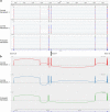
Fig. 4. FERMT1 exon 7 skipping in proband’s cDNA fibroblasts.
The first two profiles were obtained for two control fibroblasts cDNA sequencing data and the third for proband’s fibroblasts cDNA. Only exons 5 to 10 were studied. a FERMT1 cDNA alignment with human genome reference sequence. As expected, the control fibroblasts profiles showed reads aligned with the exons 5 to 10 (reverse strand) with homogeneous coverage. The proband’s profile did not present any reads aligned with the exon 7. The other exons alignments are similar to controls’. Thus, the proband’s cDNA did not contain the exon 7 sequence. b Exons 5–10 sashimi plots. As expected, the control fibroblasts cDNA did not have splice anomaly as each exon was present in mRNA. For the proband, this plot confirmed the exon 7 skipping during FERMT1 mRNA splicing: no peak could be identified for the exon 7.

References

    1. Finnegan DJ. Retrotransposons. Curr Biol. 2012;22:R432–7. doi: 10.1016/j.cub.2012.04.025. - DOI - PubMed
    1. Wang H, Xing J, Grover D, Hedges DJ, Han K, Walker JA, et al. SVA Elements: A Hominid-specific Retroposon Family. J Mol Biol. 2005;354:994–1007. doi: 10.1016/j.jmb.2005.09.085. - DOI - PubMed
    1. Raiz J, Damert A, Chira S, Held U, Klawitter S, Hamdorf M, et al. The non-autonomous retrotransposon SVA is trans-mobilized by the human LINE-1 protein machinery. Nucleic Acids Res. 2012;40:1666–83. doi: 10.1093/nar/gkr863. - DOI - PMC - PubMed
    1. Chenais B. Transposable Elements in Cancer and Other Human Diseases. Curr Cancer Drug Targets. 2015;15:227–42. doi: 10.2174/1568009615666150317122506. - DOI - PubMed
    1. Wicker T, Sabot F, Hua-Van A, Bennetzen JL, Capy P, Chalhoub B, et al. A unified classification system for eukaryotic transposable elements. Nat Rev Genet. 2007;8:973–82. doi: 10.1038/nrg2165. - DOI - PubMed