. 2022 Nov 30;8(1):18.
doi: 10.1186/s42234-022-00101-2.

A dual tracer [11C]PBR28 and [18F]FDG microPET evaluation of neuroinflammation and brain energy metabolism in murine endotoxemia

Affiliations

A dual tracer [11C]PBR28 and [18F]FDG microPET evaluation of neuroinflammation and brain energy metabolism in murine endotoxemia

Santhoshi P Palandira et al. Bioelectron Med. .

Abstract

Background: Brain metabolic alterations and neuroinflammation have been reported in several peripheral inflammatory conditions and present significant potential for targeting with new diagnostic approaches and treatments. However, non-invasive evaluation of these alterations remains a challenge.

Methods: Here, we studied the utility of a micro positron emission tomography (microPET) dual tracer ([11C]PBR28 - for microglial activation and [18F]FDG for energy metabolism) approach to assess brain dysfunction, including neuroinflammation in murine endotoxemia. MicroPET imaging data were subjected to advanced conjunction and individual analyses, followed by post-hoc analysis.

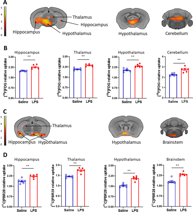
Results: There were significant increases in [11C]PBR28 and [18F]FDG uptake in the hippocampus of C57BL/6 J mice 6 h following LPS (2 mg/kg) intraperitoneal (i.p.) administration compared with saline administration. These results confirmed previous postmortem observations. In addition, patterns of significant simultaneous activation were demonstrated in the hippocampus, the thalamus, and the hypothalamus in parallel with other tracer-specific and region-specific alterations. These changes were observed in the presence of robust systemic inflammatory responses manifested by significantly increased serum cytokine levels.

Conclusions: Together, these findings demonstrate the applicability of [11C]PBR28 - [18F]FDG dual tracer microPET imaging for assessing neuroinflammation and brain metabolic alterations in conditions "classically" characterized by peripheral inflammatory and metabolic pathogenesis.

Keywords: Brain; Brain metabolism; Conjunction analysis; Microglia; Micropet imaging; Murine endotoxemia; Neuroinflammation; [11C]PBR28; [18F]FDG.


Conflict of interest statement

The authors declare no competing interests.

Figures

Fig. 1
Fig. 1
Brain regions with overlapping increases in [11C]PBR28 and [18F]FDG uptakes during endotoxemia. A Dual tracer conjunction increases in metabolism and microglial activation (TSPO binding). Brain coronal sections of statistically significant clusters (color bar represents t-value height, extent threshold T = 2.85), overlaid onto MRI atlas for visualization. Statistically significant clusters (P < 0.005) show overlapping regions of increased uptake of [11C]PBR28 and [18F]FDG in the hippocampus, the thalamus, and the hypothalamus in LPS administered (n = 6) vs saline administered (n = 6) mice (see Table 1 for stereotaxic coordinates). B Post-hoc analysis for the same statistically significant increases in metabolic activity [18F]FDG and microglial activation [11C]PBR28 (TSPO binding) in saline (n = 6) and LPS (n = 6) mice. **P = 0.002, **P = 0.004 (hippocampus - [11C]PBR28). See Methods for details
Fig. 2
Fig. 2
Brain regions with individual increases in [11C]PBR28 and [18F]FDG uptake during endotoxemia. A Increased [18F]FDG uptake in the hippocampus, the thalamus, the hypothalamus, and the cerebellum shown in coronal sections in LPS administered (n = 6) vs saline administered (n = 6) mice (see Table 1 for specific stereotaxic coordinates). SPM clusters (color bar represents t-value height, cutoff threshold T = 3.55) overlaid onto MRI atlas for visualization. Statistically significant clusters (P < 0.001). B Post-hoc analysis of [18F]FDG tracer uptake in the same groups (saline, n = 6 and LPS, n = 6) of mice. **P = 0.002. C Increased [11C]PBR28 metabolic activity in the hippocampus, the thalamus, the hypothalamus, and the brainstem shown in coronal sections in LPS administered (n = 6) vs saline administered (n = 6) mice (see Table 1 for stereotaxic coordinates). SPM clusters (color bar represents t-value height, cutoff threshold T = 3.55). Statistically significant clusters (P < 0.001) overlaid onto MRI atlas for visualization. D Post-hoc analysis of [11C]PBR28 uptake in the same groups (saline, n = 6 and LPS, n = 6) of mice. **P = 0.002, **P = 0.004 (hippocampus). See Methods for details
Fig. 3
Fig. 3
Brain regions with overlapping significantly decreased [11C]PBR28 and [18F]FDG uptake during endotoxemia. A Simultaneous (dual tracer) uptake decreases in primary and somatosensory motor cortices in LPS administered (n = 6) vs saline administered (n = 6) mice. Dual tracer decreases in metabolism and TSPO binding shown with SPM cluster overlaid onto MRI atlas for visualization (color bar represents t-value height, cutoff threshold T = 2.85). B Individual [18F]FDG and [11C]PBR28 decreases in primary and somatosensory motor cortices were subjected to post-hoc analysis in the same groups (saline, n = 6 and LPS, n = 6) of mice. **P = 0.002. See Methods for details

Similar articles

Cited by

References

    1. Alshikho MJ, Zürcher NR, Loggia ML, Cernasov P, Reynolds B, Pijanowski O, et al. Integrated magnetic resonance imaging and [(11) C]-PBR28 positron emission tomographic imaging in amyotrophic lateral sclerosis. Ann Neurol. 2018;83(6):1186–1197. doi: 10.1002/ana.25251. - DOI - PMC - PubMed
    1. Baik SH, Kang S, Lee W, Choi H, Chung S, Kim J-I, et al. A breakdown in metabolic reprogramming causes microglia dysfunction in Alzheimer's disease. Cell Metab. 2019;30(3):493–507.e6. doi: 10.1016/j.cmet.2019.06.005. - DOI - PubMed
    1. Barron AM, Tokunaga M, Zhang MR, Ji B, Suhara T, Higuchi M. Assessment of neuroinflammation in a mouse model of obesity and β-amyloidosis using PET. J Neuroinflammation. 2016;13(1):221. doi: 10.1186/s12974-016-0700-x. - DOI - PMC - PubMed
    1. Bellaver B, dos Santos JP, Leffa DT, Bobermin LD, Roppa PHA, da Silva Torres IL, et al. Systemic inflammation as a driver of brain injury: the astrocyte as an emerging player. Mol Neurobiol. 2018;55(3):2685–2695. doi: 10.1007/s12035-017-0526-2. - DOI - PubMed
    1. Betlazar C, Harrison-Brown M, Middleton RJ, Banati R, Liu GJ. Cellular sources and regional variations in the expression of the Neuroinflammatory marker translocator protein (TSPO) in the Normal brain. Int J Mol Sci. 2018;19(9):2707. - PMC - PubMed

LinkOut - more resources