

Pathological resting-state executive and language system perfusion in first-episode psychosis
- PMID: 36451364
- PMCID: PMC9668641
- DOI: 10.1016/j.nicl.2022.103261
Pathological resting-state executive and language system perfusion in first-episode psychosis
Abstract
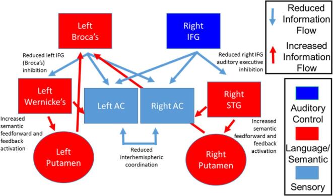
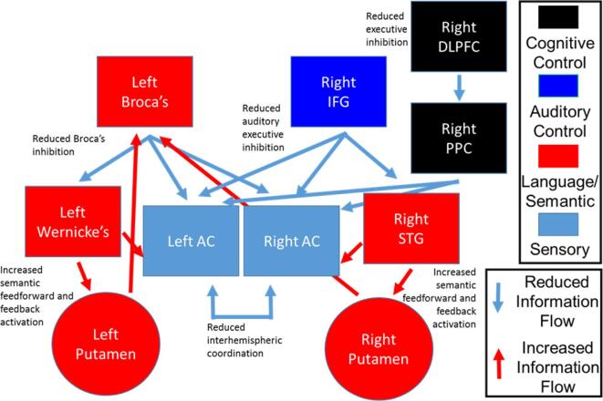
Background and hypothesis: Cortical (e.g., Broca's area and Wernicke's area) and subcortical (e.g., putamen) language-related areas and executive control areas (e.g., inferior frontal gyrus (IFG), dorsolateral prefrontal cortex (DLPFC)) show functional and structural dysconnectivity in long-term psychosis. We examined whether resting-state basal perfusion levels revealed selective pathophysiology (likely hypo- and hyper-activation) of language-related and executive areas in first-episode psychosis (FEP).
Study design: Basal resting-state perfusion was measured using pseudo-continuous Arterial Spin Labeling (pcASL). Relative cerebral blood flow (rCBF) was compared between 32 FEP and 34 matched healthy comparison (HC) individuals. Structural and functional MRI scans were acquired using a 3T Prisma scanner during the same session.
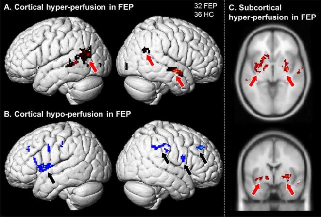
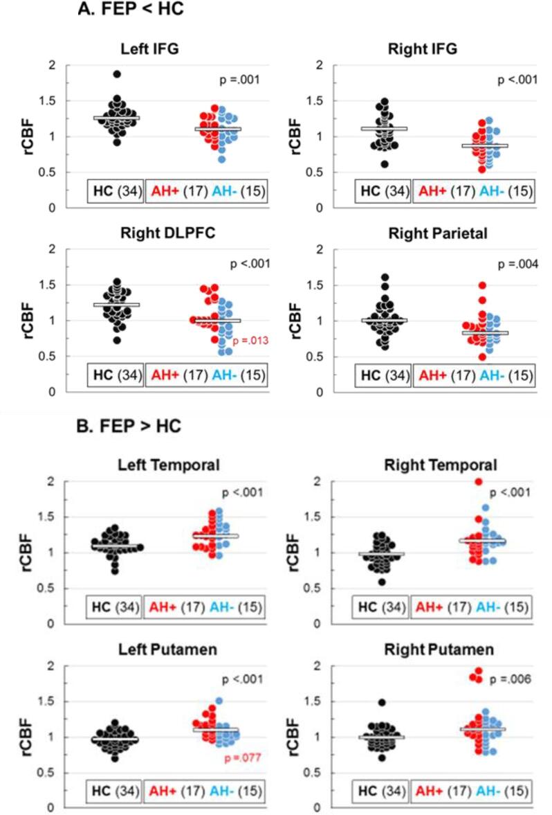
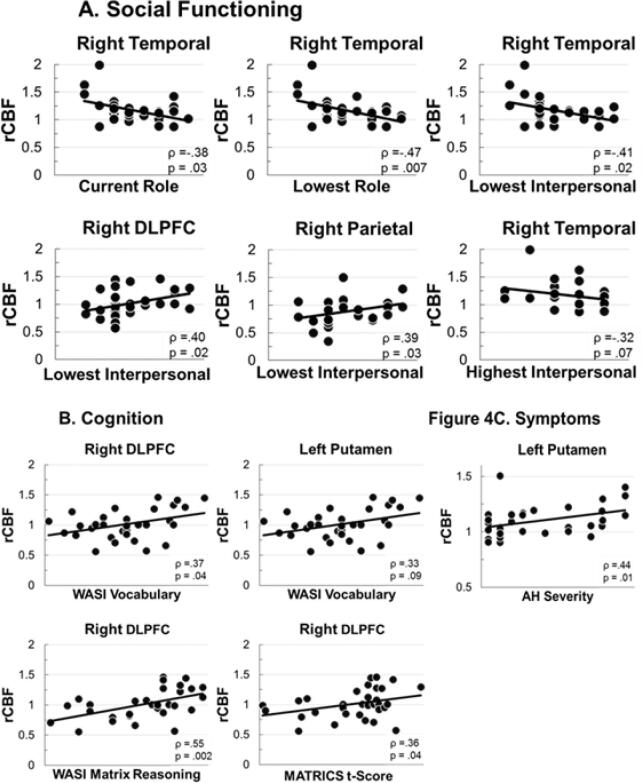
Study results: Whole-brain comparison of resting rCBF identified 8 clusters with significant between-group differences. Reduced rCBF was found in executive control areas in left and right IFG, right DLPFC, and right parietal cortex. Increased rCBF was found in left and right temporal cortex (including Wernicke's area), and left and right putamen. A positive correlation was observed between auditory hallucination severity and rCBF in the left putamen.
Conclusions: To the degree that perfusion implies activation, language and auditory processing areas in bilateral temporal lobe and putamen showed pathological hyper-activity, and cognitive control areas (IFG, DLPFC, right parietal) showed pathological hypo-activity in FEP at rest. Pathological basal activity was present across the range of symptom severity, suggesting it may be a common underlying pathology for psychosis that may be targeted with non-invasive brain stimulation to normalize resting activity levels.
Keywords: Arterial spin labeling; Auditory hallucinations; Basal perfusion; First episode psychosis.
Copyright © 2022 The Author(s). Published by Elsevier Inc. All rights reserved.
Conflict of interest statement
Declaration of Competing Interest The authors declare that they have no known competing financial interests or personal relationships that could have appeared to influence the work reported in this paper.
Figures
Similar articles
-
White Matter Microstructural Abnormalities in the Broca's-Wernicke's-Putamen "Hoffman Hallucination Circuit" and Auditory Transcallosal Fibers in First-Episode Psychosis With Auditory Hallucinations.Schizophr Bull. 2021 Jan 23;47(1):149-159. doi: 10.1093/schbul/sbaa105. Schizophr Bull. 2021. PMID: 32766733 Free PMC article.
-
Resting state auditory-language cortex connectivity is associated with hallucinations in clinical and biological subtypes of psychotic disorders.Neuroimage Clin. 2020;27:102358. doi: 10.1016/j.nicl.2020.102358. Epub 2020 Jul 22. Neuroimage Clin. 2020. PMID: 32745995 Free PMC article.
-
Cerebral blood flow and fMRI BOLD auditory language activation in temporal lobe epilepsy.Epilepsia. 2012 Apr;53(4):631-8. doi: 10.1111/j.1528-1167.2012.03403.x. Epub 2012 Feb 14. Epilepsia. 2012. PMID: 22332720 Free PMC article.
-
Precuneus and insular hypoactivation during cognitive processing in first-episode psychosis: Systematic review and meta-analysis of fMRI studies.Rev Psiquiatr Salud Ment (Engl Ed). 2020 Sep 25:S1888-9891(20)30100-2. doi: 10.1016/j.rpsm.2020.08.001. Online ahead of print. Rev Psiquiatr Salud Ment (Engl Ed). 2020. PMID: 32988773 Review. English, Spanish.
-
How Extended Is Wernicke's Area? Meta-Analytic Connectivity Study of BA20 and Integrative Proposal.Neurosci J. 2016;2016:4962562. doi: 10.1155/2016/4962562. Epub 2016 Feb 23. Neurosci J. 2016. PMID: 27006905 Free PMC article. Review.
Cited by
-
A whole-brain neuromark resting-state fMRI analysis of first-episode and early psychosis: Evidence of aberrant cortical-subcortical-cerebellar functional circuitry.Neuroimage Clin. 2024;41:103584. doi: 10.1016/j.nicl.2024.103584. Epub 2024 Feb 28. Neuroimage Clin. 2024. PMID: 38422833 Free PMC article.
-
Blood flow perfusion in visual pathway detected by arterial spin labeling magnetic resonance imaging for differential diagnosis of ocular ischemic syndrome.Front Neurosci. 2023 Feb 13;17:1121490. doi: 10.3389/fnins.2023.1121490. eCollection 2023. Front Neurosci. 2023. PMID: 36860621 Free PMC article.
-
Functional and structural connectivity correlates of semantic verbal fluency deficits in first-episode psychosis.J Psychiatr Res. 2024 Jan;169:73-80. doi: 10.1016/j.jpsychires.2023.11.032. Epub 2023 Nov 20. J Psychiatr Res. 2024. PMID: 38000187 Free PMC article.
References
-
- Addington J., Liu L., Buchy L., Cadenhead K.S., Cannon T.D., Cornblatt B.A., Perkins D.O., Seidman L.J., Tsuang M.T., Walker E.F., Woods S.W., Bearden C.E., Mathalon D.H., McGlashan T.H. North American Prodrome Longitudinal Study (NAPLS 2) The Journal of Nervous and Mental Disease. 2015;203:328–335. - PMC - PubMed
-
- Alsop D.C., Detre J.A., Golay X., Günther M., Hendrikse J., Hernandez-Garcia L., Lu H., MacIntosh B.J., Parkes L.M., Smits M., van Osch M.J., Wang D.J., Wong E.C., Zaharchuk G. Recommended implementation of arterial spin-labeled perfusion MRI for clinical applications: A consensus of the ISMRM perfusion study group and the European consortium for ASL in dementia. Magnetic Resonance Medicine. 2015;73:102–116. - PMC - PubMed
-
- Alsop D.C., Detre J.A., Golay X., Gunther M., Hendrikse J., Hernandez-Garcia L., Lu H., MacIntosh B.J., Parkes L.M., Smits M., van Osch M.J., Wang D.J., Wong E.C., Zaharchuk G. Recommended implementation of arterial spin-labeled perfusion MRI for clinical applications: A consensus of the ISMRM perfusion study group and the European consortium for ASL in dementia. Magnetic Resonance Medicine. 2015;73:102–116. - PMC - PubMed
-
- Barch D.M., Csernansky J.G. Abnormal parietal cortex activation during working memory in schizophrenia: verbal phonological coding disturbances versus domain-general executive dysfunction. American Journal of Psychiatry. 2007;164:1090–1098. - PubMed
-
- Cannon T.D., Glahn D.C., Kim J., Van Erp T.G., Karlsgodt K., Cohen M.S., Nuechterlein K.H., Bava S., Shirinyan D. Dorsolateral prefrontal cortex activity during maintenance and manipulation of information in working memory in patients with schizophrenia. Archives of General Psychiatry. 2005;62:1071–1080. - PubMed
MeSH terms
Grants and funding
LinkOut - more resources
Full Text Sources
Medical
