Case Reports
. 2022 Nov 25;101(47):e31629.
doi: 10.1097/MD.0000000000031629.

Acquired uterine arteriovenous malformation in a patient with cornual pregnancy: A case report

Affiliations
Case Reports

Acquired uterine arteriovenous malformation in a patient with cornual pregnancy: A case report

Yi Yan et al. Medicine (Baltimore). .

Abstract

Introduction: Acquired uterine arteriovenous malformation (uAVM) is a rare disease and could occur after dilation and curettage, cesarean section, or neoplastic processes.

Patient concerns: A 29-year-old female presented with acute right lower abdominal pain and positive beta human chorionic gonadotropin (β-hCG).

Diagnosis: A 6 cm ectopic right cornual pregnancy was found on ultrasound examination.

Interventions: She underwent a laparoscopic resection of the cornual ectopic pregnancy. She returned with extensive vaginal bleeding 6-month post surgery, and eventually diagnosed with arteriovenous malformation at the previous surgical site by Color Dopplor endovaginal ultrasound. Percutaneous transcatheter uterine artery embolization (UAE) was attempted, however, vaginal bleeding continued. She was taken to the operation room for a hysteroscopic ablation of uAVM.

Outcomes: Complete cessation of the bleeding was achieved without hysterectomy.

Conclusion: We report an extremely unusual case of acquired uAVM after a wedge resection of cornual pregnancy. Ultrasound evaluation of patients with post-operative persistent bleeding should be considered for evaluation of a possible arteriovenous malformation.


Conflict of interest statement

The authors have no conflicts of interest to disclose.

Figures

Figure 1.
Figure 1.
Ultrasound examination showed a live right cornual pregnancy. (A, B) Endometrial cavity is empty. An approximately 5 cm gestational sac is seen in the right cornual region as indicated with arrow. (C, D) A yolk sac seen and a fetal pole with a slow fetal heart rate of 60 beats per minute. Crown-rump length is 3.9 mm of estimated age 6 wks 1 d. Interstitial ectopic is implanted within the cornua surrounded by a thin myometrium (Arrow head).
Figure 2.
Figure 2.
Intra-operative and pathological findings of cornual pregnancy. (A, B) Partial resection of the right uterine cornus was performed. (C, D) Photomicrograph with Haematoxylin-Eosin (H&E) stain shows villous tissue (arrow) invading myometrium. Around the implantation site, hemorrhage is also noted (H&E stain, ~20×).
Figure 3.
Figure 3.
Ultrasound examination demonstrated an arteriovenous malformation (uAVM) at surgical bed. (A, B) There is a large cluster of vessels centered in the myometrium at the right fundal aspect of the uterus at the site of a previously cornual ectopic pregnancy. This occupies a volume of approximately 2.1 × 2.1 × 2.3 cm. (C, D) Pulse Doppler demonstrated a low resistance arterial as well as venous flow. An uAVM is suspected at the right fundal aspect of the uterus that site of previous cornual pregnancy.
Figure 4.
Figure 4.
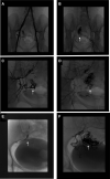
Conventional angiographic findings of uAVM. (A, B) The lower abdominal angiogram was performed followed by angiograms of both common iliac and internal iliac arteries. (C, D) Selective angiograms on the branches of the right internal iliac artery were performed followed by the left uterine artery. The malformation is demonstrated in the right upper quadrant of the uterus supplied by a large right uterine artery. In the region of the origin of the right uterine artery, the other branches of the internal iliac artery also originated. (E) There were significant difficulties in accessing the ostium of the right uterine artery. Coil embolization of at least 2 adjacent branches were performed to encourage cannulation of the right uterine artery. (F) The post-embolization arteriogram showed persistent contrast in uAVM denoting failure to occlude. uAVM = uterine arteriovenous malformation.
Figure 5.
Figure 5.
Emergency hysteroscopy with ablation of the arteriovenous malformation (A, B) Hystroscopic images of right uterine vessel showed a varicose-like structure, starting from the level of the internal OS reaching to the uterine cornu with significant pulsations. The vascular structure is likely corresponding the arteriovenous malformation that was described on imaging. Careful securing the malformation with suturing was performed and the extracorporeal knot tying technique allowed enough pressure to stop the blood supply into the abnormality. (C, D) The right uterine cornu had a bleeder that was giving arterial blood in spurts. A flat cylinder was used for coagulation with current of 80 watt. The bleeder was coagulated.

References

    1. Moawad NS, Mahajan ST, Moniz MH, et al. . Current diagnosis and treatment of interstitial pregnancy. Am J Obstet Gynecol. 2010;202:15–29. - PubMed
    1. Lau S, Tulandi T. Conservative medical and surgical management of interstitial ectopic pregnancy. Fertil Steril. 1999;72:207–15. - PubMed
    1. Grivell RM, Reid KM, Mellor A. Uterine arteriovenous malformations: a review of the current literature. Obstet Gynecol Surv. 2005;60:761–7. - PubMed
    1. Takeda A, Koyama K, Imoto S, et al. . Progressive formation of uterine arteriovenous fistula after laparoscopic-assisted myomectomy. Arch Gynecol Obstet. 2009;280:663–7. - PubMed
    1. Timor-Tritsch IE, Haynes MC, Monteagudo A, et al. . Ultrasound diagnosis and management of acquired uterine enhanced myometrial vascularity/arteriovenous malformations. Am J Obstet Gynecol. 2016;214:731 e1–e10. - PubMed

Publication types