Review
. 2022 Nov 14:16:1041853.
doi: 10.3389/fncel.2022.1041853. eCollection 2022.

Presentation and integration of multiple signals that modulate oligodendrocyte lineage progression and myelination

Affiliations
Review

Presentation and integration of multiple signals that modulate oligodendrocyte lineage progression and myelination

Christopher D Fekete et al. Front Cell Neurosci. .

Abstract

Myelination is critical for fast saltatory conduction of action potentials. Recent studies have revealed that myelin is not a static structure as previously considered but continues to be made and remodeled throughout adulthood in tune with the network requirement. Synthesis of new myelin requires turning on the switch in oligodendrocytes (OL) to initiate the myelination program that includes synthesis and transport of macromolecules needed for myelin production as well as the metabolic and other cellular functions needed to support this process. A significant amount of information is available regarding the individual intrinsic and extrinsic signals that promote OL commitment, expansion, terminal differentiation, and myelination. However, it is less clear how these signals are made available to OL lineage cells when needed, and how multiple signals are integrated to generate the correct amount of myelin that is needed in a given neural network state. Here we review the pleiotropic effects of some of the extracellular signals that affect myelination and discuss the cellular processes used by the source cells that contribute to the variation in the temporal and spatial availability of the signals, and how the recipient OL lineage cells might integrate the multiple signals presented to them in a manner dialed to the strength of the input.

Keywords: BDNF; Fyn; L1; NG2; OPC; neuron-glia communication; oligodendrocyte; trafficking.


Conflict of interest statement

The authors declare that the research was conducted in the absence of any commercial or financial relationships that could be construed as a potential conflict of interest.

Figures

Figure 1
Figure 1
Lineage progression from an NPC to a myelinating OL and examples of key signaling pathways at each stage. (A): NPC. OPCs are specified from NPCs in the ventricular and subventricular zones. Reduction in NF1 leading to enhanced activation of Ras-Mapk-Erk1/2 pathway shifts the fate of NPCs to an OPC fate and suppresses the neuronal fate. Committed OPCs upregulate Olig2 and Sox10 which in turn turns on Pdgfra transcription. (B): OPC. OPC proliferation is dependent on PDGFRα and is mediated by PI-3K activating Akt and not through Mapk activating Erk1/2. (C): NFOL. In NFOLs, PI-3K activating Akt is primarily responsible for their terminal differentiation, survival, and initiation of myelination, possibly via the transcription factor FoxO1. HDAC1/2 activity, Myrf, and upregulation of Sox10, and Nkx2.2 are also critical for terminal differentiation. (D): mOL. In myelinating OLs (mOLs), activation of the Ras-Erk1/2 pathway is critical for maintaining myelination. NF1, neurofibromin; RTK, receptor tyrosine kinase; Erk1/2-P, phosphorylated Erk1/2; PDGF, platelet-derived growth factor; PDGFRα, platelet-derived growth factor receptor alpha; PI-3K, phosphatidyl inositol-3-kinase; P-Akt, phospho-Akt; BDNF, brain-derived neurotrophic factor; NFOL, newly formed OLs; HDAC1/2, histone deacetylase 1 and 2; Myrf, myelin regulatory factor; NRG, neuregulin.
Figure 2
Figure 2
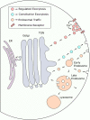
Secreted proteins are trafficked to the cell surface via multiple distinct pathways. After synthesis in the endoplasmic reticulum (ER), proteins destined for exocytosis undergo processing in the Golgi before being sorted into intracellular transport vesicles in the trans-Golgi network (TGN). Transport vesicles leaving the TGN can either be targeted directly to the plasma membrane to undergo regulated or constitutive exocytosis or be transported to the endosomal pathway, either for further sorting prior to exocytosis or to deliver endosome specific cargo. Proteins that are retrieved from the cell surface are targeted to early endosomes (EE) which act as sorting platforms in endosomal traffic to and from the membrane. EEs can mature into late endosomes (LE) which act as a final sorting station for proteins destined for lysosomal degradation. LEs can also mature into multi-vesicular bodies (MVB) which can alternatively fuse with the plasma membrane to facilitate exosome release. Small arrowheads indicate the direction of vesicle traffic.
Figure 3
Figure 3
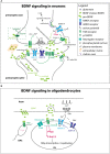
Different ways in which BDNF acts on cells (A). BDNF-TrkB function at the neuronal synapse. BDNF from the target neuron or other cells in the target area mediates the survival and axon growth of the presynaptic neuron. Some of the effect is local, while other effects may be mediated by clathrin-dependent endocytosis of BDNF-TrkB complex that could be transported retrogradely by signaling endosomes, analogous to what has been reported for NGF and its high-affinity receptor TrkA. Mature BDNF can be secreted constitutively or in a regulated manner via SNARE-dependent mechanisms. BDNF may not be immediately available to the receptor if it becomes embedded in the extracellular matrix. While the majority of the effects are mediated by mature BDNF, pro-BDNF can also be secreted and act on p75NTFR to elicit a cell death response. BDNF acting on presynaptic and postsynaptic membranes can promote LTP. At the presynaptic terminal, axon depolarizations increase Ca2+ entry through NMDA receptors, which in turn activates the SNARE-dependent release of BDNF. The neuronal activity also increases the level of TrkB at the presynaptic surface, and TrkB activation increases Ca2+ via PLC-γ activation, leading to enhanced glutamate release from presynaptic vesicles. At the postsynaptic membrane, BDNF acting on TrkB also contributes to LTP by increasing NMDA current. BDNF is secreted in an activity-dependent manner and acts in an autocrine fashion on TrkB on the same spine, which is necessary for LTP. Some of the processes indicated in one compartment also apply to the other compartment. The concept of signaling endosomes has been used to explain the function of endocytosed TrkB-BDNF complex that is transported from the internalized site on the axon back to the soma where it exerts developmental effects (Grimes et al., 1996), similar to what had been reported earlier for TrkA-NGF (Grimes et al., 1996). (B) BDNF-TrkB function at axonand OL interface. Neuronal activity triggers BDNF release and glutamate release. BDNF binds to TrkB on OLs and activates Fyn and Erk1/2, which promotes the transcription of myelin genes and myelination. NRG acting on the ErbB receptor and BDNF activating TrkB both increase Ca2+ permeation through NMDA receptors. Fyn is much less abundant in OPCs than in OLs, even though OPCs express TrkB.
Figure 4
Figure 4
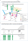
Integral membrane and extracellular matrix affecting myelination via Fyn (A). Axonal L1 forms a complex with contactin-1 on OLs. The complex becomes preferentially localized to lipid rafts, where they complex with and activate Fyn by phosphorylated Y420. Fyn in the non-raft membrane is not active (not phosphorylated on Y420 but phosphorylated on the regulatory Y531 site). In addition to the membrane-bound form, cleaved L1 also interacts with contactin-1. In addition to L1, the extracellular molecules laminin-2 and Aβ oligomer also activate Fyn on OLs. Activated Fyn also promotes TrkB function. (B) The level of L1 expression on the surface of the axon is regulated by the frequency of action potential firing. High-frequency firing increases myelination, and this effect in some cases is mediated by cAMP.

References

    1. Adams K. L., Dahl K. D., Gallo V., Macklin W. B. (2021). Intrinsic and extrinsic regulators of oligodendrocyte progenitor proliferation and differentiation. Semin. Cell Dev. Biol. 116, 16–24. 10.1016/j.semcdb.2020.10.002 - DOI - PMC - PubMed
    1. Agresti C., Meomartini M. E., Amadio S., Ambrosini E., Volonté C., Aloisi F., et al. . (2005). ATP regulates oligodendrocyte progenitor migration, proliferation and differentiation: involvement of metabotropic P2 receptors. Brain Res. Brain Res. Rev. 48, 157–165. 10.1016/j.brainresrev.2004.12.005 - DOI - PubMed
    1. Ainger K., Avossa D., Diana A. S., Barry C., Barbarese E., Carson J. H. (1997). Transport and localization elements in myelin basic protein mRNA. J. Cell Biol. 138, 1077–1087. 10.1083/jcb.138.5.1077 - DOI - PMC - PubMed
    1. Akiyama M., Hasegawa H., Hongu T., Frohman M. A., Harada A., Sakagami H., et al. . (2014). Trans-regulation of oligodendrocyte myelination by neurons through small GTPase Arf6-regulated secretion of fibroblast growth factor-2. Nat. Commun. 5:4744. 10.1038/ncomms5744 - DOI - PubMed
    1. Balkowiec A., Katz D. M. (2002). Cellular mechanisms regulating activity-dependent release of native brain-derived neurotrophic factor from hippocampal neurons. J. Neurosci. 22, 10399–10407. 10.1523/JNEUROSCI.22-23-10399.2002 - DOI - PMC - PubMed