. 2022 Nov 14:13:1035729.
doi: 10.3389/fimmu.2022.1035729. eCollection 2022.

Immunomodulatory effects of two recombinant arginine kinases in Sarcoptes Scabiei on host peripheral blood mononuclear cells

Affiliations

Immunomodulatory effects of two recombinant arginine kinases in Sarcoptes Scabiei on host peripheral blood mononuclear cells

Yanting Xu et al. Front Immunol. .

Abstract

Background: As an important zoonotic parasitic disease with global distribution, scabies causes serious public health and economic problems. Arginine kinase (AK) is involved in cell signal transduction, inflammation, and apoptosis. Two AKs were identified in Sarcoptes scabiei, but their functions in the host immune response remain unclear.

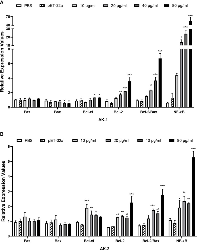
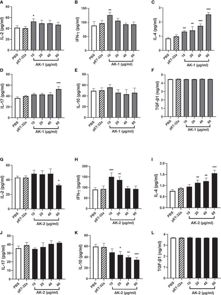
Methods: rSsAK-1 and rSsAK-2 were expressed, purified, and immunolocalized. The effects of rSsAK-1 and rSsAK-2 on rabbit PBMC proliferation, apoptosis, and migration; Bcl-2, Bcl-xl, Fas, Bax, and NF-κB transcription levels; and IL-2, IFN-γ, IL-4, IL-10, TGF-β1, and IL-17 secretion were detected.

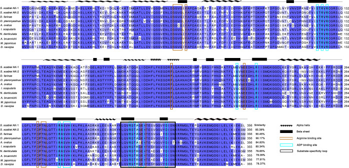
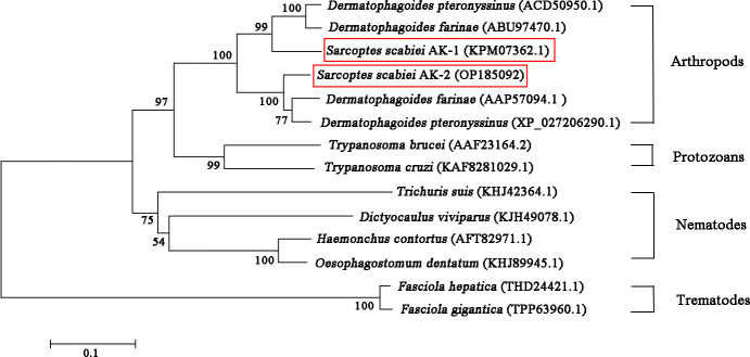
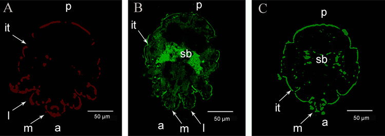
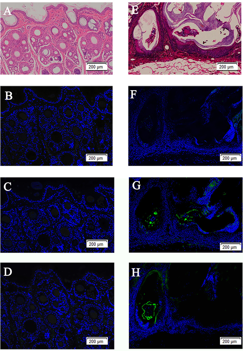
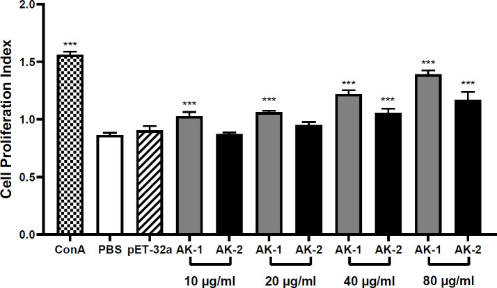
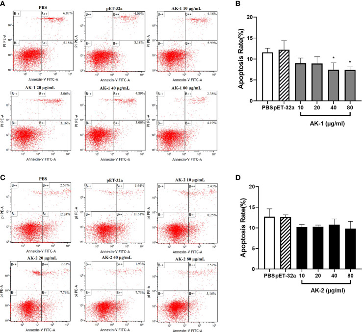
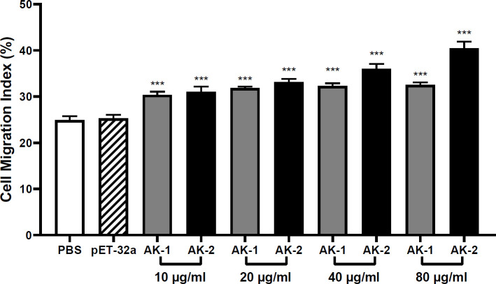
Results: rSsAK-1 and rSsAK-2 were cloned and expressed successfully. Both enzymes were ~57 kDa and contained 17-kDa tagged proteins, and had good catalytic activity and immunoreactivity. The proteins were located in the S. scabiei exoskeleton, chewing mouthparts, legs, stomach, and intestine. SsAK-1 and SsAK-2 were secreted in the pool and epidermis of the skin lesions, which may be involved in S. scabiei-host interaction. rSsAK-1 and rSsAK-2 significantly promoted cell proliferation, induced cell migration, inhibited apoptosis, and increased Bcl-2, Bcl-xl and NF-κB (p65) transcription levels concentration-dependently, and inhibited IL-2, IFN-γ, and IL-10 secretion and promoted IL-4 and IL-17 secretion.

Conclusion: rSsAK-1 and rSsAK-2 might increase Bcl-2 and Bcl-xl expression by activating the NF-κB signaling pathway to promote cell proliferation and inhibit apoptosis, which induced PBMC survival. By inducing PBMC migration to the infection site, rSsAK-1 and rSsAK-2 shifted the Th1/Th2 balance toward Th2 and changed the Th17/Treg balance, which indicated their immune role in S. scabiei allergic inflammation.

Keywords: NF-κB; PBMC; Sarcoptes scabiei; Th1/Th2; arginine kinase; inflammatory.


Conflict of interest statement

The authors declare that they do not have any commercial or associative interest that represents a conflict of interest in connection with the work submitted.

Figures

Figure 1
Figure 1
Multiple sequence alignment analysis of SsAK-1 and SsAK-2. The homology of the SsAKs to other species is shown on the right. Blue background indicates amino acids that are identical to the AKs of other species; a darker color indicates a higher consistency. Blue-green box highlights the ADP binding site. Brown box highlights the arginine binding site. The black box indicates the substrate specificity loop. The predicted secondary structure of the SsAKs is shown above the amino acid residue alignments. The AK gene accession numbers of each species are as follows: SsAK-1 (KPM07362.1), sAK-2 (OP185092), D. farina (AAP57094.1), D. pteronyssinus (ACD50950.1), A. ovatus (ABU97463.1), I. scapularis (XP_029845984.1), Neocaridina denticulate (BAH56609.1), Argiope bruennichi (KAF8790721.1), Macrophthalmus japonicus (AID47194.1), Drosophila navojoa (XP_030246642.1).
Figure 2
Figure 2
Phylogenetic relationships between SsAKs and other species. The AK amino acid sequences of 11 species were selected and the phylogenetic tree was constructed by NJ. The AK sequence accession numbers of each species are as follows: SsAK-1 (KPM07362.1), SsAK-2 (OP185092), D. farina (ABU97470.1), D. pteronyssinus (ACD50950.1), D. pteronyssinus (XP 027206290.1), D. farina (AAP57094.1), H. contortus (AFT82971.1), Trichuris suis (KHJ42364.1), Dictyocaulus viviparus (KJH49078.1), Oesophagostomum dentatum (KHJ89945.1), Fasciola hepatica (THD24421.1), F. gigantica (TPP63960.1), T. brucei (AAF23164.2), T. cruzi (KAF8281029.1).
Figure 3
Figure 3
Expression, purification and western blotting analysis of the rSsAKs. (A) rSsAKs expressed in E. coli BL21 (DE3) after induction. M: Protein molecular weight marker, lane 1: rSsAK-1; lane 2: rSsAK-2. (B) Purified rSsAKs. M: Protein molecular weight marker, lane 1: rSsAK-1; lane 2: rSsAK-2. (C) Western blotting analysis of the rSsAKs. M: Protein molecular weight marker, lanes 1-2: purified rSsAK-1 detected by rat hyperimmune serum and rat negative serum, respectively; lane 3-4: purified rSsAK-1 detected by serum from rabbits naturally infected with S. scabiei and uninfected rabbits, respectively; lane 5-6: purified rSsAK-2 detected by rat hyperimmune serum and rat negative serum, respectively; lane 7-8: purified rSsAK-2 detected by serum from rabbits naturally infected with S. scabiei and uninfected rabbits, respectively; lane 9-10: rSsAK-1from total bacterial extract detected by rat hyperimmune serum and serum from rabbits naturally infected with S. scabiei, respectively; lane 11-12: rSsAK-2from total bacterial extract detected by rat hyperimmune serum and serum from rabbits naturally infected with S. scabiei, respectively. Red arrows indicate the band of interest, i.e., the rSsAKs.
Figure 4
Figure 4
Immunolocalization of the SsAKs in S. scabiei. (A) Incubation with rat negative serum as the primary antibody. (B) Incubation with rat anti-rSsAK-1 IgG as the primary antibody. (C). Incubation with rat anti-rSsAK-2 IgG as the primary antibody. a, front end; p, rear end; m, mouthparts; l, legs; it, integument of the exoskeleton; sb, stomach and intestine.
Figure 5
Figure 5
H&E staining of skin and immunolocalization of the SsAKs in the skin. H&E staining of non-skin lesions (A) and skin lesions (E) on the toes of rabbits with S. scabiei infection. The non-skin lesions (B) and skin lesions (F) on the toes of rabbits with S. scabiei incubated with rat negative serum. The non-lesion area (C) and skin lesion area (G) of the toes of rabbits with S. scabiei infection incubated with rat anti-rSsAK-1 IgG. The non-lesion area (D) and skin lesion area (H) of the toes of rabbits with S. scabiei infection incubated with rat anti-rSsAK-2 IgG. Green fluorescence: SsAK proteins.
Figure 6
Figure 6
The effects of the rSsAKs on PBMC proliferation. CCK-8 detection of the effects of the rSsAKs on PBMC proliferation. The cell proliferation index was the optical density at 450 nm (OD450). Data are the mean ± SD of three replicates per group as compared to the PBS group. ***P < 0.001.
Figure 7
Figure 7
The effects of the rSsAKs on PBMC apoptosis. Annexin V/PI staining and flow cytometry detection of the effects of the rSsAKs on PBMC apoptosis. (A, C) Annexin V/PI staining and flow cytometry were used to detect the effects of rSsAK-1 (A) and rSsAK-2 (C) on PBMC apoptosis. (B, D) Changes in the total apoptosis rate of PBMCs stimulated by rSsAK-1 (B) and rSsAK-2 (D). The total apoptosis rate was the sum of the early and late apoptosis rates. Data are the mean ± SD of three replicates per group as compared to the PBS group. *P < 0.05.
Figure 8
Figure 8
qRT-PCR results of the effects of the rSsAKs on pro-apoptotic genes, anti-apoptotic genes, and NF-κB (p65). (A, B) The effect of rSsAK-1 (A) and rSsAK-2 (B) on the proapoptotic genes (Fas, Bax), anti-apoptotic genes (Bcl-2, Bcl-xl), and NF-κB (p65) transcription levels. Data are the mean ± SD of three replicates per group as compared to the PBS group. *P < 0.05, **P < 0.01, ***P < 0.001.
Figure 9
Figure 9
The effects of the rSsAKs on PBMC migration. The PBMC migration was counted using a Neubauer counting chamber. The migration ratio = total number of cells in the lower chamber after migration/total number of cells before migration. Data are the mean ± SD of three replicates per group as compared to the PBS group. ***P < 0.001.
Figure 10
Figure 10
ELISA results of the effects of the rSsAKs on Th1, Th2, Th17, and Treg cytokine secretion levels. (A-F) The effect of rSsAK-1 on Th1 (IL-2, IFN-γ), Th2 (IL-4), Th17 (IL-17), and Treg cytokine (IL-10, TGF-β1) secretion levels. (G-L) The effect of rSsAK-2 on Th1 (IL-2, IFN-γ), Th2 (IL-4), Th17 (IL-17), and Treg cytokine (IL-10, TGF-β1) secretion levels. Data are the mean ± SD of three replicates per group as compared to the PBS group. *P < 0.05, **P < 0.01, ***P < 0.001.

Similar articles

Cited by

References

    1. Escobar LE, Carver S, Cross PC, Rossi L, Almberg ES, Yabsley MJ, et al. . Sarcoptic mange: An emerging panzootic in wildlife. J Transboundary Emerging Dis (2021) 00:1–16. doi: 10.1111/tbed.14082 - DOI - PubMed
    1. Arlian LG, Ndate F, Morgan MS. In vivo evidence that Sarcoptes scabiei (Acari: Sarcoptidae) is the source of molecules that modulate splenic gene expression. J Med Entomol (2007) 6):1054. doi: 10.1603/0022-2585(2007)44[1054:ivetss]2.0.co;2 - DOI - PubMed
    1. Shen N, Wei W, Chen Y, Ren Y, Xiong L, Tao Y, et al. . An antibody persistent and protective two rSsCLP-based subunit cocktail vaccine against Sarcoptes scabiei in a rabbit model. Vaccines (Basel) (2020) 8(1):129. doi: 10.3390/vaccines8010129 - DOI - PMC - PubMed
    1. Bernigaud C, Samarawickrama GR, Jones MK, Robin B. Gasser, katja fischer. the challenge of developing a single-dose treatment for scabies. Trends Parasitol (2019) 35(11):931–43. doi: 10.1016/j.pt.2019.08.002 - DOI - PubMed
    1. Bhat SA, Mounsey KE, Liu X, Walton SF. Host immune responses to the itch mite, Sarcoptes scabiei, in humans. Parasit Vectors (2017) 10(1):385. doi: 10.1186/s13071-017-2320-4 - DOI - PMC - PubMed

Publication types