. 2022 Nov 7;12(18):7699-7716.
doi: 10.7150/thno.77102. eCollection 2022.

SOX4 promotes beige adipocyte-mediated adaptive thermogenesis by facilitating PRDM16-PPARγ complex

Affiliations

SOX4 promotes beige adipocyte-mediated adaptive thermogenesis by facilitating PRDM16-PPARγ complex

Huanming Shen et al. Theranostics. .

Abstract

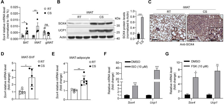
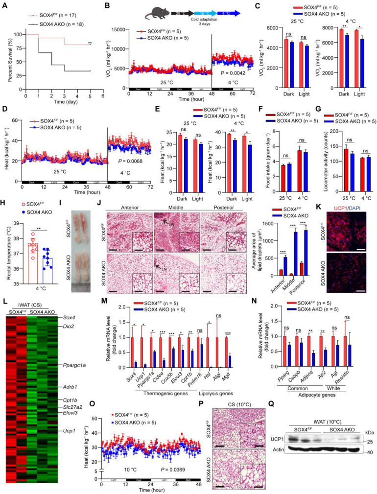
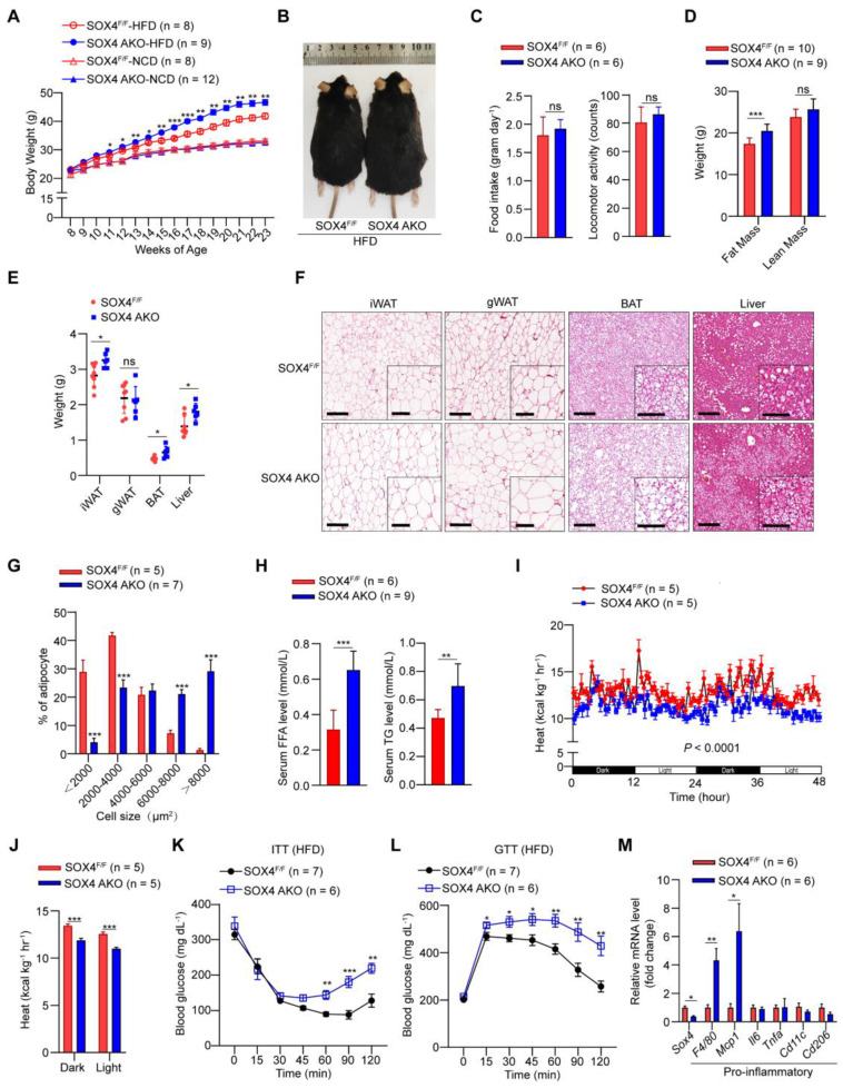
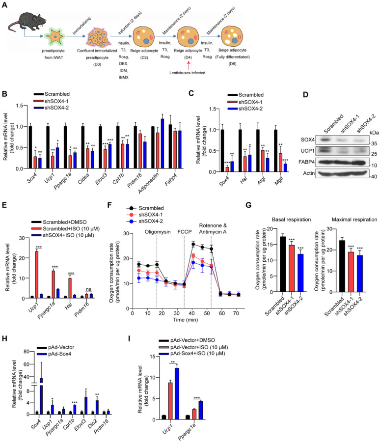
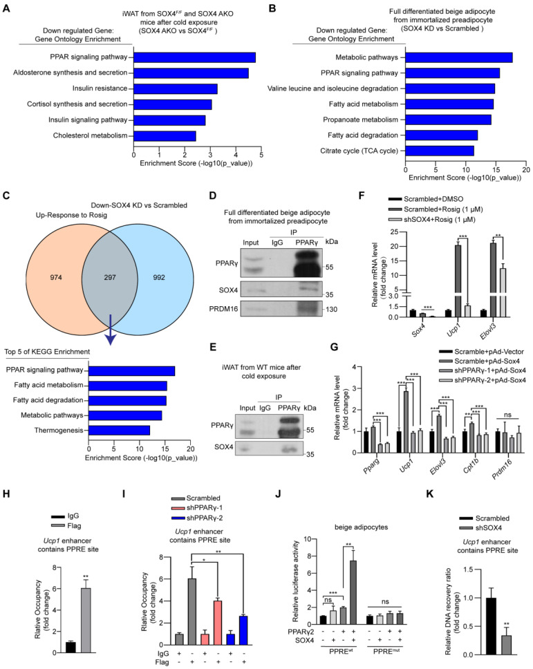
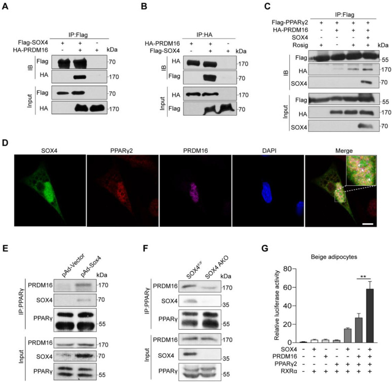
Brown and beige fat protect against cold environments and obesity by catabolizing stored energy to generate heat. This process is achieved by controlling thermogenesis-related gene expression and the development of brown/beige fat through the induction of transcription factors, most notably PPARγ. However, the cofactors that induce the expression of thermogenic genes with PPARγ are still not well understood. In this study, we explored the role of SOX4 in adaptive thermogenesis and its relationship with PPARγ. Methods: Whole transcriptome deep sequencing (RNA-seq) analysis of inguinal subcutaneous white adipose tissue (iWAT) after cold stimulation was performed to identify genes with differential expression in mice. Indirect calorimetry detected oxygen consumption rate and heat generation. mRNA levels were analyzed by qPCR assays. Proteins were detected by immunoblotting and immunofluorescence. Interaction of proteins was detected by endogenous and exogenous Co-IP. ChIP-qPCR, FAIRE assay and luciferase reporter assays were used to investigate transcriptional regulation. Results: SOX4 was identified as the main transcriptional effector of thermogenesis. Mice with either adipocyte-specific or UCP1+ cells deletion of SOX4 exhibited significant cold intolerance, decreased energy expenditure, and beige adipocyte formation, which was attributed to decreased thermogenic gene expression. In addition, these mice developed obesity on a high-fat diet, with severe hepatic steatosis, insulin resistance, and inflammation. At the cell level, loss of SOX4 from preadipocytes inhibited the development of beige adipocytes, and loss of SOX4 from mature beige adipocytes reduced the expression of thermogenesis-related genes and energy metabolism. Mechanistically, SOX4 stimulated the transcriptional activity of Ucp1 by binding to PPARγ and activating its transcriptional function. These actions of SOX4 were, at least partly, mediated by recruiting PRDM16 to PPARγ, thus forming a transcriptional complex to elevate the expression of thermogenic genes. Conclusion: SOX4, as a coactivator of PPARγ, drives the thermogenic gene expression program and thermogenesis of beige fat, promoting energy expenditure. It has important physiological significance in resisting cold and obesity.

Keywords: PPARγ; SOX4.; beige fat; obesity; thermogenesis.


Conflict of interest statement

Competing Interests: The authors have declared that no competing interest exists.

Figures

Figure 1
Figure 1
Cold stimulation induces SOX4 expression. 10-week male mice were housed at room temperature (RT) or exposed to cold exposure (cold stimulation, CS, 10 °C for one day and then 4 °C for 1 week). BAT, iWAT and gWAT were isolated and subjected to qPCR analysis (A) and Western blotting (B). The protein levels of SOX4 were quantified with image J (B, right). (C) Representative images of SOX4 immunohistochemistry (IHC) of iWATs. Scale bar, 100 µm. Insets show higher magnification, scale bar, 50 µm. (D) qPCR analysis of Sox4 mRNA expression in the SVF cells isolated from iWAT. (E) qPCR analysis of Sox4 mRNA expression in the adipocytes isolated from iWAT. (F) SVF cells isolated from iWAT were differentiated into beige adipocytes in vitro as described in method. On day 6, the differentiated beige adipocytes were treated with ISO (isoproterenol) for 4 hr, and then analyzed by qPCR. (G) Beige adipocytes differentiated from iWAT SVF cells were treated with FSK (forskolin). 4 hr later, cells were harvested for qPCR analysis.
Figure 2
Figure 2
Adipose tissue-specific SOX4 KO reduces cold tolerance, energy metabolism, and thermogenic function of beige adipocytes. (A) SOX4F/F and SOX4 AKO male mice (10-12-week) were exposed to 10 °C for one day and then to 4 °C for 5 days. Survival curves were analyzed. (B-G) 10-week male mice were exposed to 25 °C for 3 days, then 10 °C for 3 days and 4 °C for 3 days. Whole-body oxygen consumption (B, C), heat production (D, E), food intake (F) and locomotor activity (G) of mice at 25 °C and at 4 °C were analyzed. (H) SOX4F/F and SOX4 AKO male mice (10-week) were exposed to 10 °C for 3 days and to 4 °C for 3 days. The core body temperature was shown. (I-J) Representative image and H&E staining in the iWAT in (B, D) mice. Arrowhead indicated the lymph node (LN). Scale bar, 200 µm. Insets show higher magnification, scale bar, 100 µm. The size of lipid droplets was quantified with image J (J, right). (K) Immunofluorescent staining of UCP1 in the middle region of iWAT in (B, D) mice. Scale bar, 50 µm. (L-N) SOX4F/F and SOX4 AKO male mice (10-week) were exposed to 10 °C for 3 days and then to 4 °C for 3 days. iWAT was collected from each mouse, and total RNA was extracted and subjected to RNA-Seq analysis and qPCR analysis. (L) Heatmap of the RNA-Seq shows the down-regulated genes (AKO vs control) in iWAT with a cutoff of fold change ≥ 1.5 and p-value < 0.05. Thermogenic genes are indicated. (M-N) qPCR analyzed the relative mRNA levels of indicated genes in the iWAT. (O) SOX4F/F and SOX4 AKO mice were surgically removed BAT. After recovery, mice were exposed to 10 °C for 3 days. The whole-body heat production is shown. (P) H&E staining in the iWAT in (O) mice. Scale bar, 100 µm. Insets show higher magnification, scale bar, 50 µm. (Q) Western blotting showing the expression of UCP1 protein in the iWAT in (O) mice.
Figure 3
Figure 3
Adipose-specific SOX4 KO promotes high fat diet-induced obesity. (A) Growth curve of SOX4 AKO mice and control littermates fed with NCD or HFD. (B) A representative photo of control and SOX4 AKO mice after 15-weeks of HFD feeding. (C) Food intake and locomotor activity of mice in (A) after 15-weeks of HFD feeding. (D) The average fat and lean masses of control and SOX4 AKO mice after 15-weeks of HFD feeding. (E) Weights of iWAT, gWAT, BAT, and liver in control and SOX4 AKO mice after 15-weeks of HFD feeding. (F) Representative H&E staining of iWAT, gWAT, BAT, and liver from control and SOX4 AKO mice after 15-weeks of HFD feeding. Scale bar, 200 µm. Insets show higher magnification, scale bar, 100 µm. (G) Adipocyte sizes of iWAT were estimated from the H&E staining results in (F) using ImageJ. The percentage of cells with the indicated sizes was shown. (H) Serum levels of free fatty acid (FFA), triacylglycerol (TG) in control and SOX4 AKO mice after 15-weeks of HFD feeding. (I, J) Heat production of control and SOX4 AKO mice after 15-weeks of HFD. (K, L) insulin tolerance test (K) (i.p. 1.0 U/kg) and Glucose tolerance test (L) (i.p. 1.5 g/kg) of control and SOX4 AKO mice after 15-weeks of HFD feeding. (M) qPCR analysis of mRNA expression of pro-inflammatory genes in the iWAT of control and SOX4 AKO mice after 15-weeks of HFD feeding.
Figure 4
Figure 4
SOX4 regulates the thermogenic function of mature beige adipocyte. (A-D) Schematic illustration of differentiation of beige adipocytes in vitro (A). On day 4, cells were infected with lentivirus expressing Scrambled or shSox4. On day 6, cells were collected for qPCR (B-C) and Western blot (D) analyses. (E) Scrambled or shSox4 beige adipocytes (day 6) treated with or without 10 μM isoproterenol (ISO) for 4 hr. The relative mRNA levels of indicated genes were shown. (F, G) Oxygen consumption of Scrambled or shSox4 beige adipocytes (day 6) were analyzed. (H) Immortalized preadipocytes were differentiated and infected with Vector or Sox4-expression adenovirus at day 4. On day 6, cells were harvest for qPCR analyses. The relative mRNA levels of indicated genes were shown. (I) Immortalized preadipocyte were differentiated and infected with Vector or Sox4-expression adenovirus at day 4. On day 6, beige adipocytes were treated with or without 10 μM ISO for 4 hr. Real-time qPCR analysis of the indicated genes was performed.
Figure 5
Figure 5
SOX4 interacts with PPARγ2 and regulates the transcriptional activation of thermogenic genes. (A) Genes down-regulated (SOX4 AKO vs. SOX4F/F mice) in RNA-seq data from Figure 2L were subjected to gene ontology analysis. (B-C) Immortalized preadipocytes were subjected to beige adipocyte differentiation in vitro. On day 4, cells were infected with lentivirus expressing Scramble or shSox4. On day 6, cells were collected and total RNA was extracted for RNA-Seq analysis. The genes down-regulated by SOX4 knockdown were subjected to gene ontology analysis (B). Overlap (297) of genes down-regulated (992) by SOX4 knockdown in (B) and genes up-regulated (974) in Rosig-treated classic BAT (C, top, GSE144490). The overlapping genes were subjected to gene ontology analysis (C, bottom). (D-E) Beige adipocytes differentiated from immortalized preadipocyte (D) and iWATs of C57BL/6 mice exposed to 10 ℃ for 1 day and 4 ℃ for 1 week (E) were lysed and subjected to immunoprecipitation using IgG or anti-PPARγ antibody. Input and pellet fractions were analyzed by western blot using indicated antibodies. (F) Beige adipocytes differentiated from immortalized preadipocytes were infected with scrambled or shSOX4 lentiviruses on day 4. On day 6, cells were treated with or without rosig (1 μM) for 5 hr. qPCR analysis of the indicated genes were shown. The results are from 3 independent experiments. (G) Differentiated beige adipocytes were infected with scrambled or shPPARγ lentiviruses on day 4. On day 6, cells were infected with vector or SOX4-expression adenovirus (pAd-Sox4) treatment. 24 hr later, cells were harvested and subjected to qPCR analysis. The relative mRNA levels of indicated genes were shown. (H) Differentiated beige adipocytes were transfected with Flag-Sox4 on day 6. 2 days later, cells were harvested for ChIP analysis by using IgG or Flag antibody. The ChIPed DNAs were examined by qPCR for Ucp1 enhancer containing PPRE site. (I) Scramble and shPPARγ beige adipocytes (day 6) were transfected with Flag-Sox4 and 2 days later subjected to ChIP assay by using IgG or anti-Flag antibody. The ChIPed DNAs were examined by qPCR for Ucp1 enhancer containing PPRE site. (J) The Ucp1 enhancer containing PPRE site or mutated PPRE site was cloned into pGL4.26-basic vector and co-transfected into mature beige adipocytes together with β-gal in the presence or absence of PPARγ2 or SOX4 expression plasmid. After 48 hr, cells were harvested and the luciferase activity was measured. β-gal activity was used to normalize for transfection efficiency. (K) Differentiated beige adipocytes were infected with scrambled or shSOX4 lentiviruses and subjected to FAIRE assay 2 days later. The enriched DNAs were examined by qPCR for Ucp1 enhancer containing PPRE site.
Figure 6
Figure 6
SOX4 promotes the binding of PPARγ and PRDM16. (A, B) HEK293T were transfected with Flag-SOX4 and HA-PRDM16 as indicated. 48 hr after transfection, cells were lysed and subjected into immunoprecipitation with anti-Flag (A) or anti-HA (B) antibody followed by Western blotting. (C) HEK293T cells transfected with Flag-PPARγ2, SOX4, and HA-Prdm16 as indicated were treated with or without 1 μM Rosig for 4 hr. Immunoprecipitation were performed as in (A). (D) Immunofluorescence analysis showed SOX4 was colocalized with PPARγ2 and PRDM16 in the nucleus of mature beige adipocyte (D6). Scale bar, 10 µm. (E) Immortalized preadipocyte were differentiated and infected with Vector or Sox4-expression adenovirus at day 4. Mature beige adipocytes (D6) were lysed, subjected into immunoprecipitation with anti-PPARγ antibody and immunoblotted with antibodies as indicated. (F) Control and SOX4 AKO mice were exposed to 10 °C for 3 days and then 4 °C for 3 days. iWAT were isolated and lysed. Immunoprecipitation and immunoblotting were performed as in (E). (G) Fragments of 3 tandem copies of a PPARγ response element fused to a luciferase reporter vector were co-transfected into mature beige adipocytes together with β-gal, PPARγ2, RXRα, and PRDM16 in the presence or absence of SOX4 expression plasmid. Luciferase activity was corrected for corresponding β-gal activity and normalized to control activity.

Similar articles

Cited by

References

    1. Bastías-Pérez M, Zagmutt S, Soler-Vázquez MC, Serra D, Mera P, Herrero L. Impact of adaptive thermogenesis in mice on the treatment of obesity. Cells. 2020;9:316. - PMC - PubMed
    1. Chouchani ET, Kazak L, Spiegelman BM. New advances in adaptive thermogenesis: UCP1 and beyond. Cell Metab. 2019;29:27–37. - PubMed
    1. Harms M, Seale P. Brown and beige fat: development, function and therapeutic potential. Nat Med. 2013;19:1252–63. - PubMed
    1. Wang Y, Li X, Liu C, Zhou L, Shi L, Zhang Z, WTAP regulates postnatal development of brown adipose tissue by stabilizing METTL3 in mice. Life Metabolism. 2022. loac028.
    1. Wang Y, Gao M, Zhu F, Li X, Yang Y, Yan Q. et al. METTL3 is essential for postnatal development of brown adipose tissue and energy expenditure in mice. Nat Commun. 2020;11:1648. - PMC - PubMed

Publication types