

Case Reports
doi: 10.1016/j.case.2022.08.002.
eCollection 2022 Nov.
A 56-Year-Old Man with Mitral Regurgitation and Acute Pulmonary Edema: Rupture of the Papillary Muscle or Infective Endocarditis?
Affiliations
- PMID: 36451867
- PMCID: PMC9703130
- DOI: 10.1016/j.case.2022.08.002
Item in Clipboard
Case Reports
A 56-Year-Old Man with Mitral Regurgitation and Acute Pulmonary Edema: Rupture of the Papillary Muscle or Infective Endocarditis?
CASE (Phila).
.
No abstract available
Keywords: Infective endocarditis; Mitral valve prolapse; Papillary muscle rupture.
Figures


ECG standard leads showing sinus rhythm with Q waves and ST elevations in II, III and aVF. Reciprocal ST depression in aVL.

ECG precordial leads showing sinus rhythm and ST depression in V2-5.

Left coronary arteriogram still frame without occlusions or stenoses.

Subselective right coronary arteriogram still frame.

Two-dimensional TTE apical 3-chamber view systolic frame, showing the posterior mitral leaflet flailing into the left atrium.
Two-dimensional TEE intercommissural view systolic frame showing mitral leaflet tissue billowing into the left atrium resembling a prolapse.

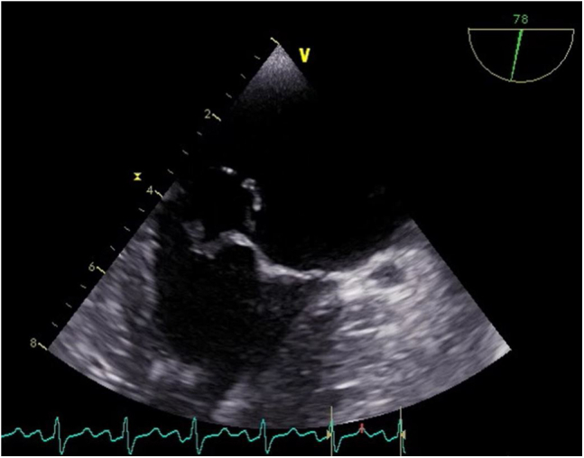
Two-dimensional TEE 3-chamber view systolic frame of the mitral valve with an echogenic mass displaying the left atrium and a possibly thickened posterior mitral leaflet compared to the anterior mitral leaflet, suggestive of IE.

Picture of the removed mitral valve showing a papillary muscle entangled in its chordae tendineae. The ruptured surface is colored in pink.
References
-
- Otto C.M., Nishimura R.A., Bonow R.O., Carabello B.A., Erwin J.P., 3rd, Gentile F., et al. 2020 ACC/AHA guideline for the management of patients with valvular heart disease: executive summary: a report of the American College of Cardiology/American Heart Association Joint Committee on clinical practice guidelines. Circulation. 2021;143:e35–e71. - PubMed
-
- Figueras J., Alcalde O., Barrabés J.A., Serra V., Alguersuari J., Cortadellas J., et al. Changes in hospital mortality rates in 425 patients with acute ST-elevation myocardial infarction and cardiac rupture over a 30-year period. Circulation. 2008;118:2783–2789. - PubMed
-
- Pant S., Patel N.J., Deshmukh A., Golwala H., Patel N., Badheka A., et al. Trends in infective endocarditis incidence, microbiology, and valve replacement in the United States from 2000 to 2011. J Am Coll Cardiol. 2015;65:2070–2076. - PubMed
-
- Jordal S., Kittang B.R., Salminen P.R., Eide G.E., Kommedal Ø., Wendelbo Ø., et al. Infective endocarditis in Western Norway: a 20-year retrospective survey. Infect Dis (Lond) 2018;50:757–763. - PubMed
-
- Ibanez B., James S., Agewall S., Antunes M.J., Bucciarelli-Ducci C., Bueno H., et al. 2017 ESC guidelines for the management of acute myocardial infarction in patients presenting with ST-segment elevation: the Task Force for the management of acute myocardial infarction in patients presenting with ST-segment elevation of the European Society of Cardiology (ESC) Eur Heart J. 2017;39:119–177. - PubMed
Publication types
LinkOut - more resources
Full Text Sources
