Review
. 2022 Nov 14:3:1058435.
doi: 10.3389/fragi.2022.1058435. eCollection 2022.

Anthracycline-induced cardiotoxicity and senescence

Affiliations
Review

Anthracycline-induced cardiotoxicity and senescence

Laura K Booth et al. Front Aging. .

Abstract

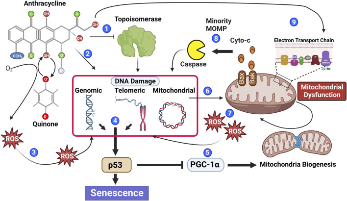
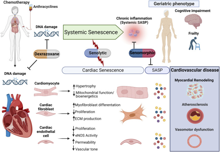
Cancer continues to place a heavy burden on healthcare systems around the world. Although cancer survivorship continues to improve, cardiotoxicity leading to cardiomyopathy and heart failure as a consequence of cancer therapy is rising, and yesterday's cancer survivors are fast becoming today's heart failure patients. Although the mechanisms driving cardiotoxicity are complex, cellular senescence is gaining attention as a major contributor to chemotherapy-induced cardiotoxicity and, therefore, may also represent a novel therapeutic target to prevent this disease. Cellular senescence is a well-recognized response to clinical doses of chemotherapies, including anthracyclines, and is defined by cell cycle exit, phenotypic alterations which include mitochondrial dysfunction, and the expression of the pro-senescent, pro-fibrotic, and pro-inflammatory senescence-associated phenotype. Senescence has an established involvement in promoting myocardial remodeling during aging, and studies have demonstrated that the elimination of senescence can attenuate the pathophysiology of several cardiovascular diseases. Most recently, pharmacology-mediated elimination of senescence, using a class of drugs termed senolytics, has been demonstrated to prevent myocardial dysfunction in preclinical models of chemotherapy-induced cardiotoxicity. In this review, we will discuss the evidence that anthracycline-induced senescence causes the long-term cardiotoxicity of anticancer chemotherapies, consider how the senescent phenotype may promote myocardial dysfunction, and examine the exciting possibility that targeting senescence may prove a therapeutic strategy to prevent or even reverse chemotherapy-induced cardiac dysfunction.

Keywords: anthracyclines; cancer; cardiac; chemotherapy; heart failure; senescence; senolytic.


Conflict of interest statement

The authors declare that the research was conducted in the absence of any commercial or financial relationships that could be construed as a potential conflict of interest.

Figures

FIGURE 1
FIGURE 1
Anthracyclines contribute to multiple biological processes that stimulate senescence. Anthracyclines can inhibit the enzymatic function of topoisomerase leading to irreversible DNA damage (1) and through DNA intercalation induce DNA damage independent of topoisomerase (2). Moreover, the quinone structure of anthracyclines can be oxidized to a semiquinone radical through the addition of an electron. Semiquinone radicals quickly react with oxygen to generate ROS causing DNA damage (3). DNA damage within the genomic or telomeric chromosomal regions leads to activation of the DNA damage response pathway, which if persistent contributes to phosphorylation of the key senescence regulator p53 (4). Phosphorylated p53 inhibits PGC-1a attenuating mitochondrial biogenesis (5). Reduced mitochondrial biogenesis in combination with anthracycline-mediated mitochondrial DNA damage (6) leads to an accumulation of mitochondrial dysfunction and increased ROS generation, contributing to additional DNA damage (7). Mitochondrial outer membrane permeability (MOMP) is also increased in dysfunctional mitochondria allowing the release of caspase-activating cytochrome C; if insufficient to induce apoptosis, the sub-lethal activation of caspase will cause further DNA damage (8). Recently, it has also been suggested that anthracyclines can cause biogenetic lesions to the electron transport chain (Tarpey et al., 2019), further contributing to increased ROS (9).
FIGURE 2
FIGURE 2
Proposed model for anthracycline-induced senescence in cardiotoxicity. We propose a model in which anthracyclines, through several interdependent mechanisms culminating in persistent DNA damage, induce systemic senescence and drive a geriatric phenotype. In the heart, senescence is induced in several cell populations which collectively contribute to age-related cardiovascular diseases, including myocardial remodeling, both directly and via expression of the senescence-associated secretory phenotype (SASP). This raises therapeutic opportunities through either elimination of senescent cells or modulation of the senescent phenotype for the management of anthracycline-induced cardiotoxicity and potentially less well-studied systemic toxicity.

Similar articles

Cited by

References

    1. Abdelgawad I. Y., Agostinucci K., Ismail S. G., Grant M. K. O., Zordoky B. N. (2022a). EA.hy926 cells and HUVECs share similar senescence phenotypes but respond differently to the senolytic drug ABT-263. Cells, 11, 1992. 10.3390/cells11131992 - DOI - PMC - PubMed
    1. Abdelgawad I. Y., Agostinucci K., Zordoky B. N. (2022b). Cardiovascular ramifications of therapy-induced endothelial cell senescence in cancer survivors. Biochim. Biophys. Acta. Mol. Basis Dis. 1868, 166352. 10.1016/j.bbadis.2022.166352 - DOI - PMC - PubMed
    1. Abdelgawad I. Y., Sadak K. T., Lone D. W., Dabour M. S., Niedernhofer L. J., Zordoky B. N. (2021). Molecular mechanisms and cardiovascular implications of cancer therapy-induced senescence. Pharmacol. Ther. 221, 107751. 10.1016/j.pharmthera.2020.107751 - DOI - PMC - PubMed
    1. Ahmad A. S., Ormiston-Smith N., Sasieni P. D. (2015). Trends in the lifetime risk of developing cancer in great ysfunc: Comparison of risk for those born from 1930 to 1960. Br. J. Cancer 112, 943–947. 10.1038/bjc.2014.606 - DOI - PMC - PubMed
    1. Ale-Agha N., Jakobs P., Goy C., Zurek M., Rosen J., Dyballa-Rukes N., et al. (2021). Mitochondrial telomerase reverse transcriptase protects from myocardial ischemia/reperfusion injury by improving complex I composition and function. Circulation 144, 1876–1890. 10.1161/CIRCULATIONAHA.120.051923 - DOI - PubMed

LinkOut - more resources