Deficiency in ST6GAL1, one of the two α2,6-sialyltransferases, has only a minor effect on the pathogenesis of prion disease
- PMID: 36452458
- PMCID: PMC9702343
- DOI: 10.3389/fmolb.2022.1058602
Deficiency in ST6GAL1, one of the two α2,6-sialyltransferases, has only a minor effect on the pathogenesis of prion disease
Abstract
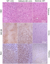
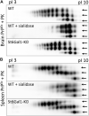
Prion diseases are a group of fatal neurodegenerative diseases caused by misfolding of the normal cellular form of the prion protein or PrPC, into a disease-associated self-replicating state or PrPSc. PrPC and PrPSc are posttranslationally modified with N-linked glycans, in which the terminal positions occupied by sialic acids residues are attached to galactose predominantly via α2-6 linkages. The sialylation status of PrPSc is an important determinant of prion disease pathogenesis, as it dictates the rate of prion replication and controls the fate of prions in an organism. The current study tests whether a knockout of ST6Gal1, one of the two mammalian sialyltransferases that catalyze the sialylation of glycans via α2-6 linkages, reduces the sialylation status of PrPSc and alters prion disease pathogenesis. We found that a global knockout of ST6Gal1 in mice significantly reduces the α2-6 sialylation of the brain parenchyma, as determined by staining with Sambucus Nigra agglutinin. However, the sialylation of PrPSc remained stable and the incubation time to disease increased only modestly in ST6Gal1 knockout mice (ST6Gal1-KO). A lack of significant changes in the PrPSc sialylation status and prion pathogenesis is attributed to the redundancy in sialylation and, in particular, the plausible involvement of a second member of the sialyltransferase family that sialylate via α2-6 linkages, ST6Gal2.
Keywords: N-glycosylation; ST6GAL1; ST6GAL2; mouse; prion; prion diseases; sialic acid; sialyltransferases.
Copyright © 2022 Makarava, Katorcha, Chang, Lau and Baskakov.
Conflict of interest statement
The authors declare that the research was conducted in the absence of any commercial or financial relationships that could be construed as a potential conflict of interest.
Figures


Similar articles
-
Role of sialylation of N-linked glycans in prion pathogenesis.Cell Tissue Res. 2023 Apr;392(1):201-214. doi: 10.1007/s00441-022-03584-2. Epub 2022 Jan 28. Cell Tissue Res. 2023. PMID: 35088180 Free PMC article. Review.
-
Analyses of N-linked glycans of PrPSc revealed predominantly 2,6-linked sialic acid residues.FEBS J. 2017 Nov;284(21):3727-3738. doi: 10.1111/febs.14268. Epub 2017 Sep 30. FEBS J. 2017. PMID: 28898525 Free PMC article.
-
Sialylation Controls Prion Fate in Vivo.J Biol Chem. 2017 Feb 10;292(6):2359-2368. doi: 10.1074/jbc.M116.768010. Epub 2016 Dec 20. J Biol Chem. 2017. PMID: 27998976 Free PMC article.
-
Majority of alpha2,6-sialylated glycans in the adult mouse brain exist in O-glycans: SALSA-MS analysis for knockout mice of alpha2,6-sialyltransferase genes.Glycobiology. 2021 Jun 3;31(5):557-570. doi: 10.1093/glycob/cwaa105. Glycobiology. 2021. PMID: 33242079
-
Multifaceted Role of Sialylation in Prion Diseases.Front Neurosci. 2016 Aug 8;10:358. doi: 10.3389/fnins.2016.00358. eCollection 2016. Front Neurosci. 2016. PMID: 27551257 Free PMC article. Review.
Cited by
-
The Multifaceted Functions of Prion Protein (PrPC) in Cancer.Cancers (Basel). 2023 Oct 13;15(20):4982. doi: 10.3390/cancers15204982. Cancers (Basel). 2023. PMID: 37894349 Free PMC article. Review.
-
Unique Glycans in Synaptic Glycoproteins in Mouse Brain.ACS Chem Neurosci. 2024 Nov 6;15(21):4033-4045. doi: 10.1021/acschemneuro.4c00399. Epub 2024 Oct 14. ACS Chem Neurosci. 2024. PMID: 39401784
-
The alteration and role of glycoconjugates in Alzheimer's disease.Front Aging Neurosci. 2024 Jun 13;16:1398641. doi: 10.3389/fnagi.2024.1398641. eCollection 2024. Front Aging Neurosci. 2024. PMID: 38946780 Free PMC article. Review.
References
-
- Ayers J. L., Schutt C. R., Shikiya R. A., Aguzzi A., Kincaid A. E., Bartz J. C. (2011). The strain-encoded relationship between PrP replication, stability and processing in neurons is predictive of the incubation period of disease. PLoS Pathog. 7 (3), e1001317. 10.1371/journal.ppat.1001317 - DOI - PMC - PubMed
Grants and funding
LinkOut - more resources
Full Text Sources
Research Materials

