Schwannoma in Soft Palate: A Rare Case Report with Review of Literature
- PMID: 36452788
- PMCID: PMC9702478
- DOI: 10.1007/s12070-020-02235-8
Schwannoma in Soft Palate: A Rare Case Report with Review of Literature
Abstract
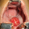
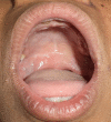
Schwannoma is a benign tumor originating from Schwann cells of the nerve sheath. Approximately 25-45% of the schwannomas are seen in the head and neck region and are found rarely in the oral cavity (only 1%). The most common intra-oral site is tongue, followed by floor of the mouth, buccal mucosa, palate, gingiva and lips. We report a rare case of schwannoma in the soft palate in a 22 years old female. She presented with 6 months history of a painless swelling in palate. The provisional diagnosis was made as some benign neoplasm of minor salivary gland. The tumour was excised intra-orally under general anesthesia. Histopathologic examination showed neural tissue arranged in predominantly Antoni A pattern and formation of verocay bodies. It is difficult to diagnose this tumor based on clinical appearance. Therefore final diagnosis can only be done after histopathological examination of the lesion. Prognosis is good and recurrence is unknown.
Keywords: Antoni A and B pattern; Schwannoma; Soft palate; Verocay bodies.
© Association of Otolaryngologists of India 2020.
Figures


Similar articles
-
Schwannoma in the midline of hard palate: a case report and review of literature.J Dent Res Dent Clin Dent Prospects. 2014 Spring;8(2):114-7. doi: 10.5681/joddd.2014.021. Epub 2014 Jun 11. J Dent Res Dent Clin Dent Prospects. 2014. PMID: 25093057 Free PMC article.
-
Plexiform (multinodular) schwannoma of soft palate. Report of a case.Folia Med (Plovdiv). 2012 Jul-Sep;54(3):62-4. doi: 10.2478/v10153-011-0099-1. Folia Med (Plovdiv). 2012. PMID: 23270209
-
Uncommon Finding of a Soft Palate Schwannoma: A Case Report.Cureus. 2023 Dec 8;15(12):e50172. doi: 10.7759/cureus.50172. eCollection 2023 Dec. Cureus. 2023. PMID: 38186499 Free PMC article.
-
Floor of mouth schwannoma mimicking a salivary gland neoplasm: a report of the case and review of the literature.BMJ Case Rep. 2021 Feb 19;14(2):e239452. doi: 10.1136/bcr-2020-239452. BMJ Case Rep. 2021. PMID: 33608339 Free PMC article. Review.
-
Schwannoma located in the palate: clinical case and literature review.Med Oral Patol Oral Cir Bucal. 2009 Sep 1;14(9):e465-8. Med Oral Patol Oral Cir Bucal. 2009. PMID: 19415055 Review.
References
-
- Dhupar A, Yadav S, Dhupar V. Schwannoma of the hard palate: a rare case. Internet J Head Neck Surg. 2011 doi: 10.5580/21f5. - DOI
LinkOut - more resources
Full Text Sources
