

CT Cisternogram Findings in Idiopathic Cerebrospinal Fluid Leaks with Emphasis on Long Term Management
- PMID: 36452803
- PMCID: PMC9702258
- DOI: 10.1007/s12070-021-02766-8
CT Cisternogram Findings in Idiopathic Cerebrospinal Fluid Leaks with Emphasis on Long Term Management
Abstract
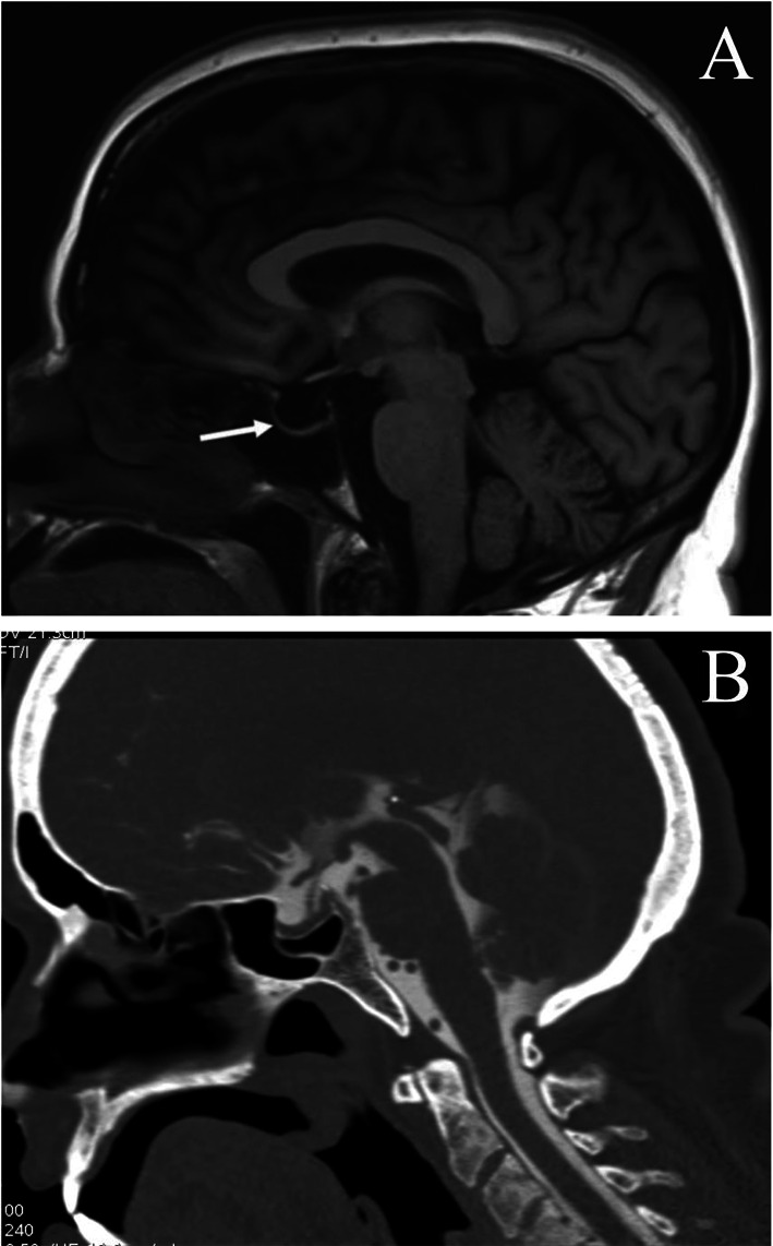
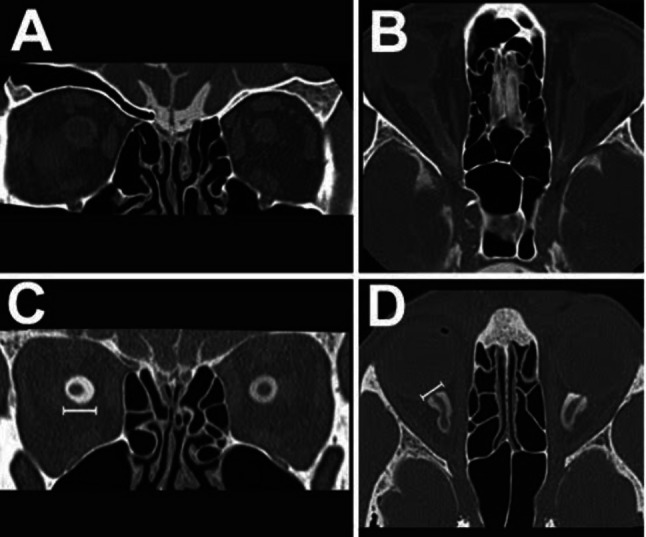
To study the various computed tomography (CT) cisternogram findings in idiopathic cerebrospinal fluid (CSF) leaks and the long term treatment modalities after surgical repair of idiopathic CSF leaks. This was a descriptive study conducted among 25 patients in MCV memorial ENT trust hospital, Pollachi between May 2014 and May 2020 amongst patients who underwent CT cisternogram for unilateral or bilateral spontaneous rhinorrhea with or without associated headache, visual disturbances and papilloedema diagnosed to be idiopathic CSF leak by investigations. These patients then underwent CSF leak repair and postoperatively were managed with weight reduction, low salt diet and diuretic therapy. Post surgery these patients were followed up for a period of 12 months and were evaluated on the basis of presence or absence of headache, rhinorrhea and papilloedema at the end of 1st month, 3rd month, 6th month and 1 year and data was collected. CT cisternogram findings were evaluated by proportion method and evaluation of long term management was done using proportion and repeated measures ANOVA for all patients. Evidence of the presence of previously mentioned CT cisternogram or contrast MRI findings at the end of 1 year of post-surgical treatment was recorded where patients were willing for the same. CT Cisternography was done for all patients and 72% patients had empty sella appearance while 28% had partially empty sella. Other findings included perioptic filling, optic blunting and arachnoid pits which were found in 11(44%), 8(32%) and 12(48%) of patients respectively. Only 3(12%) out of 25 patients had an encephalocoele. The commonest site of leak in CT cisternography was the cribriform plate (52%) followed by lateral recess of sphenoid (48%). None of the patients had multiple sites of leak in CT cisternography. On follow up post surgery maximum resolution of symptoms was found at the end of 12 months where 23 out of 25 patients improved. In our study, out of 25 only 5 patients agreed to undergo post diuretic therapy MRI scan out of which 2 patients had partially empty sella and 3 had normal sella indicating resolution of BIH. CT cisternography is an important investigation which aids in the diagnosis of CSF rhinorrhea due to idiopathic intracranial hypertension (IIH). The medical management of IIH post surgery such as weight reduction, salt restriction and diuretic therapy is also crucial to prevent recurrence of symptoms.
Keywords: Cerebrospinal fluid rhinorrhea; Computed tomography; Idiopathic intracranial hypertension; Spontaneous.
© Association of Otolaryngologists of India 2021.
Conflict of interest statement
Conflict of interestThe authors have no conflict of interest to declare that are relevant to the content of this article.
Figures
Similar articles
-
A retrospective analysis of spontaneous sphenoid sinus fistula: MR and CT findings.AJNR Am J Neuroradiol. 2000 Feb;21(2):337-42. AJNR Am J Neuroradiol. 2000. PMID: 10696020 Free PMC article.
-
Primary spontaneous cerebrospinal fluid leaks and idiopathic intracranial hypertension.J Neuroophthalmol. 2013 Dec;33(4):330-7. doi: 10.1097/WNO.0b013e318299c292. J Neuroophthalmol. 2013. PMID: 24042170 Free PMC article. Review.
-
Spontaneous cerebrospinal fluid leaks in the anterior skull base secondary to idiopathic intracranial hypertension.Eur Arch Otorhinolaryngol. 2017 May;274(5):2175-2181. doi: 10.1007/s00405-017-4455-5. Epub 2017 Feb 7. Eur Arch Otorhinolaryngol. 2017. PMID: 28175991
-
Radiographic signs of elevated intracranial pressure in idiopathic cerebrospinal fluid leaks: a possible presentation of idiopathic intracranial hypertension.Am J Rhinol. 2007 May-Jun;21(3):257-61. doi: 10.2500/ajr.2007.21.3026. Am J Rhinol. 2007. PMID: 17621805
-
Imaging of Cerebrospinal Fluid Rhinorrhea and Otorrhea.Radiol Clin North Am. 2017 Jan;55(1):167-187. doi: 10.1016/j.rcl.2016.08.005. Radiol Clin North Am. 2017. PMID: 27890184 Review.
Cited by
-
Endoscopic Endonasal Anterior Skull Base Meningoencephalocele and Cerebrospinal Fluid Leak Repair: Our Intraoperative and Postoperative Protocol and Long-term Outcomes.J Neurol Surg B Skull Base. 2024 Apr 30;86(2):173-179. doi: 10.1055/a-2297-9386. eCollection 2025 Apr. J Neurol Surg B Skull Base. 2024. PMID: 40104534 Free PMC article.
References
-
- Ommaya AK, Di Chiro, et al. Hydrocephalus and leaks. In: Wager H, et al., editors. Principles of nuclear medicine. 2. Philadelphia: Saunders; 1995.
-
- Woodworth BA, Schlosser RJ. Cerebrospinal fluid leak and encephaloceleCerebrospinal fluid leak and encephalocele. In: Kennedy DW, Hwang PH, editors. Rhinology diseases of the nose, sinuses, and skull base. 1. USA: Thieme Medical Publishers; 2012. p. 591.
LinkOut - more resources
Full Text Sources
Research Materials
Miscellaneous