Tinea versicolor: an updated review
- PMID: 36452877
- PMCID: PMC9677953
- DOI: 10.7573/dic.2022-9-2
Tinea versicolor: an updated review
Abstract
Background: Tinea versicolor is a common superficial fungal infection of the skin with various clinical manifestations. This review aims to familiarize physicians with the clinical features, diagnosis and management of tinea versicolor.
Methods: A search was conducted in July 2022 in PubMed Clinical Queries using the key terms "tinea versicolor" OR "pityriasis versicolor". The search strategy included all clinical trials, observational studies and reviews published within the past 10 years.
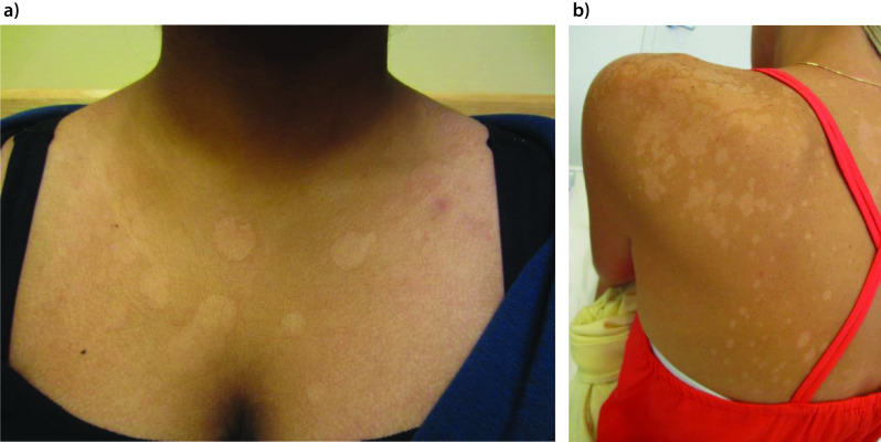
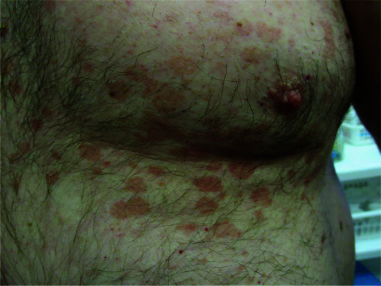
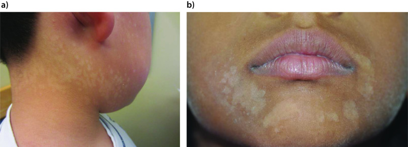
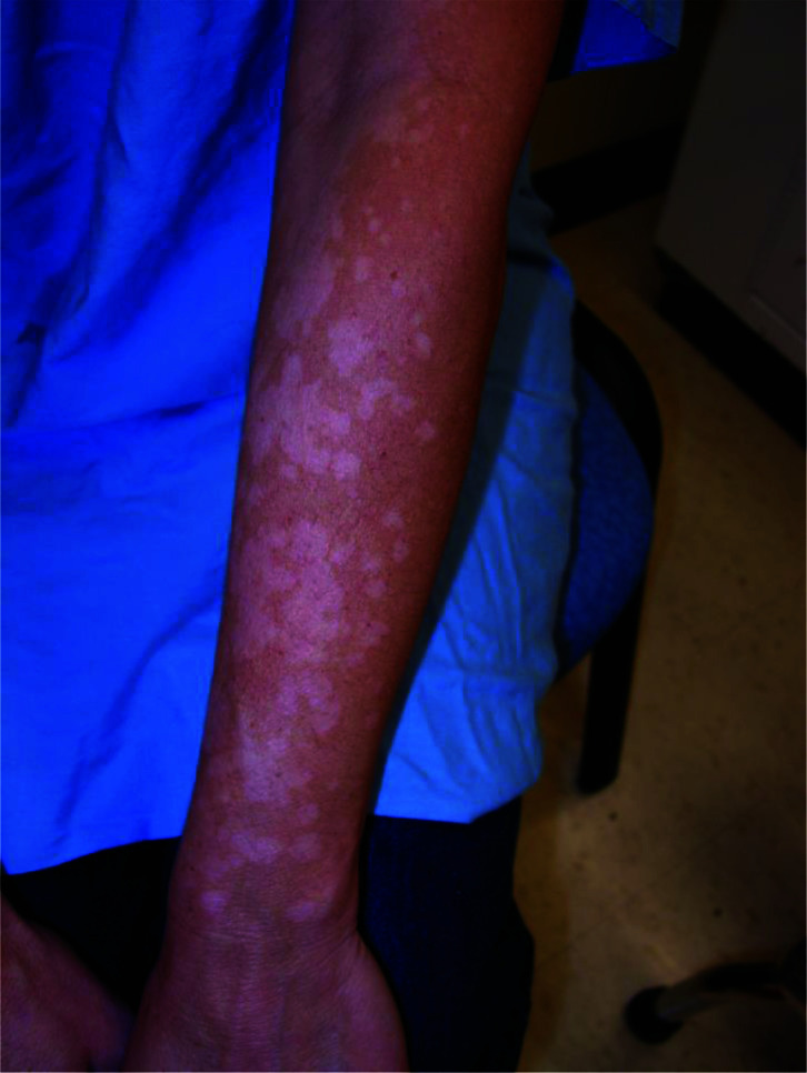
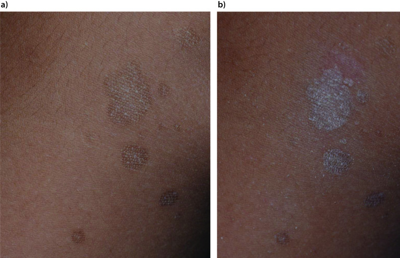
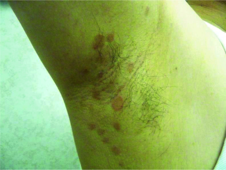
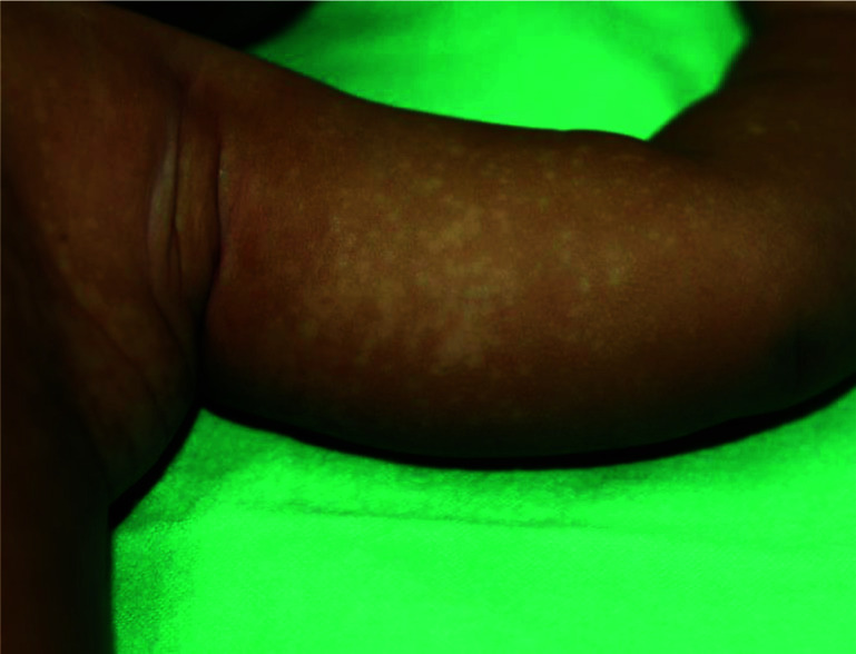
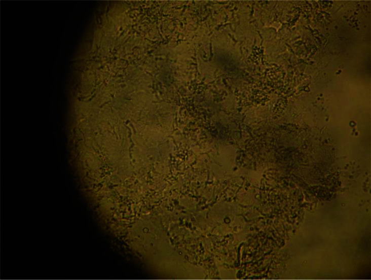
Results: Tinea versicolor is caused by Malassezia species, notably M. globosa, M. furfur and M. sympodialis. The condition is characterized by scaly hypopigmented or hyperpigmented macules/patches, primarily located on the upper trunk, neck and upper arms. The diagnosis is usually based on characteristic clinical features. If necessary, a potassium hydroxide preparation test can be performed to reveal numerous short, stubby hyphae intermixed with clusters of spores. Most patients with tinea versicolor respond to topical antifungal therapy, which has a better safety profile (fewer adverse events, fewer drug interactions) and lower cost compared to systemic treatment and is therefore the treatment of choice. Oral antifungal therapy is typically reserved for patients with extensive disease, frequent recurrences or disease that is refractory to topical therapy. Advantages of oral antifungal therapy include increased patient compliance, shorter duration of treatment, increased convenience, less time involved with therapy and reduced recurrence rates. On the other hand, oral antifungal therapy is associated with higher cost, greater adverse events and potential drug-drug interactions and is therefore not the first-line treatment for tinea versicolor. Long-term intermittent prophylactic therapy should be considered for patients with frequent recurrence of the disease.
Conclusion: Selection of antifungal agents depends on several factors, including efficacy, safety, local availability, ease of administration, likelihood of compliance and potential drug interactions of the antifungal agent.
Keywords: Malassezia species; evoked scale sign; fluconazole; itraconazole; ketoconazole; pityriasis versicolor; selenium sulfide; terbinafine; zinc pyrithione.
Copyright © 2022 Leung AKC, Barankin B, Lam JM, Leong KF, Hon KL.
Conflict of interest statement
Disclosure and potential conflicts of interest: AKCL and KLH are associate editors of Drugs in Context and confirm that this article has no other conflicts of interest otherwise. This manuscript was sent out for independent peer review. The International Committee of Medical Journal Editors (ICMJE) Potential Conflicts of Interests form for the authors is available for download at: https://www.drugsincontext.com/wp-content/uploads/2022/10/dic.2022-9-2-COI.pdf
Figures
References
-
- Goldstein BG, Goldstein AO. Tinea versicolor (pityriasis versicolor) In: Dellavalle RP, Levy ML, Rosen T, editors. Waltham, MA: UpToDate; [Accessed July 26, 2022]. https://www.uptodate.com/contents/tinea-versicolor-pityriasis-versicolor .
-
- Awad AK, Al-Ezzy AIA, Jameel GH. Phenotypic identification and molecular characterization of Malassezia Spp. isolated from pityriasis versicolor patients with special emphasis to risk factors in Diyala Province, Iraq. Open Access Maced J Med Sci. 2019;7(5):707–714. doi: 10.3889/oamjms.2019.128. - DOI - PMC - PubMed
Publication types
LinkOut - more resources
Full Text Sources
Medical
Miscellaneous