. 2023 Feb;43(2):267-285.
doi: 10.1161/ATVBAHA.122.317800. Epub 2022 Dec 1.

Neutrophils Protect Against Staphylococcus aureus Endocarditis Progression Independent of Extracellular Trap Release

Affiliations

Neutrophils Protect Against Staphylococcus aureus Endocarditis Progression Independent of Extracellular Trap Release

Severien Meyers et al. Arterioscler Thromb Vasc Biol. 2023 Feb.

Abstract

Background: Infective endocarditis (IE) is characterized by an infected thrombus at the heart valves. How bacteria bypass the immune system and cause these thrombi remains unclear. Neutrophils releasing NETs (neutrophil extracellular traps) lie at this interface between host defense and coagulation. We aimed to determine the role of NETs in IE immunothrombosis.

Methods: We used a murine model of Staphylococcus aureus endocarditis in which IE is provoked on inflamed heart valves and characterized IE thrombus content by immunostaining identifying NETs. Antibody-mediated neutrophil depletion and neutrophil-selective PAD4 (peptidylarginine deiminase 4)-knockout mice were used to clarify the role of neutrophils and NETs, respectively. S. aureus mutants deficient in key virulence factors related to immunothrombosis (nucleases or staphylocoagulases) were investigated.

Results: Neutrophils releasing NETs were present in infected thrombi and within cellular infiltrates in the surrounding vasculature. Neutrophil depletion increased occurrence of IE, whereas neutrophil-selective impairment of NET formation did not alter IE occurrence. Absence of S. aureus nuclease, which degrades NETs, did not affect endocarditis outcome. In contrast, absence of staphylocoagulases (coagulase and von Willebrand factor binding protein) led to improved survival, decreased bacteremia, smaller infiltrates, and decreased tissue destruction. Significantly more NETs were present in these vegetations, which correlated with decreased bacteria and cell death in the adjacent vascular wall.

Conclusions: Neutrophils protect against IE independent of NET release. Absence of S. aureus coagulases, but not nucleases, reduced IE severity and increased NET levels. Staphylocoagulase-induced fibrin likely hampers NETs from constraining infection and the resultant tissue damage, a hallmark of valve destruction in IE.

Keywords: Staphylococcus aureus; coagulases; endocarditis; extracellular traps; neutrophils.


Figures

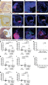
Figure 1.
Figure 1.
Histological characterization of advanced endocarditis vegetations and sterile thrombi. Various components were characterized in mice with infective endocarditis (n=11), sterile thrombi (n=7), or without thrombi (n=7), induced by the clinical strain of Staphylococcus aureus at day 3. Representative images of these components in each group are shown in A–E. A, Brightfield images of a Brown-Hopps Gram stain with bacteria seen in purple and asterisks (*) indicating large cellular infiltrates in the surrounding aortic wall. B–D, Fluorescence images of S. aureus (green) and neutrophil-specific marker Ly6G (red, B); VWF (von Willebrand factor, green, C); and platelet CD41 (red) and fibrinogen (green; D). DNA is identified by Hoechst 33342 staining, depicted in blue. Double asterisks (**) indicate large infiltrates of neutrophils in the surrounding aortic wall. E, Martius Scarlet Blue (MSB) staining that detects fibrin in red. Scale bars represent 200 μm.
Figure 2.
Figure 2.
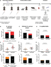
Neutrophils protect against infective endocarditis. Following neutrophil depletion, mice underwent endocarditis surgery and were monitored for 1 day. A, Schematic overview of the neutrophil depletion experiment design (made with Biorender.com). B–D, Proportions of mice that developed infective endocarditis (red) at day 1 in neutrophil-depleted (anti-Ly6G) mice compared with isotype-control mice, infected with the methicillin-resistant strain of Staphylococcus aureus USA300, a clinical endocarditis isolate S. aureus strain and a coagulase-negative strain (Staphylococcus epidermidis). E–G, Bacteremia levels at end point with corresponding median (interquartile range) in neutrophil-depleted (n=20, 14, 10) and isotype-control (n=20, 18, 10) mice. Endocarditis vegetations are depicted in red, sterile thrombi in orange, and no thrombus in black. H–J, Proportions of mice that developed sterile thrombi (orange) in neutrophil-depleted mice compared with isotype-control mice. Fisher Exact (B–D, H–J) or Mann-Whitney tests (E–G) were conducted. CFU indicates colony-forming units; and Ly6G, lymphocyte antigen 6 complex locus G6D.
Figure 3.
Figure 3.
Different strains of Staphylococcus aureus induce NETs (neutrophil extracellular traps), whereas a less virulent coagulase-negative Staphylococcus strain does not induce NETosis (the process of NET formation). A–D, In an in vitro NET release assay, isolated neutrophils (75 000 cells per well) from healthy volunteers were incubated with various bacterial strains (multiplicity of infection [MOI] 100) for 3 hours, and formed NETs were digested to smaller fragments with AluI (4 U) and measured in cell culture supernatants for H3Cit (citrullinated histone H3). A, C, H3Cit absorbances relative to nonstimulated (vehicle-treated) of digested (AluI, red) compared to nondigested (vehicle, blue) samples in the absence (n=8, A) or presence of platelets (6×10⁸ cells/mL, n=5, C). B and D, H3Cit absorbances relative to vehicle of AluI digested samples with corresponding median (interquartile range) in the absence (n=8, B) or presence of platelets (n=5, D). Each single dot represents a single donor. Significance was determined by Kruskal-Wallis test with Dunn post test (B, D). E–H, Overview scanning electron microscopy (SEM) images (secondary electron signal) of NETs formed in the in vitro NET release assay. Scale bar equals 10 μm. I–L, Detailed secondary electron images of NETs incubated with a rabbit anti-H3Cit and a gold-conjugated anti-rabbit antibody. Gold particles were identified on corresponding backscatter electron SEM images and are depicted in green. Scale bars represent 500 nm. Due to different degrees of charging, a different threshold for the gold particles was used in K than in I, J, and L. Complete P values for data presented in B and D are available in Table S1 and S2, respectively. Δnuc indicates nucleases.
Figure 4.
Figure 4.
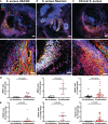
Neutrophils undergoing NET (neutrophil extracellular trap) formation and released NETs are present in mice with Staphylococcus aureus–induced endocarditis. A–C, Fluorescence microscopy images of the following NET markers: H3Cit (citrullinated histone H3, green), MPO (myeloperoxidase, red), and DNA (blue) in inflammation-induced endocarditis vegetations infected with S. aureus USA300 (n=8, A), Newman (n=6, B) or a clinical S. aureus endocarditis strain (n=25, C) at day 3. Scale bar of upper and lower panels equals 200 μm and 50 μm respectively. In C, a different color balance threshold was used for MPO than in A and B. D and E, Quantifications of MPO (D) and H3Cit (E) positive area in mice with infective endocarditis (red dots, n=8, 6, 25) compared with mice without a vegetation (black dots, n=7, 7, 18). Median (interquartile range) are represented, and Mann-Whitney tests were used to test for statistical significance (D and E).
Figure 5.
Figure 5.
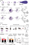
PAD4 (peptidylarginine deiminase 4)-mediated NET (neutrophil extracellular traps) release neither protects against nor promotes infective endocarditis. A–E, In vitro assay: neutrophils were isolated from control (MRP8+ or PAD4fl/fl) or neutrophil-selective PAD4 knockout mice (MRP8Cre+×PAD4fl/fl), incubated with Staphylococcus aureus USA300 or ionomycin, and the different stages of NETosis were assessed according to nuclear morphology. A, Schematic overview of the different stages of NET formation. B, Percentage of cells at various NETosis stages in control (average of n=8) and neutrophil-selective PAD4 knockout mice (average of n=9). C and D, Percentage of NETs induced by ionomycin (C) and S. aureus USA300 (D) in neutrophil-selective PAD4 knockout (n=9) mice compared with controls (n=8). Medians (interquartile ranges) are shown. E, Schematic overview (created with Biorender.com) of endocarditis experiments performed in neutrophil-selective PAD4 knockout and control mice. F and G, Proportions of mice that developed inflammation-induced endocarditis (red, F) or sterile thrombi (orange, G) at day 3 in neutrophil-selective PAD4 knockout mice compared with control mice. H, Bacteremia levels at end point with corresponding median (interquartile range; n=18, 16). I, Survival graph of neutrophil-selective PAD4 knockout (black, n=23) and control mice (gray, n=25). Mann-Whitney (C and D, H), Fisher Exact (F and G) or log-rank (Mantel-Cox) test (I). CFU indicates colony-forming units; MRP8, myeloid related protein 8; and NETosis, the process of NET formation.
Figure 6.
Figure 6.
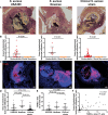
Large cellular infiltrates in the surrounding aortic wall promote tissue destruction. A, Brown-Hopps Gram stain of endocarditis vegetations induced by Staphylococcus aureus USA300 (n=19), S. aureus Newman (n=6), or the clinical S. aureus strain (n=42), depicting large cellular infiltrates (*) in the surrounding vasculature. B, Quantifications of the extraluminal leukocyte infiltration area in infected (red dots, n=19, 6, 42) compared with sterile (orange dots, n=18, 9, 19, respectively) thrombi. C, Click-iT Plus TUNEL (terminal deoxynucleotidyl transferase dUTP nick-end labeling) assay stained images of endocarditis vegetations (n=18, 6, 19, respectively), depicting apoptotic cells in red. DNA is stained with Hoechst 33342 in blue. D, Comparison of the extraluminal leukocyte infiltration area between the 3 different strains of S. aureus (n=19, 6, 42, respectively). E, Quantification of the TUNEL-positive area outside the thrombus in mice with infected vegetations caused by the 3 different strains of S. aureus (n=18, 6, 19, respectively). F, Spearman correlation between the leukocyte infiltration area and TUNEL-positive area located outside the infected vegetation caused by the 3 different strains of S. aureus (n=43). Scale bar represents 200 μm. Median (interquartile range) are shown and significance level is evaluated by Mann-Whitney (B) or Kruskal-Wallis test with Dunn post tests (D and E).
Figure 7.
Figure 7.
Absence of staphylocoagulases, but not of nucleases, improves endocarditis outcome. A–D, Proportions of mice that developed inflammation-induced endocarditis (A) or sterile thrombi (B) at day 3, bacteremia levels at end point (n=9, 10, C), and survival rates (n=12, 12, D) of mice infected with the nuclease mutant (Δnuc [nucleases]) compared to wild-type (WT) Staphylococcus aureus USA300. E–H, Proportions of mice that developed inflammation-induced endocarditis (E) or sterile thrombi (F) at day 3, bacteremia levels at endpoint (n=23, 26, G), and survival rates (n=27, 26, H) of mice infected with a mutant doubly deficient in ΔcoaΔvwb (coagulase and von Willebrand factor binding protein) compared to WT S. aureus USA300. Median (interquartile range) are represented in C and G. Fisher Exact (A and B, E and F), Mann-Whitney test (C and G), and log-rank (Mantel-Cox; D and H) tests were conducted to determine significance. CFU indicates colony-forming units.
Figure 8.
Figure 8.
Staphylocoagulases prevent NETs (neutrophil extracellular traps) from constraining bacterial infection and promoting tissue destruction. A, Brightfield images of a Brown-Hopps Gram stain (n=10, 10, 4) with bacteria represented in purple. B, Click-iT Plus TUNEL (terminal deoxynucleotidyl transferase dUTP nick-end labeling) assay stained images (n=10,10, 4), depicting apoptotic cells in red. C and D, Fluorescence microscopy images (n=10, 10, 4) of Staphylococcus aureus in green and neutrophil-specific marker Ly6G in red (C), and MPO (myeloperoxidase) in red and H3Cit (citrullinated histone H3) in green (D). DNA is stained with Hoechst 33342 in blue. Scale bars represent 200 μm. E–L, Quantifications of extraluminal leukocyte infiltration (E), TUNEL (F), bacteria (H), Ly6G (K), and MPO (L)-positive area situated outside infected thrombi and H3Cit inside the thrombus (I) in control (C57Bl/6J) mice with infective endocarditis induced by wild-type (WT; black, n=10) S. aureus, and in control (C57Bl/6J and PAD4 (peptidylarginine deiminase 4)fl/fl, dark gray, n=10) and neutrophil-selective PAD4 knockout mice (MRP8Cre+×PAD4fl/fl, light gray, n=4) with infective endocarditis induced by S. aureus coaΔvwb (coagulase and von Willebrand factor binding protein). G, Spearman correlation between the H3Cit-positive area and TUNEL-positive area, both located outside the infected vegetations caused by WT (n=10, C57Bl/6J) and S. aureus ΔcoaΔvwb (n=10, C57Bl/6J and PAD4fl/fl). J, Spearman correlation between bacteria located outside the vegetation and H3Cit inside the thrombus infected with WT (n=10, C57Bl/6J) and S. aureus ΔcoaΔvwb (n=10, C57Bl/6J and PAD4fl/fl). Median (interquartile range) are represented in E and F, H and I, K and L. Significance was determined by Kruskal-Wallis test with Dunn post test (E and F, H and I, K and L). MRP8Cre+ indicates myeloid related protein 8, Cre recombinase expressing.

References

    1. Vogkou CT, Vlachogiannis NI, Palaiodimos L, Kousoulis AA. The causative agents in infective endocarditis: a systematic review comprising 33,214 cases. Eur J Clin Microbiol Infect Dis. 2016;35:1227–1245. doi: 10.1007/s10096-016-2660-6 - PubMed
    1. Yang X, Chen H, Zhang D, Shen L, An G, Zhao S. Global magnitude and temporal trend of infective endocarditis, 1990-2019: results from the global burden of disease study. Eur J Prev Cardiol. 2021;29:1227. doi: 10.1093/eurjpc/zwab184 - PubMed
    1. Talha KM, Baddour LM, Thornhill MH, Arshad V, Tariq W, Tleyjeh IM, Scott CG, Hyun MC, Bailey KR, Anavekar NS, et al. . Escalating incidence of infective endocarditis in Europe in the 21st century. Open Heart. 2021;8:e001846. doi: 10.1136/openhrt-2021-001846 - PMC - PubMed
    1. Dayer MJ, Jones S, Prendergast B, Baddour LM, Lockhart PB, Thornhill MH. Incidence of infective endocarditis in England, 2000-13: a secular trend, interrupted time-series analysis. Lancet. 2015;385:1219–1228. doi: 10.1016/S0140-6736(14)62007-9 - PMC - PubMed
    1. Miro JM, Anguera I, Cabell CH, Chen AY, Stafford JA, Corey GR, Olaison L, Eykyn S, Hoen B, Abrutyn E, et al. ; International Collaboration on Endocarditis Merged Database Study Group. Staphylococcus aureus native valve infective endocarditis: report of 566 episodes from the International Collaboration on Endocarditis Merged Database. Clin Infect Dis. 2005;41:507–514. doi: 10.1086/431979 - PubMed

Publication types