Review
. 2023 Jun;18(6):1249-1256.
doi: 10.4103/1673-5374.355741.

The lymphatic system: a therapeutic target for central nervous system disorders

Affiliations
Review

The lymphatic system: a therapeutic target for central nervous system disorders

Jia-Qi Xu et al. Neural Regen Res. 2023 Jun.

Abstract

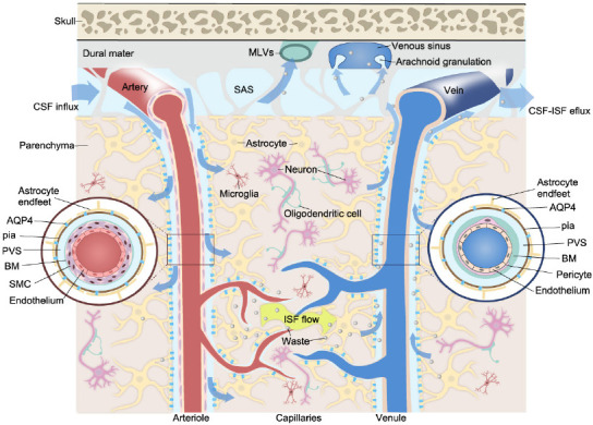
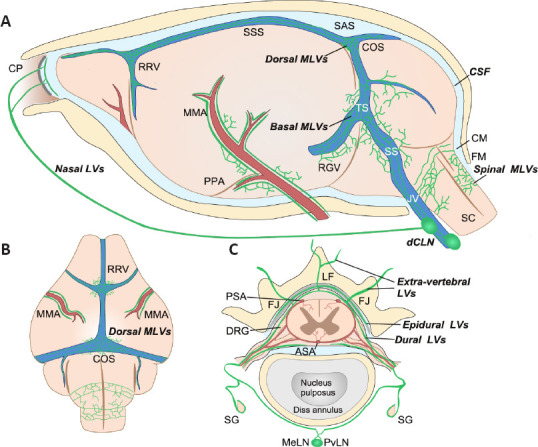
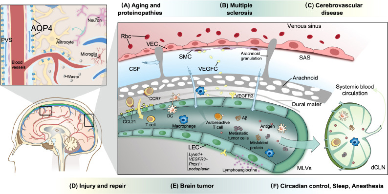
The lymphatic vasculature forms an organized network that covers the whole body and is involved in fluid homeostasis, metabolite clearance, and immune surveillance. The recent identification of functional lymphatic vessels in the meninges of the brain and the spinal cord has provided novel insights into neurophysiology. They emerge as major pathways for fluid exchange. The abundance of immune cells in lymphatic vessels and meninges also suggests that lymphatic vessels are actively involved in neuroimmunity. The lymphatic system, through its role in the clearance of neurotoxic proteins, autoimmune cell infiltration, and the transmission of pro-inflammatory signals, participates in the pathogenesis of a variety of neurological disorders, including neurodegenerative and neuroinflammatory diseases and traumatic injury. Vascular endothelial growth factor C is the master regulator of lymphangiogenesis, a process that is critical for the maintenance of central nervous system homeostasis. In this review, we summarize current knowledge and recent advances relating to the anatomical features and immunological functions of the lymphatic system of the central nervous system and highlight its potential as a therapeutic target for neurological disorders and central nervous system repair.

Keywords: central nervous system; central nervous system injury; glymphatic system; lymphatic vessels; meninges; neurodegenerative disorders; neuroinflammatory diseases; vascular endothelial growth factor C.


Conflict of interest statement

None

Figures

Figure 1
Figure 1
Schematic illustration of the CSF-ISF flow in the glymphatic system. CSF flows in the PVS and moves inward along penetrating arteries and arterioles, and outward along capillaries and parenchymal venules. The PVS around the arteriole and venule forms based on the presence of different vascular unit components (inserts). The PVS surrounds vascular SMCs or the basement membrane and is bounded by perivascular astrocyte end-feet. AQP4 channels on the astrocyte end-feet allow fluid to pass between the PVS and brain parenchyma in both directions. Metabolic waste and macromolecules are drained to the SAS and are further absorbed via arachnoid granulations or MLVs. Created with Vectornator. AQP4: Aquaporin 4; BM: basement membrane; CSF: cerebrospinal fluid; ISF: interstitial fluid; MLVs: meningeal lymphatic vessels; PVS: perivascular space; SAS: subarachnoid space; SMC: smooth muscle cell.
Figure 2
Figure 2
Schematic illustration of the anatomical position of meningeal lymphatic vessels around the mouse brain and spinal cord. (A) On the lateral side of the mouse brain, MLVs run along the artery (red) and venous sinus (blue). Basal MLVs running along the TS and SS have been identified as the main route for CSF uptake and drainage. Parasinusoidal MLVs develop from LVs along the PPA, the JV, and cranial nerves. Spinal MLVs covering the FM connect to MLVs in the cervical spinal canal. (B) On the dorsal side of the mouse brain, dorsal MLVs run along the RRV, COS, and MMA. (C) Spinal MLVs (or vertebral LVs) are present in the epidural space around the spinal cord and the dura mater. On the dorsal side of the spinal canal, they connect at the midline and extend to the CM. However, they do not form a circuit on the ventral side. Spinal LVs further connect with extra-vertebral peripheral LVs at the ventral border of the LF or the level of the transverse FJ. They cover the dura mater of DRG and contact SG. Longitudinal connecting LVs between vertebral units also exist (not shown). Created with Vectornator. ASA: Anterior spinal artery; CM: cisterna magna; COS: the confluence of the sinuses; CP: cribriform plate; dCLNs: deep cervical lymph nodes; DRG: dorsal root ganglia; FJ: facet joint; FM: foramen magnum; JV: jugular vein; LF: ligamentum flavum; MeLN: mediastinal lymph node; MMA: middle meningeal artery; PPA: pterygopalatine artery; PSA: posterior spinal artery; PvLN: posterior vertebral lymph node; RGV: retroglenoid vein; RRV: rostral rhinal vein; SC: spinal cord; SG: sympathetic ganglia; SS: sigmoid sinus; SSS: superior sagittal sinus; TS: transverse sinus.
Figure 3
Figure 3
The role of the lymphatic system in neurological diseases. The expression of AQP4 and MLV-mediated drainage play critical roles in the maintenance of CNS homeostasis. VEGF-C secreted by myeloid cells and SMCs promotes MLV development via VEGFR-3 present on LECs. Antigens and meningeal immune cells (lymphocytes, DCs, macrophages, and neutrophils, among others) cross the lymphatic vessel wall and are transported to the draining lymph nodes, where they activate inflammatory responses. Meningeal T cells are transported to the dCLNs via a CCR7/CCL21-dependent pathway. Mural LECs in leptomeninges or in the brain parenchyma, which have large vacuoles or inclusions, do not form tubular structures. (A) The clearance of neurotoxic proteins, such as Aβ, tau, and α-synuclein, via the glymphatic system and MLVs, is impaired in the aging brain. The abnormal accumulation of neurotoxic proteins leads to degenerative diseases. (B) Autoreactive encephalitogenic T cells are activated and invade the brain parenchyma via lymphatic pathways. (C) Enlargement of the PVS, impairment of the glymphatic pathway, and disruption of the BBB are characteristic of cerebrovascular diseases, such as SVD. (D) Brain-to-CLN signaling via VEGFR-3 is involved in the systemic inflammatory response after stroke and brain injury. Meanwhile, the loss of perivascular AQP4 polarization and the disruption of lymphatic drainage resulting from an increase in ICP in TBI both lead to impaired clearance. Moreover, lymphoangiocrine signals produced by LECs might help injury repair. (E) Glioblastoma and melanoma cells can promote lymphangiogenesis. Tumor cells can metastasize via MLVs. Meanwhile, tumor antigen from the brain reaches dCLNs and activates intracranial immunosurveillance. (F) Clearance rates are different during sleep, wakefulness, and under anesthesia. CSF influx exhibits a pattern of circadian control. Created with Vectornator. AQP4: Aquaporin 4; CCR7: C-C chemokine receptor 7; CCL21: C-C chemokine ligand 21; CSF: cerebrospinal fluid; DC: dendritic cell; dCLN: deep cervical lymph node; ICP: intracranial pressure; LEC: lymphatic endothelial cell; MLVs: meningeal lymphatic vessels; PVS: perivascular space; Rbc: red blood cell; SAS: subarachnoid space; SMC: smooth muscle cell; VEC: vascular endothelial cell; VEGF-C: vascular endothelial growth factor C; VEGFR-3: vascular endothelial growth receptor 3.

References

    1. Absinta M, Ha SK, Nair G, Sati P, Luciano NJ, Palisoc M, Louveau A, Zaghloul KA, Pittaluga S, Kipnis J, Reich DS. Human and nonhuman primate meninges harbor lymphatic vessels that can be visualized noninvasively by MRI. Elife. 2017;6:e29738. - PMC - PubMed
    1. Achariyar TM, Li B, Peng W, Verghese PB, Shi Y, McConnell E, Benraiss A, Kasper T, Song W, Takano T, Holtzman DM, Nedergaard M, Deane R. Glymphatic distribution of CSF-derived apoE into brain is isoform specific and suppressed during sleep deprivation. Mol Neurodegener. 2016;11:74. - PMC - PubMed
    1. Ahn JH, Cho H, Kim JH, Kim SH, Ham JS, Park I, Suh SH, Hong SP, Song JH, Hong YK, Jeong Y, Park SH, Koh GY. Meningeal lymphatic vessels at the skull base drain cerebrospinal fluid. Nature. 2019;572:62–66. - PubMed
    1. Albargothy NJ, Johnston DA, MacGregor-Sharp M, Weller RO, Verma A, Hawkes CA, Carare RO. Convective influx/glymphatic system:tracers injected into the CSF enter and leave the brain along separate periarterial basement membrane pathways. Acta Neuropathol. 2018;136:139–152. - PMC - PubMed
    1. Alitalo K. The lymphatic vasculature in disease. Nat Med. 2011;17:1371–1380. - PubMed