. 2022 Dec 21;10(6):e0240922.
doi: 10.1128/spectrum.02409-22. Epub 2022 Dec 1.

Cec4-Derived Peptide Inhibits Planktonic and Biofilm-Associated Methicillin Resistant Staphylococcus epidermidis

Affiliations

Cec4-Derived Peptide Inhibits Planktonic and Biofilm-Associated Methicillin Resistant Staphylococcus epidermidis

Chengju Mao et al. Microbiol Spectr. .

Abstract

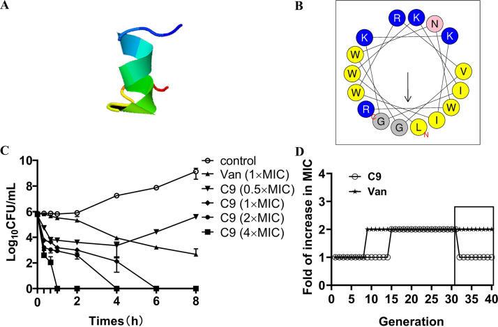
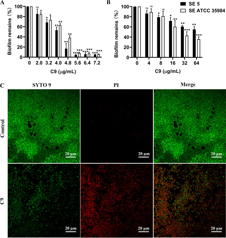
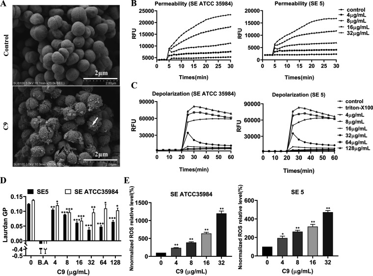
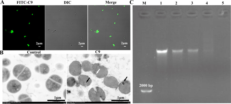
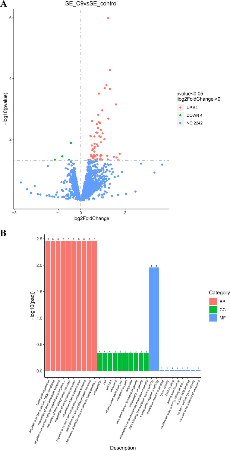
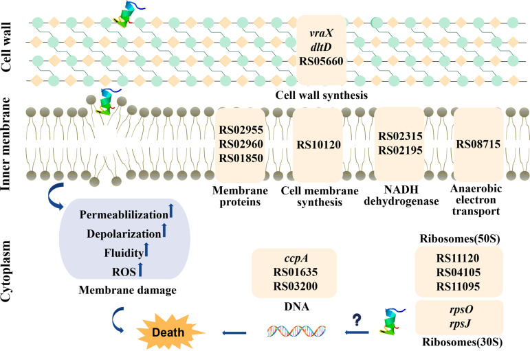
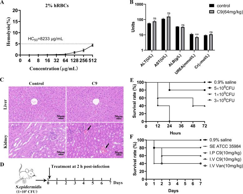
Staphylococcus epidermidis is part of the normal microbiota that colonizes the skin and mucosal surfaces of human beings. Previous studies suggested that S. epidermidis possessed low virulence, but recent studies confirmed that it can acquire high virulence from Staphylococcus aureus and with the increasing detection of methicillin-resistant S. epidermidis. It has become a major pathogen of graft-associated and hospital-acquired infections. In previous studies, we modified the antimicrobial peptide Cec4 (41 amino acids) and obtained the derived peptide C9 (16 amino acids) showing better antimicrobial activity against S. epidermidis with an MIC value of 8 μg/mL. The peptide has rapid bactericidal activity without detectable high-level resistance, showing certain inhibition and eradication ability on S. epidermidis biofilms. The damage of cell membrane structures by C9 was observed by scanning emission microscopy (SEM) and transmission electron microscopy (TEM). In addition, C9 altered the S. epidermidis cell membrane permeability, depolarization levels, fluidity, and reactive oxygen species (ROS) accumulation and possessed the ability to bind genomic DNA. Analysis of the transcriptional profiles of C9-treated cells revealed changes in genes involved in cell wall and ribosome biosynthesis, membrane protein transport, oxidative stress, and DNA transcription regulation. At the same time, the median lethal dose of C9 in mice was more than 128 mg/kg, and the intraperitoneal administration of 64 mg/kg was less toxic to the liver and kidneys of mice. Furthermore, C9 also showed a certain therapeutic effect on the mouse bacteremia model. In conclusion, C9 may be a candidate drug against S. epidermidis, which has the potential to be further developed as an antibacterial therapeutic agent. IMPORTANCE S. epidermidis is one of the most important pathogens of graft-related infection and hospital-acquired infection. The growing problem of antibiotic resistance, as well as the emergence of bacterial pathogenicity, highlights the need for antimicrobials with new modes of action. Antimicrobial peptides have been extensively studied over the past 30 years as ideal alternatives to antibiotics, and we report here that the derived peptide C9 is characterized by rapid bactericidal and antibiofilm activity, avoiding the development of resistance by acting on multiple nonspecific targets of the cell membrane or cell components. In addition, it has therapeutic potential against S. epidermidis infection in vivo. This study provides a rationale for the further development and application of C9 as an effective candidate antibiotic.

Keywords: S. epidermidis; antibacterial activity; antibacterial mechanism; antimicrobial peptide.


Conflict of interest statement

The authors declare no conflict of interest.

Figures

FIG 1
FIG 1
C9 shows bactericidal activity against S. epidermidis without detectable resistance development. (A) Three-dimensional structure projections of C9. (B) The helical wheel projection of C9; yellow circles denote hydrophobic amino acids, and blue circles denote hydrophilic amino acids. (C) Time-kill curve of C9 and vancomycin against S. epidermidis ATCC 35984; the experiments were conducted triplicate and presented as the mean ± SD. (D) Drug resistance development of S. epidermidis ATCC 35984 after subinhibitory doses of C9 and vancomycin (Van) treatment over 30 generations. The MIC changes of last 10 generations after passing the induced strain in the absence of the drug.
FIG 2
FIG 2
Effects of C9 on S. epidermidis biofilms. (A and B) Effects of C9 on biofilm formation (A) and established biofilm (B) of S. epidermidis ATCC 35984 and S. epidermidis 5. The adherent biofilm was stained with crystal violet and presented as the percentage of biofilm remaining in comparison with the untreated control (0× MIC). The statistics reflect the comparison between S. epidermidis 5 and S. epidermidis ATCC 35984 at different C9 concentrations with their respective controls (0 μg/mL). Results were presented as the mean ± SD. *, P < 0.05; **, P < 0.01; ***, P < 0.001. (C) Bactericidal effects of C9 (32 μg/mL) on mature biofilm of S. epidermidis ATCC 35984 observed in CLSM using SYTO 9 and PI staining. Bacterial cells stained with green fluorescence (SYTO 9) are viable, and those with red fluorescence (PI) are dead. Scale bar = 20 μm.
FIG 3
FIG 3
Membrane active mechanism of actions by C9. (A) Scanning electron microscopy (SEM) images of S. epidermidis ATCC 35984 treated with C9 (2× MIC) for 2 h. Black arrows represent perforations and white arrows represent bubbles or small protrusions. The scale bar is 2 μm. (B and C) The dynamic curves of the membrane-impermeable fluorescent dye PI (B) and the membrane potential-sensitive dye DISC3-5 (C) reflect the changes of membrane permeability and membrane depolarization after C9 treatment of S. epidermidis. PBS was used as a negative control. Individual data points (n = 3) and means are shown; error bars not shown for clarity. (D) Membrane fluidity of S. epidermidis ATCC 35984 (SE ATCC 35984) and S. epidermidis 5 (SE 5) after treatment with C9 was evaluated based on Laurdan generalized polarization (Laurdan GP). Benzyl alcohol (B.A) served as the control for fluidization. (E) Total ROS accumulation in S. epidermidis ATCC 35984 and S. epidermidis 5 was determined by DCFH-DA; before the fluorescence assay, probe-labeled cells were treated with C9 at 37°C for 1 h. Results are presented as the mean ± SD. *, P < 0.05; **, P < 0.01; ***, P < 0.001.
FIG 4
FIG 4
Interaction of C9 with intracellular material. (A) CLSM shows the localization of C9 in S. epidermidis ATCC 35984 after treatment with FITC-C9 at 1× MIC for 1 h. Scale bar = 2 μm. (B) Transmission electron microscopy (TEM) images of S. epidermidis ATCC 35984 treated with C9 (2× MIC) for 2 h. Scale bar = 1 μm. (C) Gel retardation analysis of the binding of C9 to S. epidermidis ATCC 35984 DNA. Lane M, DL2000 DNA marker; lanes 1 to 5, the genomic DNA treated with C9 at 0, 8, 16, 32, and 64 μg/mL, respectively, at 37°C for 1 h.
FIG 5
FIG 5
Transcriptome analysis of S. epidermidis ATCC 35984 after exposure to C9. (A) Volcano plot annotation analysis of the differential expression genes (DEGs) in S. epidermidis ATCC 35984 after exposure to C9 (4 μg/mL) for 3 h. Significantly differentially expressed genes were treated with red dots (upregulated) or green dots (downregulated). The abscissa represents the fold change, and the ordinate represents the statistical significance. (B) Gene Ontology (GO) annotation analysis of the DEGs in S. epidermidis ATCC 35984, broadly separated into biological processes (BP), cellular components (CC), and molecular function (MF).
FIG 6
FIG 6
Schematic representation for the mechanism of action of C9 against S. epidermidis. It is possible that C9 kills the S. epidermidis by crossing the cell wall and destroying cell membrane integrity, further resulting in membrane dysfunction, or acting on intracellular targets, to affect the transcriptional regulation of DNA and ribosomal biosynthesis, which in turn affects the normal physiological activities of bacteria. Only the DEGs relevant to this study are shown in Table S3. Some genes do not have corresponding gene names assigned. In these cases, “RS05660” at the top of the figure corresponds to gene ID EQW00_RS05660.
FIG 7
FIG 7
Toxicity assessment of C9 and the therapeutic efficacy on mouse bacteremia. (A) Hemolysis of C9 against human red blood cells. Triton X-100 (0.1%) and PBS were used as positive and negative controls, respectively. HC50 is the concentration of the peptides that resulted in 50% lysis of red blood cells. (B) Blood biochemical assays of mice for 24 h of C9 treatment (64 mg/kg). Serum levels of alanine aminotransferase (ALT), aspartate aminotransferase (AST), albumin (ALB), urea nitrogen (UREA), and blood creatinine (Cr) were determined. Data are presented as means ± SD (5 mice per group), and the statistical analysis was assayed by the independent samples t test (C) Histopathology observations of the liver and kidney after C9 (64 mg/kg) treatment, kidney venous congestion is indicated by a black arrow. (D) Scheme of the experimental protocol for the mouse bacteremia model. (E) The survival rate changes after infection with S. epidermidis (5 mice per group). (F) The survival rate changes after C9 and vancomycin treatment (5 mice per group).

Similar articles

Cited by

References

    1. Byrd AL, Belkaid Y, Segre JA. 2018. The human skin microbiome. Nat Rev Microbiol 16:143–155. doi: 10.1038/nrmicro.2017.157. - DOI - PubMed
    1. Chon JW, Lee UJ, Bensen R, West S, Paredes A, Lim J, Khan S, Hart ME, Phillips KS, Sung K. 2020. Virulence characteristics of mecA-positive multidrug-resistant clinical coagulase-negative staphylococci. Microorganisms 8:659. doi: 10.3390/microorganisms8050659. - DOI - PMC - PubMed
    1. Rasoul M, Rokhsareh M, Mohammad SM, Sajad K, Ahmadreza M. 2019. The human immune system against Staphylococcus epidermidis. Crit Rev Immunol 39:151–163. doi: 10.1615/CritRevImmunol.2019031282. - DOI - PubMed
    1. Du X, Larsen J, Li M, Walter A, Slavetinsky C, Both A, Sanchez Carballo PM, Stegger M, Lehmann E, Liu Y, Liu J, Slavetinsky J, Duda KA, Krismer B, Heilbronner S, Weidenmaier C, Mayer C, Rohde H, Winstel V, Peschel A. 2021. Staphylococcus epidermidis clones express Staphylococcus aureus-type wall teichoic acid to shift from a commensal to pathogen lifestyle. Nat Microbiol 6:757–768. doi: 10.1038/s41564-021-00913-z. - DOI - PubMed
    1. Shrestha LB, Bhattarai NR, Khanal B. 2018. Comparative evaluation of methods for the detection of biofilm formation in coagulase-negative staphylococci and correlation with antibiogram. Infect Drug Resist 11:607–613. doi: 10.2147/IDR.S159764. - DOI - PMC - PubMed

Publication types

LinkOut - more resources