. 2022 Dec 1:11:e79543.
doi: 10.7554/eLife.79543.

Urine-derived exosomes from individuals with IPF carry pro-fibrotic cargo

Affiliations

Urine-derived exosomes from individuals with IPF carry pro-fibrotic cargo

Sharon Elliot et al. Elife. .

Abstract

Background: MicroRNAs (miRNA) and other components contained in extracellular vesicles may reflect the presence of a disease. Lung tissue, sputum, and sera of individuals with idiopathic pulmonary fibrosis (IPF) show alterations in miRNA expression. We designed this study to test whether urine and/or tissue derived exosomal miRNAs from individuals with IPF carry cargo that can promote fibrosis.

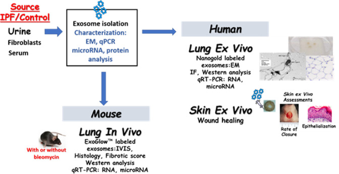
Methods: Exosomes were isolated from urine (U-IPFexo), lung tissue myofibroblasts (MF-IPFexo), serum from individuals with IPF (n=16) and age/sex-matched controls without lung disease (n=10). We analyzed microRNA expression of isolated exosomes and their in vivo bio-distribution. We investigated the effect on ex vivo skin wound healing and in in vivo mouse lung models.

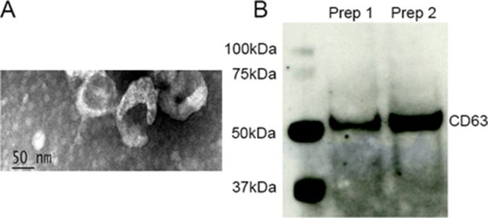
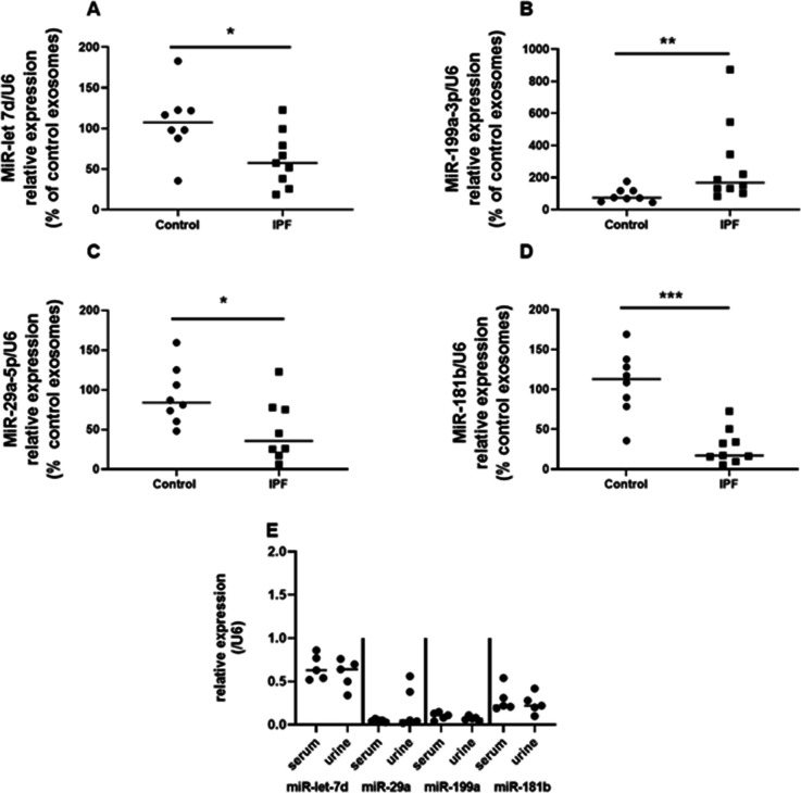
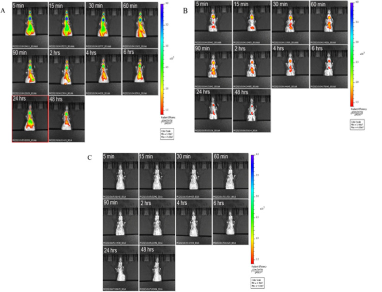
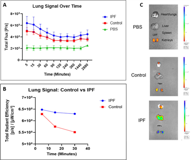
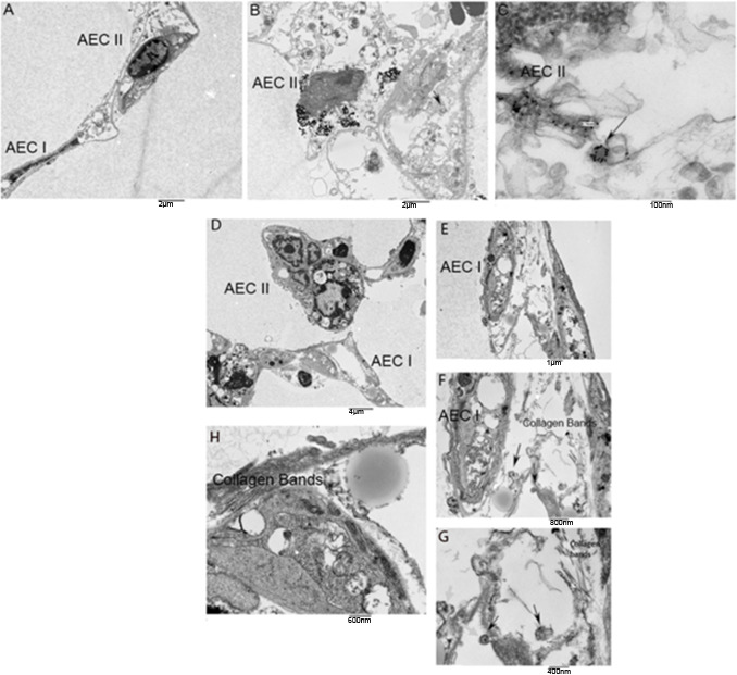
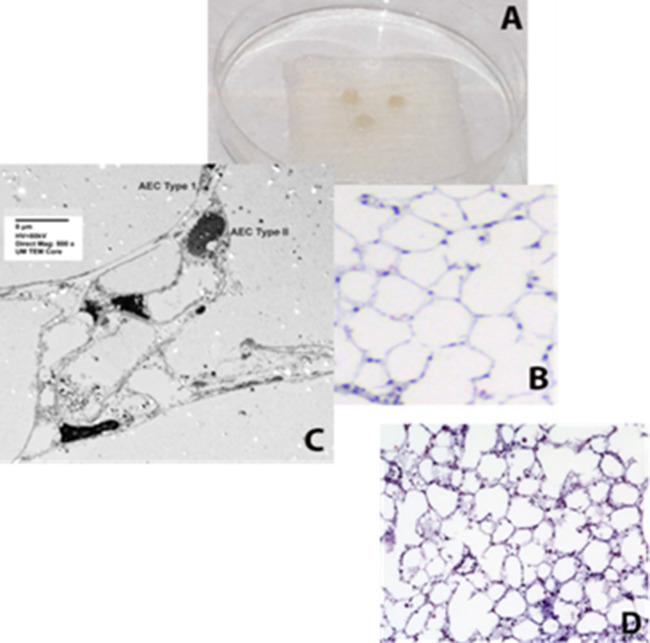
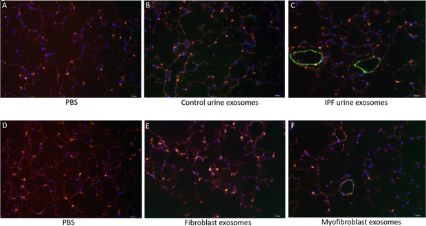
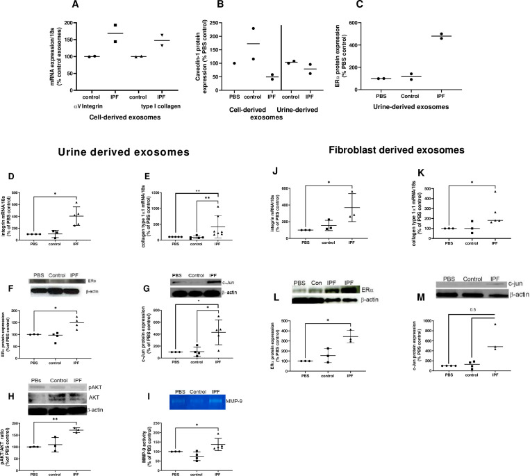
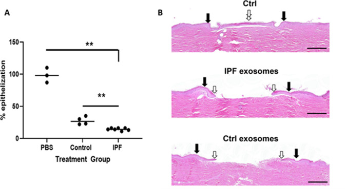
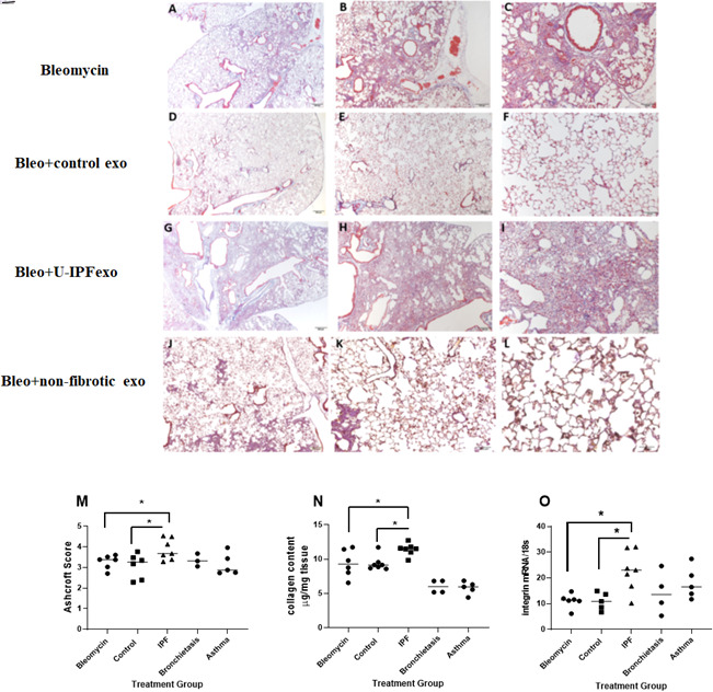
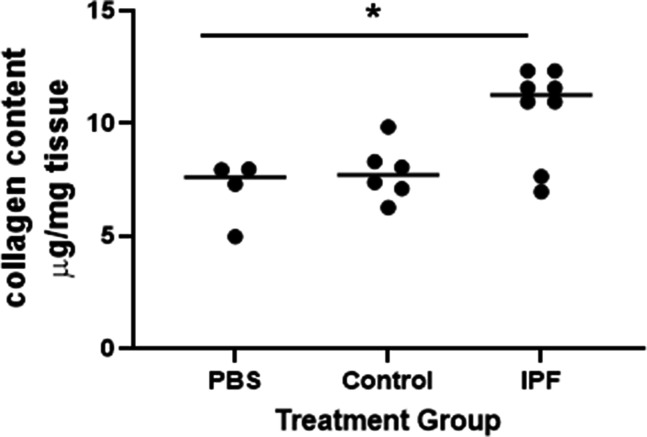
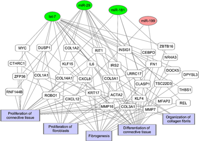
Results: U-IPFexo or MF-IPFexo expressed miR-let-7d, miR-29a-5p, miR-181b-3p and miR-199a-3p consistent with previous reports of miRNA expression obtained from lung tissue/sera from patients with IPF. In vivo bio-distribution experiments detected bioluminescent exosomes in the lung of normal C57Bl6 mice within 5 min after intravenous infusion, followed by distribution to other organs irrespective of exosome source. Exosomes labeled with gold nanoparticles and imaged by transmission electron microscopy were visualized in alveolar epithelial type I and type II cells. Treatment of human and mouse lung punches obtained from control, non-fibrotic lungs with either U-IPFexo or MF-IPFexo produced a fibrotic phenotype. A fibrotic phenotype was also induced in a human ex vivo skin model and in in vivo lung models.

Conclusions: Our results provide evidence of a systemic feature of IPF whereby exosomes contain pro-fibrotic miRNAs when obtained from a fibrotic source and interfere with response to tissue injury as measured in skin and lung models.

Funding: This work was supported in part by Lester and Sue Smith Foundation and The Samrick Family Foundation and NIH grants R21 AG060338 (SE and MKG), U01 DK119085 (IP, RS, MTC).

Keywords: Fibrosis; Urine; cell biology; exosomes; human; medicine; microRNA; mouse.


Conflict of interest statement

SE holds pending patent applications for Family - Mesenchymal stem cell-derived extracellular vesicles and uses thereof for treating and diagnosing fibrotic diseases (30309-001*), Family - Diagnostic and therapeutic uses of compositions comprising purified, enriched potent exosomes containing disease-based and therapy based signature cargo (130309-003*), and Family - Urine-derived exosomes from individuals with IPF carry pro-fibrotic cargo and impair tissue repair (130309-004*), PC, SP, XX, SS, ER, SH, JP, RS, IP No competing interests declared, JL ZenBio, SD participated on a paid role on the Scientific Advisory Board for Akron Biotech and roles on the Scientific Advisory Board for ICN2 and on the Board of Trustees for BIST. SD also received payments/stock options from Berg Pharma and stock options from Aanika Biosciences. The author has no other competing interests to declare, MT DSMB, provisional patent, NIH support, MG holds pending patent applications for Family - Mesenchymal stem cell-derived extracellular vesicles and uses thereof for treating and diagnosing fibrotic diseases (30309-001*), Family - Diagnostic and therapeutic uses of compositions comprising purified, enriched potent exosomes containing disease-based and therapy based signature cargo (130309-003*), and Family - Urine-derived exosomes from individuals with IPF carry pro-fibrotic cargo and impair tissue repair (130309-004*). MKG has a role as Chair, DMSB, Medical College of South Carolina, Mesenchymal Stem Cells in Type I Diabetes (T1D) Phase 1 trial (July 2019-present). The author has no other competing interests to declare

Figures

Figure 1.
Figure 1.. Overview of experimental details and design.
Figure 2.
Figure 2.. A Transmission electron microscopy of isolated exosomes.
Image magnification Scale bar = 50 nm. 1B. Isolated exosomes express CD63.
Figure 3.
Figure 3.. Expression of miR-let-7d (A), miR-29a-5p (B), miR 181b-3p (C) and miR-199a-3p (D) in urine-derived exosomes reveals a pattern corresponding to that reported in serum and whole lung of individuals with IPF.
PCR was performed on extracted urine-derived exosomes as described in methods. Data are graphed as relative miRNA expression normalized to U6 and percent of control expression. * p<0.05, **p<0.01, *** p<0.001 compared to control exosomes. Each point represents an individual patient exosome sample. n=5–14 individual samples/group, P values were calculated by Mann-Whitney U test. E. Urine and serum-derived exosomes isolated from the same individuals with IPF have similar miRNA expression. Exosome isolation, RNA preparation and PCR performed as described in methods. n=5 individual samples of urine and serum-derived exosomes. Paired T test analysis was performed. Figure 3—source data 1.
Figure 4.
Figure 4.. Biodistribution of circulating urine-derived exosomes.
Shown are representative in vivo bioluminescence images to study the biodistribution of ExoGlow labeled urine-derived exosomes in mice (n=3/group) at the indicated time points. Panel A=mouse injected with labeled U-IPF exo; Panel B=mouse injected with labeled urine-derived exosomes from age and sex-matched control individuals without lung disease. Panel C=mouse injected with PBS. Intensity of luminescence seen in bar from lowest (red) to highest (blue). n=3 individual exosome preparations/group.
Figure 4—figure supplement 1.
Figure 4—figure supplement 1.. Lung fluorescence intensity over time of mice injected with urine-derived exosomes from individuals with IPF (U-IPFexo), urine-derived exosomes from individuals without IPF (Control exosomes), and PBS.
C.Ex-vivo fluorescence imaging of isolated organs at 48 hours following exosome treatment in micee.
Figure 5.
Figure 5.. Representative TEM photos of lung punches.
Panels A-C show mouse lung punches injected with gold nanoparticle labeled urine-derived exosomes from age and sex-matched control subjects (without lung disease) or U-IPFexo (panels D-H). TEM revealed exosomes in alveolar epithelial cells (AEC) type I and type II. Arrows in panels C, F, and G highlight exosomes containing nanoparticles. n=2 individual exosome preparations/group.
Figure 5—figure supplement 1.
Figure 5—figure supplement 1.. Histology and trichrome staining of lung punches from C57BL6 mice.
Lung punches from control lungs shown in tissue culture dish (S1A) have normal histology (S1B, Trichrome staining ×10 mag) and structure by TEM (S1C, ×500 mag). Histology of non-injected lung punch (SA1D).
Figure 6.
Figure 6.. Immunofluorescence staining of lung punches injected with exosomes derived from urine (B–C) or myofibroblasts or fibroblasts (E–F).
Lung punches were fixed four days post injection with either PBS (panels A or D) or control urine-derived exosomes (panel B), U-IPFexo (panel C), control fibroblast (panel E) or MF-IPF exosomes (panel F). Shown are representative merged photographs at 20 x, surfactant protein C (SPC, red), αSMC actin (green) and DAPI (blue). n=3 individual exosome preparations/group. Scale bar 50 µm.
Figure 7.
Figure 7.. Fibrotic pathways are activated in lung punches after injection with urine (U-IPFexo) or myofibroblast-derived (MF-IPFexo) exosomes.
Human (A–C) and mouse lung (D–M) punches were injected with PBS alone, U-IPFexo (D–I) or MF-IPFexo (J–M) or age and sex-matched control urine exosomes or lung fibroblast exosomes from control subjects (without lung disease). Punches were collected 4 days later and processed as described in Methods. Human lung punches were injected with MF-IPFexo or fibroblast cell derived exosomes (panels A and B) or urine-derived control exosomes and U-IPFexo (panels B and C). n=2 human lung punch isolates, 2 biological exosome preparations/group. Panels D-M, n=3 mouse lung replicates/group, n=3–5 biological exosome isolates/group Data are graphed as percent PBS control. αv-integrin (panels A, D and J, Figure 7—source data 1) and collagen type 1 (panels A, E and K, Figure 7—source data 1) mRNA expression increased in punches injected with IPFexo (derived from urine or myofibroblasts). Downstream fibrotic pathways; ERα (C, F and L), activated AKT (H), c-Jun (G and M), protein expression and MMP-9 activity (I) were also stimulated by exosomes from individuals with IPF. * p<0.05, **p<0.01. p Values were calculated by Mann Whitney U test.
Figure 8.
Figure 8.. Epithelization in ex vivo wound healing is decreased by urine-derived IPF exosomes (U-IPFexo).
Human skin was wounded, injected with U-IPFexo or control (age and sex-matched from individuals without lung disease) exosomes and maintained at the air-liquid interface. Wound healing was assessed at day 4 post-wounding, a time point when exponential epithelialization occurs. (A) Data are graphed as mean with each data point representing a single wound. Experiments were performed using triplicate technical replicates and two to three biological replicates (Figure 7—source data 1). p<0.005 PBS and control compared to IPF, PBS vs control = 0.05 p values were calculated by Mann Whitney U test. (B). Photos of gross skin show visual signs of closure and correspond to the histology assessments. Black arrows point to the initial site of wounding, while white arrows point to the wound edge of the migrating epithelial tongue. Scale bars, 500 µm proportional to the image size.
Figure 9.
Figure 9.. Assessment of fibrosis in Bleomycin (Bleo) treated mice intravenously infused with exosomes derived from the urine of individuals with IPF (U-IPFexo) compared to infusion with urine exosomes derived from age and sex- matched control subjects without lung disease or urine exosomes derived from subjects with non-CF bronchiectasis or asthma (non-fibrotic lung disease).
Histological sections of lung tissue were stained with Masson’s-Trichrome as described in Materials and Methods. Representative photomicrographs (4 x, 10 x, and 20 x) of lung sections from Bleo +vehicle (panels A-C), Bleo +control exosome injected mice (panels D-F), from Bleo +U-IPFexo injected mice (panels G-I) or from non-fibrotic inducing exosomes (Bronchiectasis, panels J-L). Fibrotic score (M), collagen content (N), αvintegrin (O) increased after Bleo +U-IPFexo treatment. (M) Ashcroft scores were used to evaluate the degree of fibrosis. Data are graphed as the mean score of 32 fields/section of lung. (N) Collagen content was estimated by hydroxyproline assay as described in Methods. Data are graphed as μg/mg of lung tissue. (O) αv-integrin mRNA expression was determined by RT-PCR as a marker of fibrosis. Data are graphed normalized for 18 S content. Each data point represents an individual mouse, n=4–11 technical replicates/group and two biological replicates/group (Figure 9—source data 1) *p<0.05 compared to control exosome treatment or compared to Bleo +vehicle treatment. Data were analyzed using one-way analysis of variance (ANOVA) and Mann-Whitney U test. Scale bar panels A, D, G, J, 200 µm; panels B, E,H, K,100 µm; panels C, F,I,L, 50 µm.
Figure 9—figure supplement 1.
Figure 9—figure supplement 1.. Collagen content increases in mice receiving urine derived exosomes from individuals with IPF.
Naïve mice were treated with PBS, control or IPF urine-derived exosomes. Mice were sacrificed 21 days later as described in methods. Data are graphed as mean ± SEM. Each data point represents an individual mouse (n=2 exosome preps/group, Figure 9—figure supplement 1—source data 1). p<0.05 IPF compared to control and PBS, Data were analyzed using Mann Whitney test.
Figure 10.
Figure 10.. Potential microRNA regulated pathways leading to fibrosis.
The genes and biological processes in the network are generated from the IPF vs Control Lung dataset from NCBI GEO (GS21369) of 11 IPF samples and 6 healthy lung samples.

References

    1. Aranda JF, Canfrán-Duque A, Goedeke L, Suárez Y, Fernández-Hernando C. The mir-199-dynamin regulatory axis controls receptor-mediated endocytosis. Journal of Cell Science. 2015;128:3197–3209. doi: 10.1242/jcs.165233. - DOI - PMC - PubMed
    1. Ashcroft T, Simpson JM, Timbrell V. Simple method of estimating severity of pulmonary fibrosis on a numerical scale. Journal of Clinical Pathology. 1988;41:467–470. doi: 10.1136/jcp.41.4.467. - DOI - PMC - PubMed
    1. Batagov AO, Kurochkin IV. Exosomes secreted by human cells transport largely mrna fragments that are enriched in the 3’-untranslated regions. Biology Direct. 2013;8:12. doi: 10.1186/1745-6150-8-12. - DOI - PMC - PubMed
    1. Berrondo C, Flax J, Kucherov V, Siebert A, Osinski T, Rosenberg A, Fucile C, Richheimer S, Beckham CJ. Expression of the long non-coding RNA HOTAIR correlates with disease progression in bladder cancer and is contained in bladder cancer patient urinary exosomes. PLOS ONE. 2016;11:e0147236. doi: 10.1371/journal.pone.0147236. - DOI - PMC - PubMed
    1. Betzer O, Perets N, Angel A, Motiei M, Sadan T, Yadid G, Offen D, Popovtzer R. In vivo neuroimaging of exosomes using gold nanoparticles. ACS Nano. 2017;11:10883–10893. doi: 10.1021/acsnano.7b04495. - DOI - PubMed

Publication types