Review
. 2023 Apr 1;103(2):1565-1644.
doi: 10.1152/physrev.00059.2021. Epub 2022 Dec 1.

CGRP physiology, pharmacology, and therapeutic targets: migraine and beyond

Affiliations
Review

CGRP physiology, pharmacology, and therapeutic targets: migraine and beyond

Andrew F Russo et al. Physiol Rev. .

Abstract

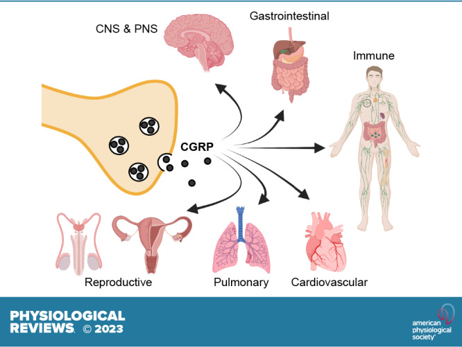
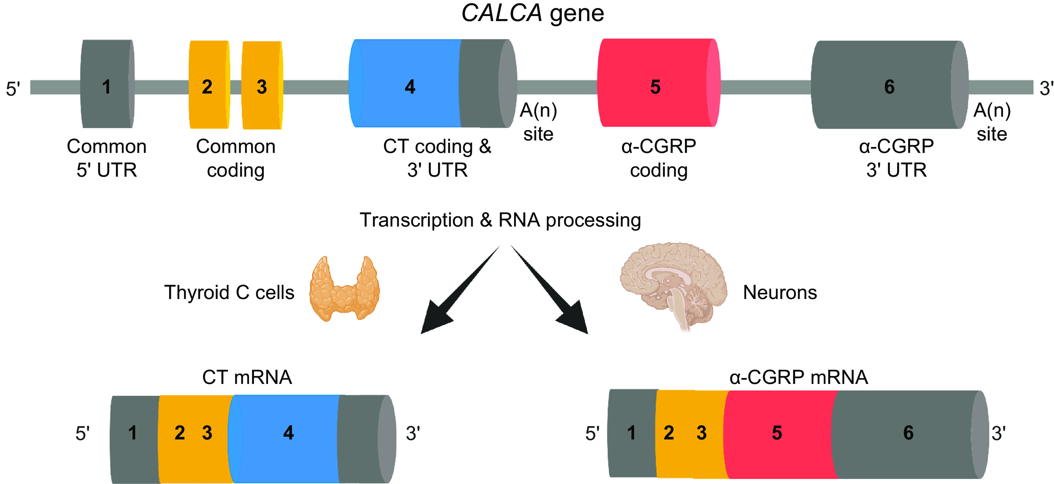
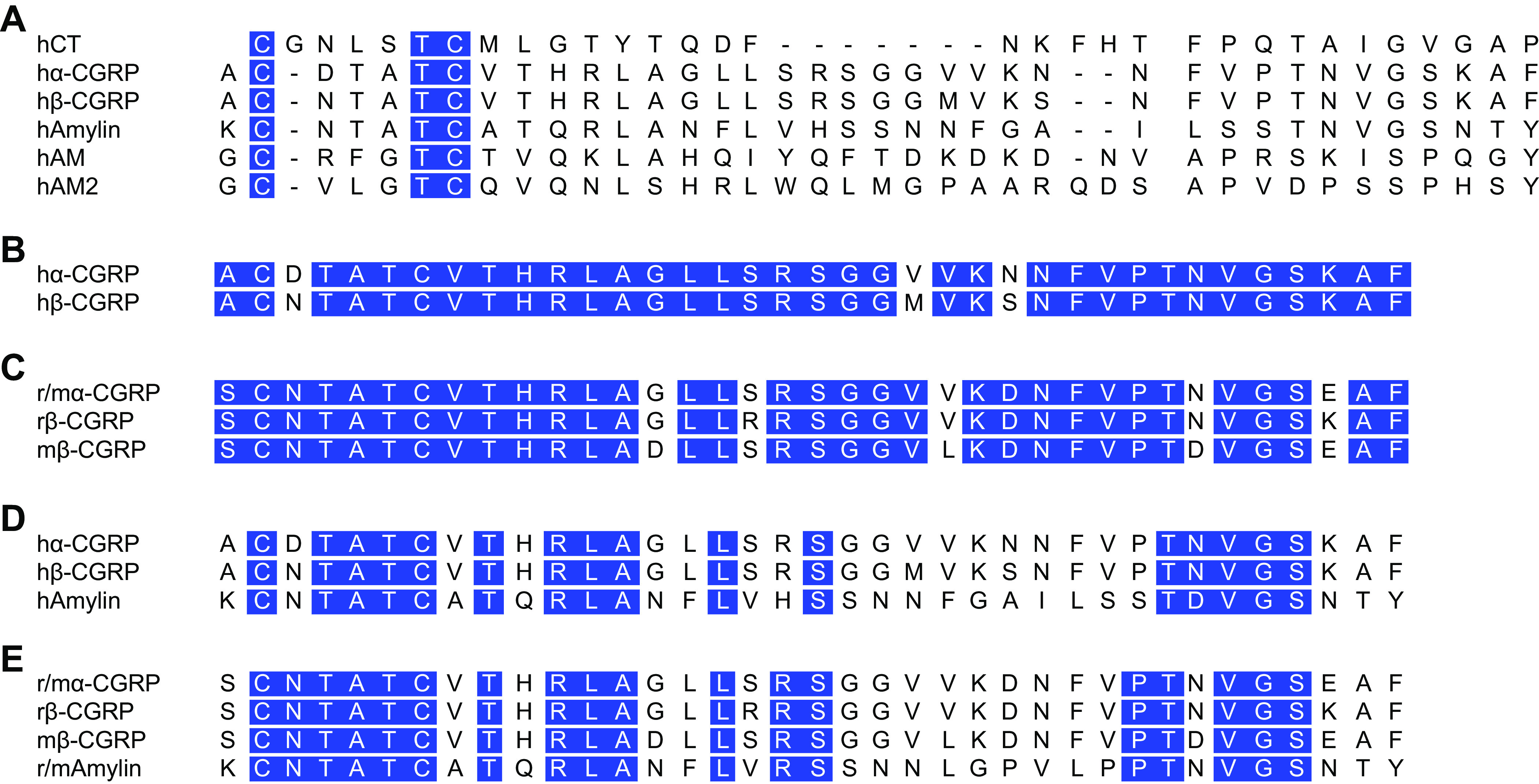
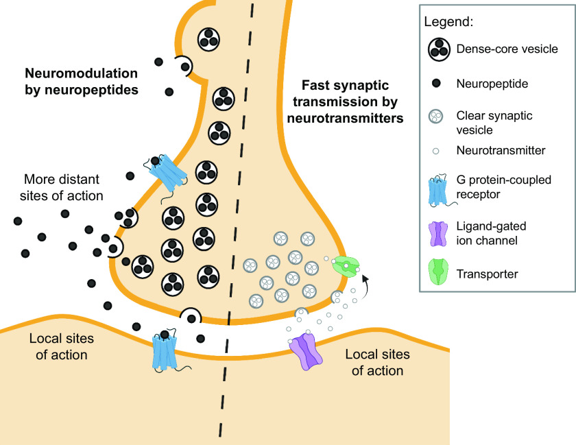
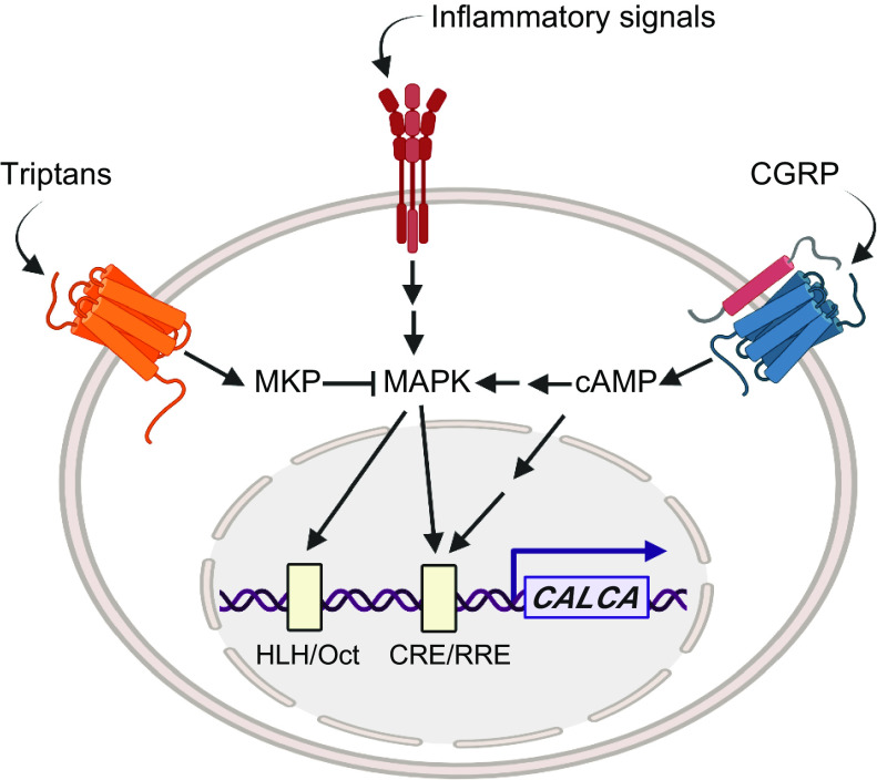
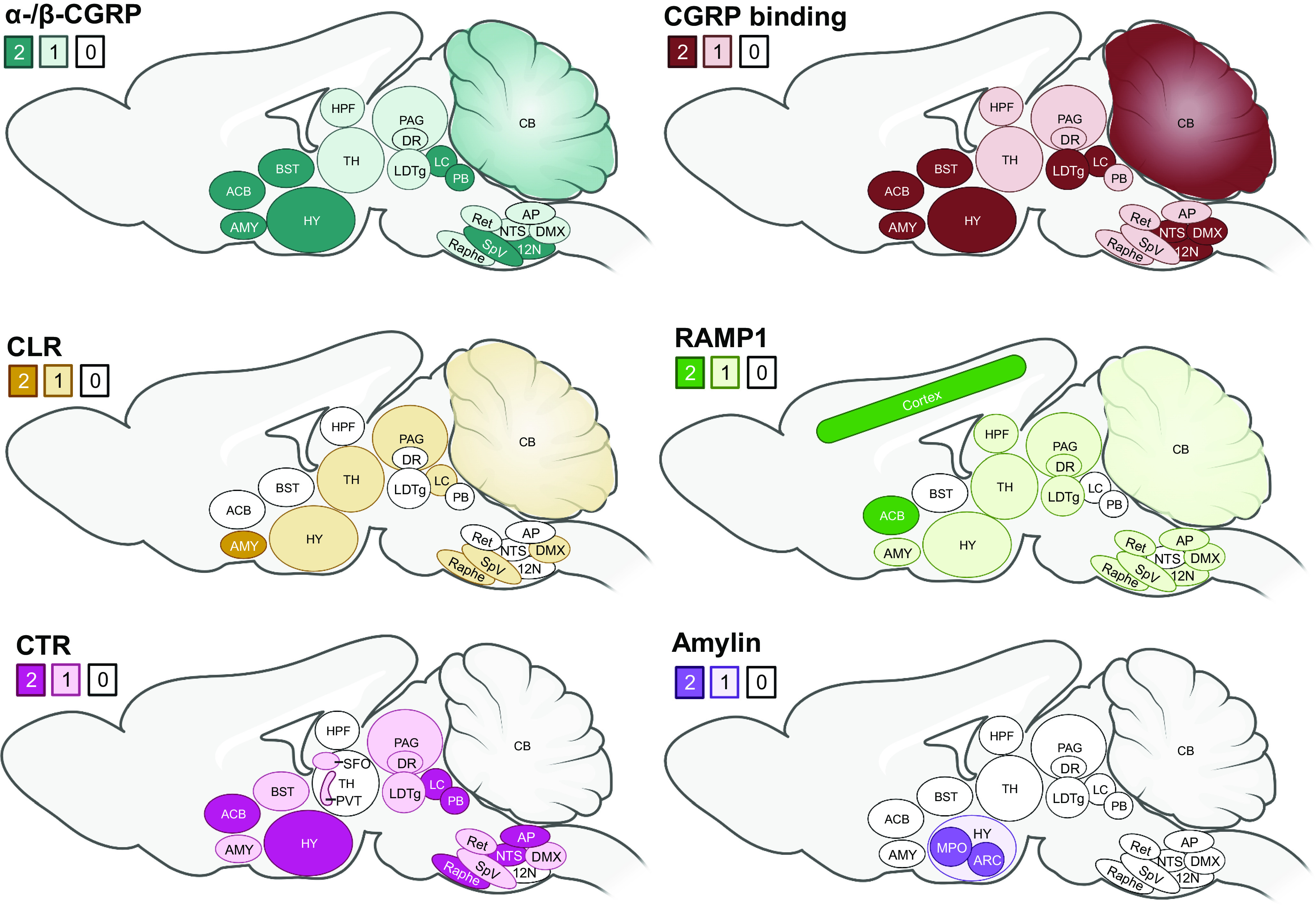
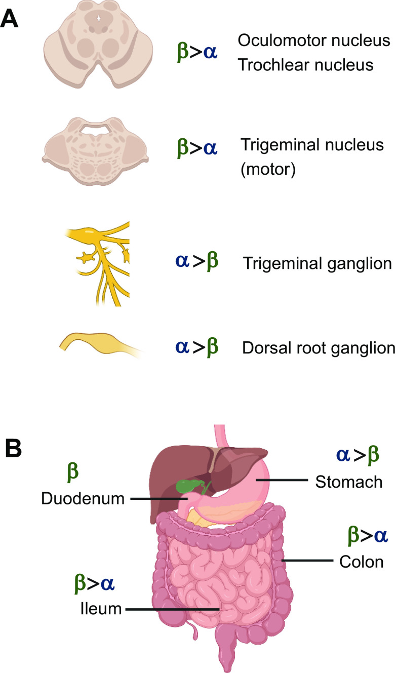
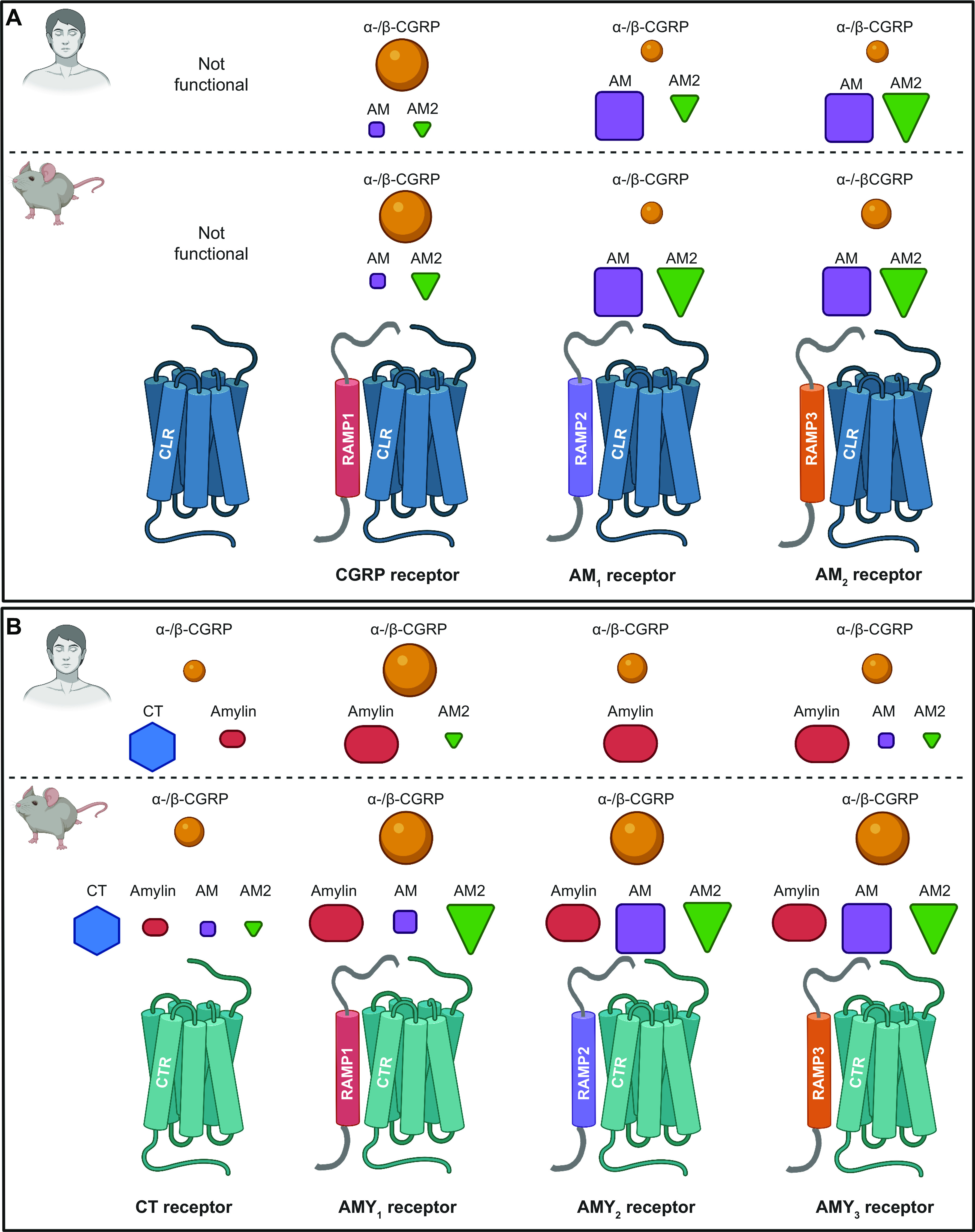
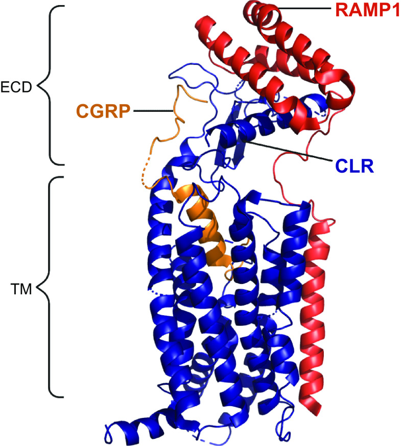
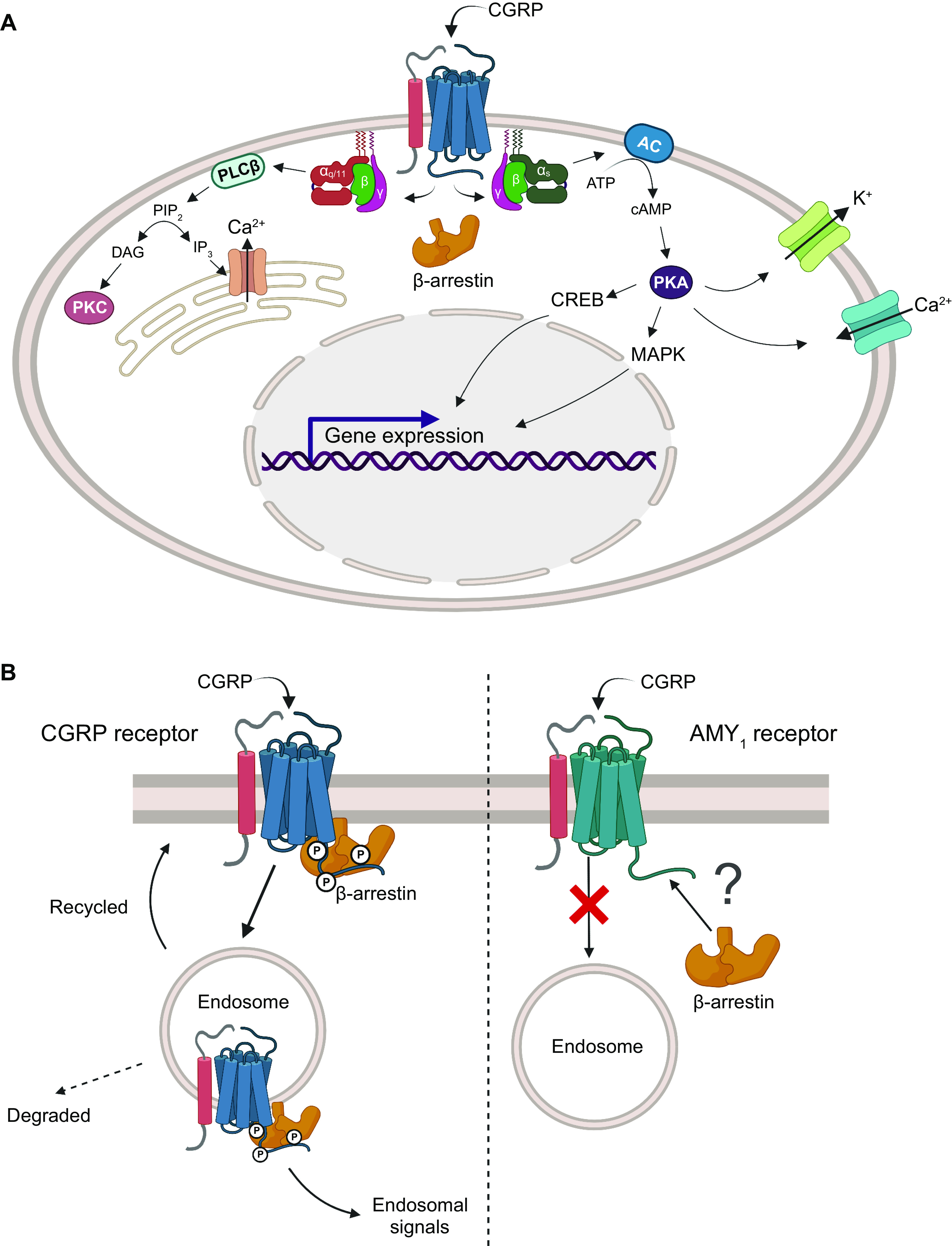
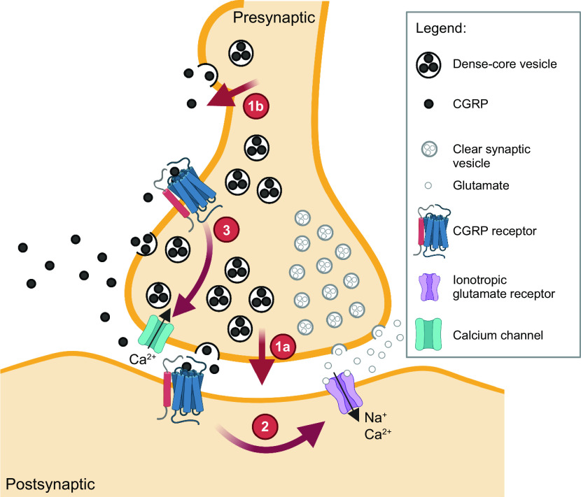
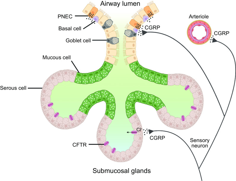
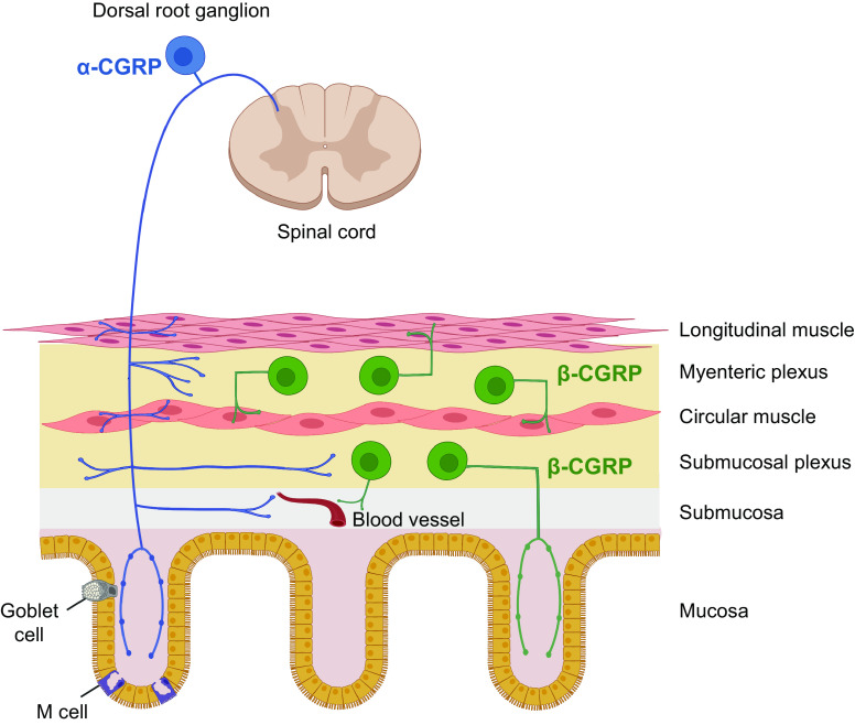
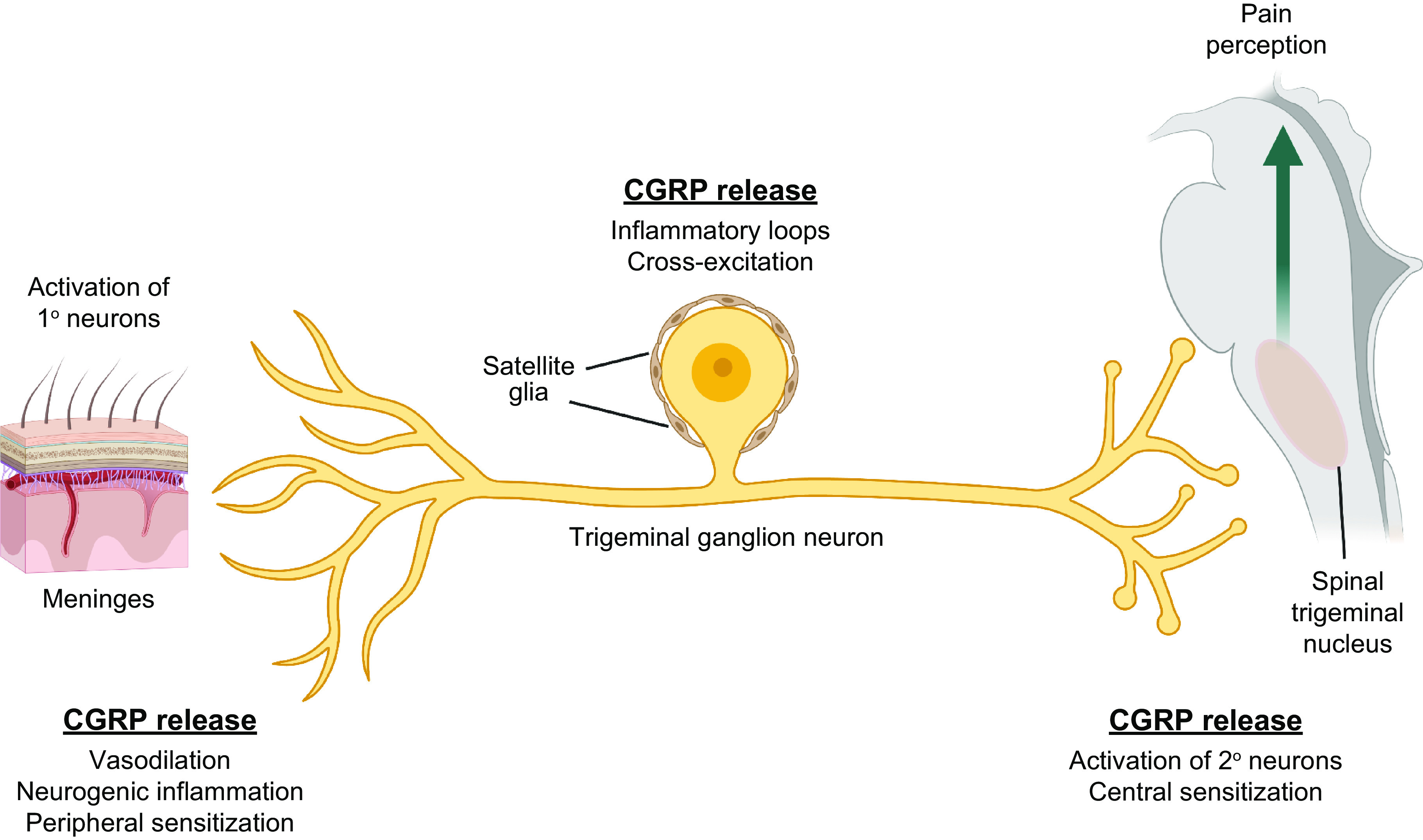
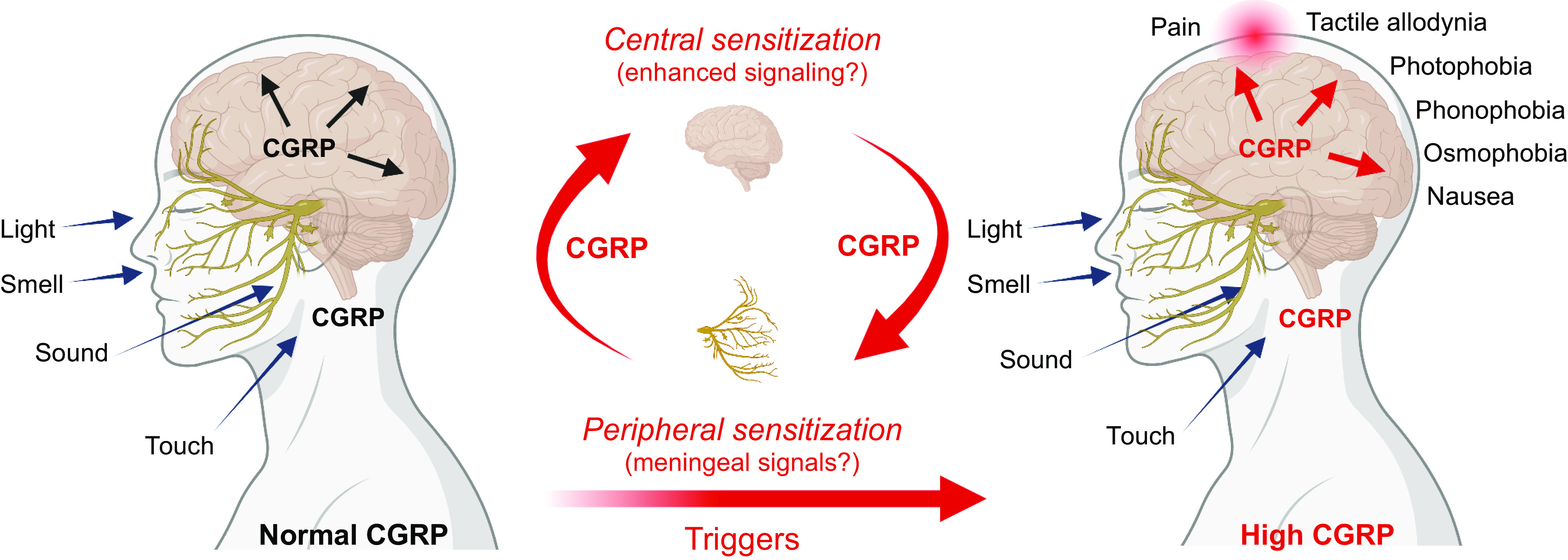
Calcitonin gene-related peptide (CGRP) is a neuropeptide with diverse physiological functions. Its two isoforms (α and β) are widely expressed throughout the body in sensory neurons as well as in other cell types, such as motor neurons and neuroendocrine cells. CGRP acts via at least two G protein-coupled receptors that form unusual complexes with receptor activity-modifying proteins. These are the CGRP receptor and the AMY1 receptor; in rodents, additional receptors come into play. Although CGRP is known to produce many effects, the precise molecular identity of the receptor(s) that mediates CGRP effects is seldom clear. Despite the many enigmas still in CGRP biology, therapeutics that target the CGRP axis to treat or prevent migraine are a bench-to-bedside success story. This review provides a contextual background on the regulation and sites of CGRP expression and CGRP receptor pharmacology. The physiological actions of CGRP in the nervous system are discussed, along with updates on CGRP actions in the cardiovascular, pulmonary, gastrointestinal, immune, hematopoietic, and reproductive systems and metabolic effects of CGRP in muscle and adipose tissues. We cover how CGRP in these systems is associated with disease states, most notably migraine. In this context, we discuss how CGRP actions in both the peripheral and central nervous systems provide a basis for therapeutic targeting of CGRP in migraine. Finally, we highlight potentially fertile ground for the development of additional therapeutics and combinatorial strategies that could be designed to modulate CGRP signaling for migraine and other diseases.

Keywords: CGRP; GPCR; migraine; neuropeptide; trigeminal nerve.


Conflict of interest statement

D.L.H. has received research support from Living Cell Technologies and AbbVie and has acted as an advisor, speaker, or consultant for Lilly, Teva, Merck, and Amgen. A.F.R. has received research support from Lundbeck and has acted as an advisor or consultant for Lilly, AbbVie, Amgen, Novartis, Lundbeck, and Schedule One Therapeutics.

Figures

None
Graphical abstract
Figure 1.
Figure 1.
RNA processing of CALCA RNA to yield calcitonin (CT) and α-calcitonin gene-related peptide (CGRP). The CALCA gene contains 6 exons. Exon 1 is a 5′ untranslated region (UTR) common to both calcitonin and α-CGRP mRNAs. Exons 2 and 3 encode NH2-terminal sequences common to both calcitonin and α-CGRP propeptides. Exon 4 encodes calcitonin-specific sequences and 3′ UTR, followed by a cleavage and polyadenylation [A(n)] site. Exon 5 encodes α-CGRP-specific sequences that are spliced onto exon 3. Exon 6 is a 3′ UTR specific to α-CGRP mRNA and is followed by an A(n) site. Calcitonin mRNA is predominantly made in thyroid C cells, and α-CGRP mRNA is predominantly made in neurons of the central and peripheral nervous system, as well as in neuroendocrine and other cells. β-CGRP is encoded by a separate gene (CALCB), not shown. Image created with BioRender.com, with permission.
Figure 2.
Figure 2.
Amino acid sequence alignment of calcitonin gene-related peptide (CGRP) and related peptides from human and rat. In A, the human (h) calcitonin (CT) peptide family are shown, omitting the NH2-terminal extensions of adrenomedullin (AM) and adrenomedullin 2 (AM2). Note that the early nomenclature referred to human α- and β-CGRP as CGRP I and CGRP II, respectively. B and C align the CGRPs to illustrate their similarities and differences. D and E compare the CGRPs to amylin. Mouse and rat α-CGRP/amylin are identical and are therefore noted as “r/m” in the figure. In all peptides, a disulfide bond is formed between the 2 NH2-terminal cysteines, and they each have a COOH-terminal amide (not shown). In all panels, identical amino acids between sets of sequences are highlighted in blue.
Figure 3.
Figure 3.
Neuropeptides versus neurotransmitters. Neuromodulation by volume transmission of neuropeptides compared with fast synaptic transmission by classical neurotransmitters. Release of neuropeptides from cell bodies, axonal varicosities, and synapses allows a broad diffusion or volume transmission of neuropeptides (released from dense core vesicles) to more distant sites of action than the more locally restricted classical neurotransmitters (released from clear synaptic vesicles). Image created with BioRender.com, with permission.
Figure 4.
Figure 4.
Regulation of CALCA gene expression. CALCA transcription is stimulated by cAMP and mitogen-activated protein kinase (MAPK) signaling pathways that are activated and repressed by extracellular signals. Calcitonin gene-related peptide (CGRP) and other ligands that act via GPCRs coupled to Gαs increase cAMP, which activates a kinase cascade leading to activation of factors at the CRE/RRE enhancer. The cAMP cascade can also activate MAPK signaling pathways that lead to activation of factors at the helix-loop-helix (HLH)/Oct enhancer. Inflammatory signals, such as tumor necrosis factor α (TNF-α) and other cytokines, activate MAPK signaling pathways leading to activation of factors at the HLH/Oct and CRE/RRE enhancers. Triptans, which act via inhibitory GPCRs, can activate MKP-1, which inhibits MAPK and hence reduces enhancer activity. Glucocorticoids, retinoic acid, and vitamin D3 also repress the gene by less defined mechanisms that inhibit the enhancers (not shown). The CGRP receptor shown represents a generic CGRP-responsive receptor, not specifically the canonical receptor or AMY1. Image created with BioRender.com, with permission.
Figure 5.
Figure 5.
Sites of α-/β-calcitonin gene-related peptide (CGRP), amylin, CGRP binding (125I-CGRP), and receptor subunit expression in the brain. Calcitonin is not shown as it is not detected in the brain. CLR, calcitonin receptor-like receptor; CTR, calcitonin receptor; HY, hypothalamus; LDTg, laterodorsal tegmental nucleus; RAMP1, receptor activity-modifying protein 1; TH, thalamus; see TABLE 1 for other abbreviations. The shaded numbers reflect approximate differences in abundance or number of binding sites, with 2 reflecting the most and 0 the least. Much data are not clear-cut. For binding, this largely relates to radiolabeled forms of CGRP in autoradiography. For peptide and receptor subunits, this is a composite of mRNA and immunoreactivity across multiple studies. The data are mostly from rodent and primate from the following citations: rodent CGRP (see TABLE 1), primate CGRP (–175), CGRP binding (, , –182), CLR (, , , , , –194), RAMP1 (, , , , , , , , , , –200), CTR (, , , , –211), and amylin (, –219). Image created with BioRender.com, with permission.
Figure 6.
Figure 6.
Selected sites of expression of α-calcitonin gene-related peptide (CGRP) and β-CGRP, with relative expression indicated. A: sites of expression in the nervous system. B: sites of expression in the gastrointestinal tract, including expression from extrinsic and intrinsic nerves. In many locations, such as the trigeminal ganglion and dorsal root ganglion, α-CGRP expression exceeds β-CGRP expression, whereas in other locations the reverse is true. Note that human anatomy is shown for simplicity but the majority of supporting literature is rat and mouse (30, 31, 232). Image created with BioRender.com, with permission.
Figure 7.
Figure 7.
Members of the calcitonin family of receptors, showing their molecular composition and relative preferences for endogenous agonist peptides, comparing human and mouse pharmacology. A: calcitonin receptor-like receptor (CLR)-based receptors with associated receptor activity-modifying proteins (RAMPs). B: calcitonin receptor (CTR)-based receptors with associated RAMPs. For A and B, CLR or CTR combines with either RAMP1, RAMP2, or RAMP3 to form receptors for calcitonin gene-related peptide (CGRP), adrenomedullin (AM), adrenomedullin 2 (AM2), calcitonin (CT), or amylin. In A and B, the agonist pharmacology at human receptors is shown at top above the dashed line and at mouse receptors at bottom. The relative size of the peptide ligand symbol indicates the relative potency. A large symbol indicates that this is the most potent/cognate ligand or is approximately equal in potency to the cognate ligand, a medium-sized symbol indicates a ligand that is approximately ≤10-fold less potent than the cognate/most potent ligand for that receptor, and the smallest-sized symbol represents a ligand that is between 10- and 100-fold less potent than the cognate/most potent ligand for that receptor. In all cases, other ligands have been tested, but their potency is >100-fold less potent than the cognate/most potent ligand for each receptor at which they have been tested and they are therefore not shown. A broad consensus is given, using species-matched ligands where possible and using data from multiple studies, as summarized in www.guidetopharmacology.org. Thus, at the human CGRP receptor, CGRP (α-CGRP and β-CGRP) is between 10- and 100-fold more potent than AM or AM2. Mouse receptors are activated by more ligands. CLR is functional only with RAMP, as it cannot reach the cell surface without it (286, 287). CTR can act as a receptor for calcitonin without RAMP. Splice variants of CTR can also partner with RAMPs to create additional receptor subtypes; the figure represents CT(a). Image created with BioRender.com, with permission.
Figure 8.
Figure 8.
Structural model of the active calcitonin gene-related peptide (CGRP) receptor, comprising calcitonin receptor-like receptor (CLR) and receptor activity-modifying protein (RAMP)1, with CGRP bound. The extracellular domain (ECD) and transmembrane bundle (TM) are both involved in binding CGRP. Gαs was bound in the structure [PDB:6e3y (342)] but is not shown in this figure. Image created with BioRender.com, with permission.
Figure 9.
Figure 9.
Acute calcitonin gene-related peptide (CGRP) signaling and regulation. A: an overview of pathways commonly activated by CGRP is shown. In many cases, activation of a signaling pathway by CGRP cannot be specifically assigned to the CGRP or AMY1 receptor. Signaling from the cell surface receptors, not endosomal receptors, is shown. B: an overview of receptor fate, after cell surface stimulation. The CGRP receptor (left) recruits β-arrestin and is internalized into endosomes. For the AMY1 receptor (right) internalization is not clearly evident, and it is unclear whether the receptor is phosphorylated or recruits β-arrestin. Image created with BioRender.com, with permission. AC, adenylate cyclase; DAG, diacylglycerol; IP3, inositol 1,4,5-trisphosphate; MAPK, mitogen-activated protein kinase; PIP2, phosphatidylinositol 4,5-bisphosphate; PKA, protein kinase A; PLC, phospholipase C.
Figure 10.
Figure 10.
Modulation of neuronal signaling by calcitonin gene-related peptide (CGRP). CGRP is released from a neuron at the synapse (1a) and from nonsynaptic sites (e.g., a varicosity) (1b). The released CGRP binds postsynaptic receptors to produce modulatory effects at the synapse by increasing glutamate receptor signaling (2) and binds presynaptic receptors in a paracrine or autocrine manner to activate calcium channels (3). The CGRP receptor shown represents a generic CGRP-responsive receptor, not specifically the canonical receptor or AMY1. Image created with BioRender.com, with permission.
Figure 11.
Figure 11.
Calcitonin gene-related peptide (CGRP) in the cardiovascular system. Overview of CGRP’s actions in the cardiovascular system, with local vasodilatory mechanisms on left and systemic actions that are generally protective on right. For vasodilation, CGRP released from perivascular sensory neurons activates signaling pathways in vascular smooth muscle cells (VSMCs) that activate K+ channels to cause relaxation and endothelial cells to release nitric oxide (NO), which also causes VSMC relaxation. The systemic cardiovascular protective functions include vasodilation and other actions. AC, adenylate cyclase; eNOS, endothelial nitric oxide synthase; PKA, protein kinase A; PKG, protein kinase G; sGC, soluble guanylate cyclase. Image created with BioRender.com, with permission.
Figure 12.
Figure 12.
Calcitonin gene-related peptide (CGRP) in the lung. Sensory nerves containing CGRP (black circles) lie within the submucosa, where they innervate blood vessels and the airway epithelium, including submucosal glands. CGRP is also released from pulmonary neuroendocrine cells (PNECs). Among the actions of CGRP is CFTR-dependent glandular fluid secretion that likely protects the airway from secondary infection, stimulation of mucus secretion from goblet cells, and stimulation of glandular stem cell progenitors, such as basal cells, to proliferate to become transient amplifying cells that help ameliorate epithelial injuries. Image created with BioRender.com, with permission.
Figure 13.
Figure 13.
Extrinsic and intrinsic calcitonin gene-related peptide (CGRP)-containing neurons of the intestine. Extrinsic sensory neurons from the dorsal root ganglion (DRG) contain primarily α-CGRP and innervate all layers of the intestinal wall: intrinsic enteric neurons in myenteric and submucosal plexuses, blood vessels in the submucosal plexus, circular and longitudinal smooth muscle layers, and the mucosa, including microfold (M) cells in Peyer’s patches and mucus-producing goblet cells in the colon. Intrinsic enteric neurons contain primarily β-CGRP and are found in both the myenteric and submucosal plexuses. Both α- and β-CGRP neurons innervate the blood vessels. Image created with BioRender.com, with permission.
Figure 14.
Figure 14.
Calcitonin gene-related peptide (CGRP) in the trigeminovascular system. CGRP acts at 3 distinct regions within the trigeminovascular system. Upon activation, afferent trigeminal fibers release CGRP in the dura and pia layers of the meninges. CGRP actions on blood vessels, resident immune cells, glial Schwann cells, and trigeminal fibers can cause vasodilation and neurogenic inflammation and potentially lead to further CGRP release and peripheral sensitization. Within the ganglia, CGRP can act on satellite glia and neural cell bodies to initiate inflammatory loops and cross-signaling that could further CGRP release and excitation of nociceptive cell bodies. In the spinal trigeminal nucleus, CGRP modulates glutamatergic signals that can lead to activation of second-order neurons and central sensitization. These second-order neurons project to higher brain regions, leading to pain perception. Image created with BioRender.com, with permission.
Figure 15.
Figure 15.
Proposed roles of central and peripheral calcitonin gene-related peptide (CGRP) in migraine. Normal levels of CGRP in the central nervous system (CNS) and peripheral nervous system (PNS) are proposed to modulate sensory inputs. When central or peripheral CGRP levels are elevated, perhaps in response to CNS signals, such as repeated cortical spreading depression (CSD) events, or peripheral signals, such as an altered trigeminovascular microenvironment, peripheral and central sensitization can occur, possibly in a reinforcing loop of positive feedback and feedforward signals between the PNS and CNS as indicated. The increased signaling leads to heightened sensitivity to sensory input that is manifested as photophobia and other migraine symptoms, with pain occurring possibly as a protective response to drive the person to reduce sensory input.

Similar articles

Cited by

References

    1. Rosenfeld MG, Amara SG, Roos BA, Ong ES, Evans RM. Altered expression of the calcitonin gene associated with RNA polymorphism. Nature 290: 63–65, 1981. doi:10.1038/290063a0. - DOI - PubMed
    1. Amara SG, Jonas V, Rosenfeld MG, Ong ES, Evans RM. Alternative RNA processing in calcitonin gene expression generates mRNAs encoding different polypeptide products. Nature 298: 240–244, 1982. doi:10.1038/298240a0. - DOI - PubMed
    1. Rosenfeld MG, Mermod JJ, Amara SG, Swanson LW, Sawchenko PE, Rivier J, Vale WW, Evans RM. Production of a novel neuropeptide encoded by the calcitonin gene via tissue-specific RNA processing. Nature 304: 129–135, 1983. doi:10.1038/304129a0. - DOI - PubMed
    1. Grunditz T, Ekman R, Håkanson R, Rerup C, Sundler F, Uddman R. Calcitonin gene-related peptide in thyroid nerve fibers and C cells: effects on thyroid hormone secretion and response to hypercalcemia. Endocrinology 119: 2313–2324, 1986. doi:10.1210/endo-119-5-2313. - DOI - PubMed
    1. Uddman R, Luts A, Sundler F. Occurrence and distribution of calcitonin gene-related peptide in the mammalian respiratory tract and middle ear. Cell Tissue Res 241: 551–555, 1985. doi:10.1007/BF00214575. - DOI - PubMed

Publication types

MeSH terms

Substances