. 2022 Dec 6;119(49):e2123487119.
doi: 10.1073/pnas.2123487119. Epub 2022 Dec 1.

Comprehensive evaluation of human-derived anti-poly-GA antibodies in cellular and animal models of C9orf72 disease

Affiliations

Comprehensive evaluation of human-derived anti-poly-GA antibodies in cellular and animal models of C9orf72 disease

Melanie Jambeau et al. Proc Natl Acad Sci U S A. .

Abstract

Hexanucleotide G4C2 repeat expansions in the C9orf72 gene are the most common genetic cause of amyotrophic lateral sclerosis and frontotemporal dementia. Dipeptide repeat proteins (DPRs) generated by translation of repeat-containing RNAs show toxic effects in vivo as well as in vitro and are key targets for therapeutic intervention. We generated human antibodies that bind DPRs with high affinity and specificity. Anti-GA antibodies engaged extra- and intra-cellular poly-GA and reduced aggregate formation in a poly-GA overexpressing human cell line. However, antibody treatment in human neuronal cultures synthesizing exogenous poly-GA resulted in the formation of large extracellular immune complexes and did not affect accumulation of intracellular poly-GA aggregates. Treatment with antibodies was also shown to directly alter the morphological and biochemical properties of poly-GA and to shift poly-GA/antibody complexes to more rapidly sedimenting ones. These alterations were not observed with poly-GP and have important implications for accurate measurement of poly-GA levels including the need to evaluate all centrifugation fractions and disrupt the interaction between treatment antibodies and poly-GA by denaturation. Targeting poly-GA and poly-GP in two mouse models expressing G4C2 repeats by systemic antibody delivery for up to 16 mo was well-tolerated and led to measurable brain penetration of antibodies. Long-term treatment with anti-GA antibodies produced improvement in an open-field movement test in aged C9orf72450 mice. However, chronic administration of anti-GA antibodies in AAV-(G4C2)149 mice was associated with increased levels of poly-GA detected by immunoassay and did not significantly reduce poly-GA aggregates or alleviate disease progression in this model.

Keywords: C9orf72; amyotrophic lateral sclerosis; dipeptide repeat proteins; frontotemporal dementia; immunotherapy.


Conflict of interest statement

The authors declare a competing interest. K.D.M., N.C., P.B., C.H., R.M.N., F.M., and J.G. are employees of Neurimmune and S.N., Y.G., S.D., I.D.-L., M.W.K., and M.R. are employees of Biogen.

Figures

Fig. 1.
Fig. 1.
chα-GA3 is internalized via vesicular compartments in human neurons, but does not alter GA50-GFP vesicular localization. (A) Confocal images of human neural cultures expressing inducible GA50-GFP, treated for 3 d with chα-GA3 or IgG control and stained with a secondary α-human antibody (red). White arrows show antibody/GA50-GFP double-positive cells. Arrowheads show antibody uptake in cells not expressing GA50-GFP. (Scale, 20 µm.) (B) Quantification of the percentage of GA50-GFP expressing cells with internalized antibody. Four replicates, unpaired t test, *** P ≤ 0.001. (C) Confocal images after staining with either α-RAB7 (late endosomes) or α-LAMP1 (lysosomes). Chimeric antibodies are detected with α-mouse antibody (red). (D) Deconvoluted STED image from a neuron expressing GA50-GFP treated with chα-GA3 and stained with RAB7. Inset of the STED image (Upper panel) and the created 2D surface (Lower panels) are enlarged from the original site (white box). (E) The percentage of GA50-GFP particles containing a vesicle within a 100 nm radius were considered as colocalized with the indicated vesicle. Each dot represents a field of view. Mann and Whitney U test (Rab7 P = 0.087 and Lamp1 P = 0.544).
Fig. 2.
Fig. 2.
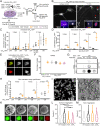
Poly-GA and antibodies form large heterocomplexes in long-term treated neuronal cultures. (A) Experimental strategies to test the effect of antibody treatment on human neurons expressing GA50-GFP. (B) Immunofluorescence showing GA50-GFP in neurons treated for 21 d with either an IgG control (Left panel) or chα-GA1 (Right panel). The presence of large irregular extracellular GA50-GFP structures only in samples treated with chα-GA1 antibody (red arrowheads and Bottom Right Inset). Insets illustrate different GA50-GFP intracellular structures observed across all conditions, including preinclusions (white arrowheads). (Scale, 10 and 5 µm.) (C) Quantification of intracellular GA50-GFP structures normalized to the number of nuclei, 36 images per well, and 3–6 wells per condition. (D) Quantification of GA50-GFP extracellular structures normalized to the number of nuclei, 5 images per well, and 3–6 wells per condition. Means +/− SD, beta-binomial test. (E) Confocal imaging of an chα-GA1 extracellular structure colocalizing with GA50-GFP (Upper row), and an IgG control extracellular structure, not colocalizing with GA50-GFP (Lower row). Antibodies were detected with α-mouse-Alexa 647. (F) Cell viability of human neurons expressing GA50-GFP treated with chα-GA1,3 or control antibody for 3 d. (G) Representative blots and (H) quantification of a filter retardation assay for human neurons treated during 3, 7, or 21 d with chα-GA1 or IgG control. Intensity of each replicate was normalized to the 3 d IgG control samples. Unpaired t test is used on the log10(x+1) transformed data. (I) Epi-fluorescent image of GA50-GFP aggregates isolated from transiently transfected HEK293T cells. (J) Isolated GA50-GFP aggregates visualized via SEM imaging. (K) Representative SEM and corresponding IF images of nontreated (NT) or Alexa Fluor 647-labeled IgG control-, α-GA1-, and α-GA3-treated GA50-GFP aggregates. (L and M) Surface porosity (L) and total area (M) quantifications of the antibody-treated GA50-GFP aggregates. NT n = 139; IgG control n = 125; α-GA1 n = 183; α-GA3 n = 193. One-way ANOVA followed by Tukey’s test. P > 0.05 (no indication), ** P ≤ 0.01, *** P ≤ 0.001.
Fig. 3.
Fig. 3.
Immunoassay with sample denaturation identifies elevated levels of poly-GA in brains of mice treated with α-GA antibodies. (A) Scheme of chronic antibody treatment in C9450 mice receiving intraperitoneal injection of PBS, chα-GA1, chα-GA3, or chα-GP1 antibodies from 3 to 19 mo of age. (B) Scheme showing drug–antibody interference in the measurement of poly-GA protein levels using a sandwich-ELISA assay (Left panel). The right panel illustrates the effect of sample denaturation prior to measurements. (C) Adaptation of a mouse brain fractionation protocol to denature samples and avoid interference of the antibody treatment by ELISA. Fractions highlighted in bold were analyzed. (D, E) Poly-GA immunoassay from the supernatant S1 (D) and after ultracentrifugation (supernatant S2 and pellet P2) (E) of brains from 7-mo-old C9450 (n ≥ 8 per group). (F) Scheme of chronic antibody treatment by intraperitoneal injection of chα-GA1 or IgG control to AAV-(G4C2) mice from 2 to 12 mo of age. (GJ) Poly-GA immunoassay from the supernatant S1 (G, I) and after ultracentrifugation (supernatant S2 and pellet P2) (H, J) of brains from AAV-(G4C2) mice at 4 mo (n ≥ 3 per group for AAV-(G4C2)2 and n ≥ 6 for AAV-(G4C2)149) (G, H) and 12 mo (n ≥ 12 per group for AAV-(G4C2)2 and n ≥ 25 for AAV-(G4C2)149) (I, J). (K, L) Poly-GP immunoassay from the supernatant S1 (K) and after ultracentrifugation (supernatant S2 and pellet P2) (L) of brains from AAV-(G4C2) mice at 12 mo of age. Mean ± SD, one-way ANOVA followed by Tukey’s test. P > 0.05 (ns for not significant), ** P ≤ 0.01, *** P ≤ 0.001, and **** P ≤ 0.0001.
Fig. 4.
Fig. 4.
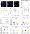
Chronic administration of α-GA antibodies did not reduce poly-GA aggregates load and impacted only a subset of phenotypes in one of two C9orf72 mouse models. (A) Immunofluorescence of poly-GA in the motor cortex of 12-mo-old AAV-(G4C2) mice treated with chα-GA1 or IgG control. (B) Percent area occupied by poly-GA aggregates detected with an N-terminal-poly-GA antibody in the cortex (n ≥ 3 per group for AAV-(G4C2)2 and n ≥ 7 for AAV-(G4C2)149). (Scale, 25 μm.) Mean ± SD, one-way ANOVA followed by Tukey’s test. (C) Survival Kaplan–Meier curves of C9450 and wild-type mice receiving injections of PBS, chα-GA1, chα-GA3and chα-GP1 antibodies for 16 mo. (D) Distance traveled in the open-field by 18-mo-old males (n ≥ 8 per group). Mean ± SD, Kruskal–Wallis test followed by Dunnett’s test. (E) Nuclei quantification in the hippocampal CA1 region in 19-mo-old mice (n ≥ 4 mice per group; n ≥ 3 matched sections per mouse). Mean ± SD, one-way ANOVA followed by Dunnett’s test. (F) Survival Kaplan–Meier curve of AAV-(G4C2) mice receiving injections of chα-GA1 or IgG control for 10 mo. (GI) Distance traveled (G), velocity of movement (H) and time spent moving (I) in the open-field (n ≥ 16 per group for AAV-(G4C2)2 and n ≥ 24 for AAV-(G4C2)149). (J) Time taken to fall from inverted grid by 9-mo-old AAV-(G4C2) female mice (n ≥ 8 per group). (K) Brain weights of AAV-(G4C2) mice treated for 10 mo. Mean ± SD, one-way ANOVA followed by Dunnett’s test. P > 0.05 (no indication or ns for not significant), * P ≤ 0.05, ** P ≤ 0.01, *** P ≤ 0.001.

References

    1. Renton A., et al. , A hexanucleotide repeat expansion in C9orf72 is the cause of chromosome 9p21-linked Als-Ftd. Neuron 72, 257–268 (2011). - PMC - PubMed
    1. DeJesus-Hernandez M., et al. , Expanded Ggggcc hexanucleotide repeat in noncoding region of C9orf72 causes chromosome 9p-linked Ftd and Als. Neuron 72, 245–256 (2011). - PMC - PubMed
    1. Taylor J. P., Brown R. H. Jr., Cleveland D. W., Decoding als: From genes to mechanism. Nature 539, 197–206 (2016). - PMC - PubMed
    1. Ash P. E., et al. , Unconventional translation of C9orf72 Ggggcc expansion generates insoluble polypeptides specific to C9ftd/Als. Neuron 77, 639–646 (2013). - PMC - PubMed
    1. Mori K., et al. , The C9orf72 Ggggcc repeat is translated into aggregating dipeptide-repeat proteins in Ftld/Als. Science 339, 1335–1338 (2013). - PubMed

Publication types

LinkOut - more resources