. 2022 Dec 6;119(49):e2209531119.
doi: 10.1073/pnas.2209531119. Epub 2022 Dec 1.

A novel post-developmental role of the Hox genes underlies normal adult behavior

Affiliations

A novel post-developmental role of the Hox genes underlies normal adult behavior

Abdul Raouf Issa et al. Proc Natl Acad Sci U S A. .

Abstract

The molecular mechanisms underlying the stability of mature neurons and neural circuits are poorly understood. Here we explore this problem and discover that the Hox genes are a component of the genetic program that maintains normal neural function in adult Drosophila. We show that post-developmental downregulation of the Hox gene Ultrabithorax (Ubx) in adult neurons leads to substantial anomalies in flight. Mapping the cellular basis of these effects reveals that Ubx is required within a subset of dopaminergic neurons, and cell circuitry analyses in combination with optogenetics allow us to link these dopaminergic neurons to flight control. Functional imaging experiments show that Ubx is necessary for normal dopaminergic activity, and neuron-specific RNA-sequencing defines two previously uncharacterized ion channel-encoding genes as potential mediators of Ubx behavioral roles. Our study thus reveals a novel role of the Hox system in controlling adult behavior and neural function. Based on the broad evolutionary conservation of the Hox system across distantly related animal phyla, we predict that the Hox genes might play neurophysiological roles in adult forms of other species, including humans.

Keywords: Drosophila; Hox genes; flight; neuron; post-mitotic.


Conflict of interest statement

The authors declare no competing interest.

Figures
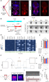
Fig. 1.
Fig. 1.
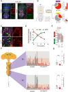
Ubx is necessary for normal flight maintenance in adult Drosophila. (A) Confocal images of the whole VNC of adult Drosophila showing expression of Ubx protein in specific regions of the adult VNC (T1-T3, indicate the respective thoracic segments). (B) Strategy for conditional neuronal downregulation of Ubx in the adult. Flies expressing UbxRNAi under temperature-sensitive Gal80ts repression, were exposed to elevated temperatures (30°C) upon hatching to allow active downregulation of Ubx. At permissive temperature (18°C) Gal80ts protein is functional and represses expression of UbxRNAi (‘Ubx ON’); conversely, expression of UbxRNAi is activated in cells at temperatures above 30°C (‘Ubx OFF’). Behavioral experiments were performed on adult males and females in Ubx ON/OFF modes. (CE) Evaluation of flight performance on tethered Drosophila. (C) Representative flight duration is indicated by video snapshots of tethered WT, pan-neuronal (Tub-Gal80ts;Elav-Gal4, UbxRNAi), and dopaminergic (Tub-Gal80ts;TH-Gal4, UbxRNAi) UbxRNAi expressing flies, at 30°C. The orange arrow indicates the induction stimulus (air-puff), and WT flies are able to fly above 10 min compared to UbxRNAi expressing flies. (D and E) Flight duration represented as a heat map, and as averages of flight duration at week 2, for flies expressing UbxRNAi in different nerve cells (Ubx/elav/Vglut/TH/Cha>UbxRNAi) compared to controls (UAS-UbxRNAi) under Gal80ts repression (week 1 = 6–7 d; week 2 = 8–10 d; week 3 = 14–15 d; n > 20). (F) Cartoon illustrating the forced flight test procedure (for details, see Materials and Methods). Flies are shown as brown dots on the cylinder and membrane. (G) Average forced flight index at week 2 of flies expressing Tub-Gal80ts; UbxRNAi in different neuronal populations and reared at 30°C compared to controls (Tub-Gal80ts; UbxRNAi) (Each data point represents a group of n > 12 flies). (H) Representative raw images of GCaMP fluorescence (warmer color) in flight muscles. (I) Representative calcium signal traces in dorsal longitudinal flight muscle (Dlm) from control (Mef2LexA,GCaMP6mLexAOP/Tub-Gal80ts;TH-Gal4/FLP) and experimental (Mef2LexA,GCaMP6mLexAOP/Tub-Gal80ts; TH-Gal4/UbxRNAi) flies. (J) Normalized mean of GCaMP signals maximum during sustained flight (n = 5–10). All error bars represent SEM. Significant values in all figures based on Mann–Whitney U test or one-way ANOVA with the post hoc Tukey–Kramer test: P < 0.05, ∗∗P < 0.01, ∗∗∗P < 0.001, and ∗∗∗∗P < 0.0001.
Fig. 2.
Fig. 2.
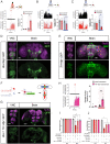
Ubx is expressed in adult VNC Dopaminergic (TH) neurons. (A) Confocal images of the VNC of adult Drosophila showing expression of Ubx in TH neurons. TH neurons are labeled by GFP driven by TH-Gal4. (B) Ubx positive TH neurons in the VNC. At the Left, a cartoon showing the presence of Ubx positive TH neurons (red dot circles) in both ventral (yellow) and dorsal (purple) sides of VNC. Ubx is expressed in ~29% (5 ± 0.9) (ventral) and 70% (17 ± 0.4) (dorsal) of dopamine neurons in the VNC (n = 8–10) (Right). (CE) Conditional reduction of Ubx expression by UbxRNAi in TH neurons. (C) Confocal imaging of a region of the adult VNC with high Ubx protein expression, showing the effects of UbxRNAi treatment at 30°C on Ubx expression. Genotypes: control (TH-Gal4; UAS-Nls::GFP, UAS-FLP); UbxRNAi (TH-Gal4; UAS-Nls::GFP,Tub-Gal80ts; UAS-UbxRNAi). Note the apparent decrease in red signal within TH neurons (white circles) (flies were 8–10 d old, n = 5–6). (D) Kinetics of Ubx and GFP expression in adult flies expressing UbxRNAi in TH neurons (TH-Gal4; UAS-NLS::GFP,Tub-Gal80ts; UAS-UbxRNAi) in non-permissive conditions (30°C) over time (1–3 wk). Note that as time elapses the reduction in Ubx expression becomes more pronounced, and, in contrast, expression of GFP becomes stronger. (E) Quantification of Ubx expression in normal and UbxRNAi conditions at permissive (18°C) and non-permissive (30°C) temperatures for Gal80ts repression (at week 2). Note that, at low temperature, Ubx levels are not different between control and UbxRNAi lines; in contrast, GFP expression is highly reduced due to Gal80ts repression. At high temperatures, Ubx expression is significantly downregulated. (F) Quantification of Ubx in individual nuclei in the adult VNC. (Left) Diagram of the adult VNC (anterior to the top) illustrating the approximate positions of TH neurons (green). (Middle) Quantification of Ubx expression in individual nuclei in T2 (Top) and T3 (Bottom) of control (grey) and TH>UbxRNAi (red) dopaminergic neurons, showing a clear reduction of Ubx expression in the population of TH nuclei expressing the UbxRNAi construct. (G) Expression levels of Ubx protein in individual TH neurons with normal (grey) or down-regulated (red) Ubx expression in T2 (top) and T3 (Bottom) regions of the adult VNC demonstrating a significant reduction in Ubx expression under UbxRNAi treatment in both segments. Error bars represent SEM. Significant values in all figures based on Mann–Whitney U test: P < 0.05, ∗∗∗P < 0.001, and ∗∗∗∗P < 0.0001.
Fig. 3.
Fig. 3.
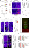
Modulation of neural activity of Ubx+ TH neurons affects flight. (A) Optogenetic activation (red shade) of TH neurons expressing channelrhodopsin CsChrimson (TH>CsChrimson) induces spontaneous flight (Top: Cartoon representation of experimental results) (B and C). Optogenetic inhibition (blue shade) of TH neurons (TH>GtACR2) (b) and Ubx+ cells (Ubx>GtACR2) (c) by expressing GtACR2 reduces flight. In each figure, Left panels represent wing flapping frequency, and Right panels depict a histogram of average frequencies. Control flies are UAS-CsChrimson and UAS-GtACR (n = 6). (and E) Confocal images showing TH (d) and Ubx cells (e) in VNC and brain. (F) Schematic illustration of the Split-Gal4 system based on the complementation between the two functional domains of Gal4, the DNA-binding (DBD) and transcription-activation (AD) domains. Each domain is fused to a heterodimerizing leucine zipper (Zip+ or Zip) that promotes the fusion of the two domains when expressed in the same cell reconstituting transcriptional activity. This technique was used to generate UbxTH-Gal4 lines. (G) Confocal images of the fly VNC (ventral side) and brain (anterior side) showing the UAS-Myr::GFP expression pattern driven by UbxGal4.DBD ∩ pleGal4.AD (UbxTH-Gal4) defines a subset of TH neurons that express Ubx. (H) Flight patterns of flies before and after optogenetic activation of Ubx+ TH cells, (n = 8–9). (I and J) Flies with Ubx positive neurons inhibited by expression of the potassium channel encoded by the Homo sapiens KCNJ2 gene (Kir2.1) show reduced flight duration (I) and forced flight index (J); a reduction is also observed when Kir2.1 is expressed in the TH-Gal4 domain, and, notably, in the UbxTH intersectional domain. See also Movie S4 (related to Fig. 3A), Movie S5 (related to Fig. 3B), Movie S6 (related to Fig. 3C), and Movie S7 (related to Fig. 3H). Error bars represent SEM. Significant values in all figures based on Mann–Whitney U test or one-way ANOVA with the post hoc Tukey–Kramer test: P < 0.05, ∗∗P < 0.01, and ∗∗∗P < 0.001.
Fig. 4.
Fig. 4.
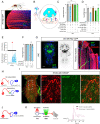
Levels of neural activity of Ubx+ TH neurons during flight. (A) Schematic illustration of the CaLexA system, which records neuronal activity based on Ca2+-NFAT interaction. Cartoon representation of the CaLexA system in the absence (Top panel) and presence of neural activity (Bottom panel); in the latter, Ca2+ accumulation dephosphorylates NFAT, triggering the transport of the transcription factor mLexA-VP16-NFAT into the nucleus, where the chimeric transcription factor LexA binds to the LexA operator (LexAop), and subsequently induces expression of the GFP reporter gene. (B) Experimental protocol. Resting and flying Drosophila were collected for dissection and their VNCs were immunostained and imaged. (C) Representative images of TH neurons in the VNCs of resting and flying wild-type Drosophila (TH>CaLexA), immunolabelled with GFP (warmer color). Note that resting flies show low GFP intensity/CaLexA signals while flying flies show very high signal intensity. (D and E) Quantification of GFP positive TH neurons (D) and GFP intensity (E) of the VNC thoracic segments 2 (T2) and 3 (T3) in TH>CaLexA flies in different conditions (resting: n = 7; flying: n = 8). (F) Increase in GFP intensity is correlated with flight (n = 21). (G) Representative images of TH neurons in the VNCs of control WT Drosophila (TH>CaLexA) and TH neurons expressing UbxRNAi (TH>CaLexA; UbxRNAi). (H and I) Quantification of GFP positive TH neurons (H) and GFP intensity (I) of VNC in segments T2 and T3 in TH>CaLexA and TH>CaLexA; UbxRNAi (n > 5). (J) GFP-positive TH neurons are also Ubx positive. (K) Representative images of the UbxTH intersectional domain in the VNC of resting and flying Drosophila (Ubx∩TH>CaLexA). (L and M) Quantification of GFP positive cells (L) and GFP intensity (M) of VNC in T2 and T3 segments in Ubx∩TH>CaLexA (n = 5). (N and O) Increase in TH neurons activity generated by conditionally expressing the voltage-gated bacterial Na+ channel (NaChBac) rescues the flight deficit resulting from Ubx downregulation. Averages of flight duration (N) and forced flight index (O) of flies after heat shock (at 30°C). Error bars in figures represent SEM. Significant values in all figures based on Mann–Whitney U test or one-way ANOVA with the post hoc Tukey–Kramer test: P < 0.05, ∗∗P < 0.01, ∗∗∗P < 0.001.
Fig. 5.
Fig. 5.
TH neurons modulate flight through direct contact with flight motoneurons. (A) Confocal image of thoracic muscles of fly expressing myristoylated GFP in dopaminergic neurons (TH>Myr:GFP). No GFP signal can be observed. (B) Schematics of a dopaminergic synapse and receptors. (C and D) Average flight duration (C) and force flight (D) at 30°C of flies with downregulated expression of dopamine receptors (DAMBRNAi) in specific flight motor neurons. (E) Red light-evoked (red shade) activation of  Dlm motoneuron expressing CsChrimson (Dlm-Mn>CsChrimson) induces spontaneous flight (n ≥ 15). See also Movie S8. (F) Flies with Dlm neurons inhibited by expression of Kir2.1 show reduced flight duration. (G) Representative confocal images showing the projections of Dlm motoneurons within the VNC (Left panel) and longitudinal flight muscles (Right panel). (H) Cartoon illustrating the principle underlying GFP Reconstitution Across Synaptic Partners (GRASP). GFP is reconstituted when two complementary segments of GFP associate on the extracellular surfaces of adjacent neurons. (I) Confocal image of GRASP reconstitution in the VNC of Drosophila TH;Dlm-Mn>GRASP (TH-LexA>LexAOP:RFP,LexAOP:spGFP11 & Dlm-Mn-Gal4>UAS:spGFP1-10). GRASP fluorescence reveals structural links between TH and Dlm neurons in the VNC. (J) Cartoon illustrating the experimental approach to determine a functional connection between TH and Dlm neurons. (K) Red light-evoked (red shade) activation of TH neuron expressing CsChrimson (TH>CsChrimson) induces spontaneous activity of Dlm motoneurons in Drosophila TH;Dlm-Mn>CsChrimson;GCAMP6 (TH-LexA,LexAOP:CsChrimson;Dlm-Mn-Gal4,UAS:GCAMP6m) compared to controls. See also Movie S9. Error bars represent SEM. Significant values in all figures based on Mann–Whitney U test or one-way ANOVA with the post hoc Tukey–Kramer test: *P < 0.01, ∗∗∗P < 0.001.
Fig. 6.
Fig. 6.
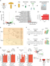
SLC gene symporter genes are differentially expressed in response to Ubx downregulation in TH neurons. (A) Experimental design of our neuron-specific transcriptomic experiment. (B) Volcano plot depicting differentially expressed genes (DEGs). Red and green dots represent down- and upregulated genes, respectively. (C) Gene Ontology (GO) enrichment analysis. GO molecular functions of DEGs show the top four genes (red bar) predicted to have symporter activity (involvement in calcium, potassium, sodium and/or solute co-transport). (D) The four differentially expressed symporter genes are strongly downregulated. None of these genes has been previously characterized in flies. (E) Dendrogram of the differentially expressed symporter genes detected in Drosophila showing their relation to other gene families across the animal kingdom. The analysis suggests that the differentially expressed symporter genes belong to the human SLC5 and SLC24 protein families. Abbreviations are: Saccharomyces cerevisiae (Sc), Arabidopsis thaliana (At), Danio rerio (Dr), Gallus gallus (Gg), Homo sapiens (Hs), Macaca mulatta (Mm), Oryctolagus cuniculus (Oc), Rattus norvegicus (Rn), Mus musculus (Mmu) Drosophila melanogaster (Dm), Caenorhabditis elegans (Ce) (values near/above internodes correspond average of branch lengths from T-Coffee analyses). (F) Structural predictions for the symporter proteins encoded by differentially expressed symporter genes based on human SLC5A8 and SLC24A5. Left panel: SACS MEMSAT2 graphs predicting 13 and 12 transmembrane domains for symporter proteins SLC5 and SLC24, respectively. Right panel: predicted 3D structures of SLC5 and SLC24 proteins. (G and H) Conditional downregulation of symporter genes CG1090 and CG6723 in TH neurons affects flight maintenance in tethered flies (G) and ability in forced flight experiments (H). (I) Proposed cellular and molecular model of Ubx-dependent control of flight behavior in Drosophila. Error bars in figures represent SEM. Significant values in all figures based on Mann–Whitney U test or one-way ANOVA with the post hoc Tukey–Kramer test: P < 0.05.

Similar articles

Cited by

References

    1. Deneris E. S., Hobert O., Maintenance of postmitotic neuronal cell identity. Nat. Neurosci. 17, 899–907 (2014). - PMC - PubMed
    1. Holmberg J., Perlmann T., Maintaining differentiated cellular identity. Nat. Rev. Genet. 13, 429–439 (2012). - PubMed
    1. Picao-Osorio J., Johnston J., Landgraf M., Berni J., Alonso C., MicroRNA-encoded behavior in Drosophila. Science 350, 815–820 (2015). - PMC - PubMed
    1. Picao-Osorio J., Lago-Baldaia I., Patraquim P., Alonso C., Pervasive behavioral effects of microRNA regulation in Drosophila. Genetics 206, 1535–1548 (2017). - PMC - PubMed
    1. Issa A., Picao-Osorio J., Rito N., Chiappe M., Alonso C., A single microRNA-Hox gene module controls equivalent movements in biomechanically distinct forms of Drosophila. Curr. Biol. 29, 2665–2675.e2664 (2019). - PMC - PubMed

Publication types

LinkOut - more resources