. 2022 Dec 1;18(12):e1010956.
doi: 10.1371/journal.ppat.1010956. eCollection 2022 Dec.

The E46K mutation modulates α-synuclein prion replication in transgenic mice

Affiliations

The E46K mutation modulates α-synuclein prion replication in transgenic mice

Sara A M Holec et al. PLoS Pathog. .

Abstract

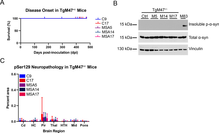
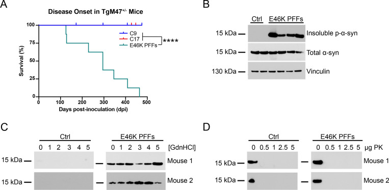
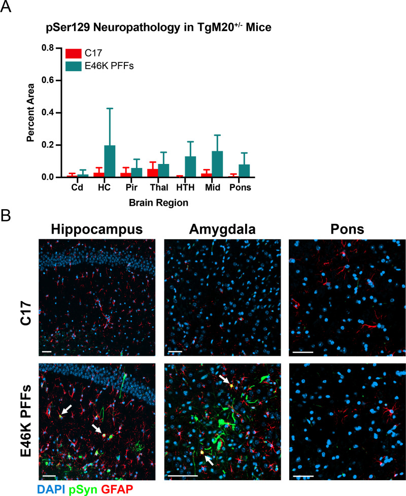
In multiple system atrophy (MSA), the α-synuclein protein misfolds into a self-templating prion conformation that spreads throughout the brain, leading to progressive neurodegeneration. While the E46K mutation in α-synuclein causes familial Parkinson's disease (PD), we previously discovered that this mutation blocks in vitro propagation of MSA prions. Recent studies by others indicate that α-synuclein adopts a misfolded conformation in MSA in which a Greek key motif is stabilized by an intramolecular salt bridge between residues E46 and K80. Hypothesizing that the E46K mutation impedes salt bridge formation and, therefore, exerts a selective pressure that can modulate α-synuclein strain propagation, we asked whether three distinct α-synuclein prion strains could propagate in TgM47+/- mice, which express human α-synuclein with the E46K mutation. Following intracranial injection of these strains, TgM47+/- mice were resistant to MSA prion transmission, whereas recombinant E46K preformed fibrils (PFFs) transmitted neurological disease to mice and induced the formation of phosphorylated α-synuclein neuropathology. In contrast, heterotypic seeding following wild-type (WT) PFF-inoculation resulted in preclinical α-synuclein prion propagation. Moreover, when we inoculated TgM20+/- mice, which express WT human α-synuclein, with E46K PFFs, we observed delayed transmission kinetics with an incomplete attack rate. These findings suggest that the E46K mutation constrains the number of α-synuclein prion conformations that can propagate in TgM47+/- mice, expanding our understanding of the selective pressures that impact α-synuclein prion replication.


Conflict of interest statement

The authors have declared that no competing interests exist.

Figures

Fig 1
Fig 1. TgM47+/- mice are resistant to MSA prion transmission.
Eight-week old TgM47+/- mice were inoculated with either control (C9 and C17) or MSA patient samples (MSA5, MSA14, and MSA17). (A) Kaplan-Meier plot of disease onset in TgM47+/- mice following inoculation. TgM47+/- mice did not develop overt motor signs by 475 days postinoculation (dpi; P > 0.05). (B) Two brain homogenates from TgM47+/- mice inoculated with either control or with each of the MSA patient samples were assayed for the presence of insoluble α-synuclein following detergent extraction. An MSA-inoculated TgM83+/- mouse brain, which we have previously shown is positive for phosphorylated α-synuclein, was included as a positive control (EP1536Y primary antibody). None of the homogenates from the inoculated mice contained aggregated α-synuclein, though total α-synuclein levels (MJFR1 primary antibody) were consistent across all homogenates tested. Vinculin shown as a loading control. (C) Fixed half-brains from the same mice were analyzed for phosphorylated α-synuclein neuropathology in the caudate (Cd), hippocampus (HC), piriform cortex and amygdala (Pir), thalamus (Thal), hypothalamus (HTH), midbrain (Mid), and pons. None of the inoculated animals developed detectable pathology (P > 0.05).
Fig 2
Fig 2. E46K PFFs transmit neurological disease to TgM47+/- mice.
Eight-week-old TgM47+/- mice were inoculated with 30 μg of E46K α-synuclein PFFs or 30 μL of 1% brain homogenate from control patient samples. (A) Kaplan-Meier plot shows E46K PFFs induced neurological disease 286 ± 122 days postinoculation (dpi; ****P < 0.0001). (B) Two brain homogenates from TgM47+/- mice inoculated with control patient samples and four homogenates from mice inoculated with E46K PFFs were assayed for the presence of insoluble α-synuclein following detergent extraction. All four homogenates from E46K PFF–inoculated mice contained phosphorylated α-synuclein (EP1536Y primary antibody). All mice tested expressed similar levels of total α-synuclein (MJFR1 primary antibody). Vinculin shown as a loading control. (C) Guanidine hydrochloride (GdnHCl) denaturation was performed using brain homogenates from two control-inoculated (left) and two E46K PFF–inoculated TgM47+/- mice (right) using 0, 1, 2, 3, 4, and 5 M GdnHCl. After detergent extraction, α-synuclein in the resulting protein pellet was detected via Western blot with the total α-synuclein antibody MJFR1. (D) Proteinase K (PK) digestion was performed using brain homogenates from two control-inoculated (left) and two E46K PFF–inoculated TgM47+/- mice (right) using 0, 0.5, 1, 2.5, and 5 μg PK. After detergent extraction, total α-synuclein in the resulting protein pellets was detected via Western blot with the antibody MJFR1.
Fig 3
Fig 3. E46K PFFs induce phosphorylated α-synuclein neuropathology in TgM47+/- mice.
Fixed tissues from TgM47+/- mice inoculated with the control patient sample C17 or E46K PFFs were analyzed for phosphorylated α-synuclein inclusions (EP1536Y primary antibody). (A) Quantification of neuropathology showed the caudate (Cd) was unaffected by E46K PFF transmission. The hippocampus (HC), piriform cortex and amygdala (Pir), and thalamus (Thal) developed inclusions that were not statistically different from control (P > 0.05). However, E46K PFFs induced robust inclusion formation in the hypothalamus (HTH; ***P < 0.001), midbrain (Mid; ****P < 0.0001), and pons (****P < 0.0001). (B) Representative images from TgM47+/- mice inoculated with control patient samples (left column) or E46K PFFs (middle column) as well as an MSA-inoculated TgM83+/- mouse sample shown for comparison (left column). From top to bottom, images of the dentate gyrus, periaqueductal gray, and pons are shown. Region denoted with a white box in the pons is shown at higher magnification in the bottom row. Phosphorylated α-synuclein in green, glial fibrillary acidic protein (GFAP) in red, and DAPI in blue. Scale bar: 50 μm.
Fig 4
Fig 4. TgM47+/- mice propagate WT α-synuclein PFFs.
Eight-week-old TgM47+/- mice were inoculated with 30 μg of WT human α-synuclein PFFs or 30 μL of 1% brain homogenate from control patient samples. (A) Kaplan-Meier plot shows mice inoculated with WT PFFs did not develop neurological disease 531 days postinoculation (dpi; P > 0.05). (B) Two brain homogenates from TgM47+/- mice inoculated with control patient samples and four homogenates from mice inoculated with WT PFFs were assayed for the presence of insoluble α-synuclein following detergent extraction. An MSA-inoculated TgM83+/- mouse brain, which we have previously shown is positive for phosphorylated α-synuclein, was included as a positive control (EP1536Y primary antibody). None of the samples from inoculated TgM47+/- mice contained aggregated α-synuclein, though total α-synuclein levels (MJFR1 primary antibody) were consistent across all mice tested. Vinculin shown as a loading control. (C) Guanidine hydrochloride (GdnHCl) denaturation was performed using brain homogenates from two control-inoculated (left) and two WT PFF–inoculated TgM47+/- mice (right) using 0, 1, 2, 3, 4, and 5 M GdnHCl. After detergent extraction, α-synuclein in the resulting protein pellet was detected via Western blot with the total α-synuclein antibody MJFR1. (D) Proteinase K (PK) digestion was performed using brain homogenates from one control-inoculated (top) and one WT PFF–inoculated TgM47+/- mouse (bottom) using 0, 0.5, 1, 2.5, and 5 μg PK. After detergent extraction, total α-synuclein in the resulting protein pellets was detected via Western blot with the antibody MJFR1.
Fig 5
Fig 5. TgM20+/- mice propagate E46K α-synuclein PFFs.
Eight-week-old TgM20+/- mice were inoculated with 30 μg of E46K human α-synuclein PFFs or 30 μL of 1% brain homogenate from control patient samples. (A) Kaplan-Meier plot shows five of eight mice inoculated with E46K PFFs developed neurological disease by 475 days postinoculation (dpi; P > 0.05). Incubation times for control-inoculated mice previously reported in Holec, et al., 2022 [46]. (B) Two brain homogenates from TgM20+/- mice inoculated with control patient samples and three homogenates from mice inoculated with E46K PFFs were assayed for the presence of insoluble α-synuclein following detergent extraction (EP1536Y primary antibody). Some of the samples from E46K PFF–inoculated TgM20+/- mice contained aggregated α-synuclein (EP1536Y primary antibody), though soluble α-synuclein was present in all mice tested. Vinculin shown as a loading control. (C) Proteinase K (PK) digestion was performed using brain homogenates from control-inoculated and E46K PFF–inoculated TgM20+/- mice using 0, 0.5, 1, 2.5, and 5 μg PK. After detergent extraction, phosphorylated and total α-synuclein in the resulting protein pellets were detected via Western blot using both EP1536Y (left) and MJFR1 (right) primary antibodies, respectively.
Fig 6
Fig 6. TgM20+/- mice inoculated with E46K PFFs develop mild phosphorylated α-synuclein neuropathology.
Fixed tissues from TgM20+/- mice inoculated with control patient sample C17 or E46K PFFs were immunostained for phosphorylated α-synuclein (EP1536Y primary antibody) and glial fibrillary acidic protein (GFAP: astrocytes). (A) Quantification of stained brain slices showed no significant phosphorylated α-synuclein inclusions were present in the caudate (Cd), hippocampus (HC), piriform cortex and amygdala (Pir), thalamus (Thal), hypothalamus (HTH), midbrain (Mid), or pons (P > 0.05). Neuropathology data for control-inoculated mice was previously reported in Holec, et al., 2022 [46]. (B) Representative images of the HC, amygdala, and pons from mice inoculated with either control patient sample C17 (top) or E46K PFFs (bottom). Notably, some mice did develop some inclusions in the HC and amygdala. While these inclusions appeared to be mostly neuronal, overlap with GFAP staining suggests there may also be colocalization with astrocytes. Phosphorylated α-synuclein in green, GFAP in red, and DAPI in blue. Scale bar: 50 μm.

Similar articles

Cited by

References

    1. Spillantini MG, Schmidt ML, Lee VM-Y, Trojanowski JQ, Jakes R, Goedert M. α-Synuclein in Lewy bodies. Nature. 1997;388:839–40. - PubMed
    1. Gilman S, Low PA, Quinn N, Albanese A, Ben-Shlomo Y, Fowler CJ, et al.. Consensus statement on the diagnosis of multiple system atrophy. J Neurol Sci. 1999;163:94–8. doi: 10.1016/s0022-510x(98)00304-9 - DOI - PubMed
    1. Gilman S, May SJ, Shults CW, Tanner CM, Kukull W, Lee VM-Y, et al.. The North American Multiple System Atrophy Study Group. J Neural Transm (Vienna). 2005;112:1687–94. doi: 10.1007/s00702-005-0381-6 - DOI - PubMed
    1. Gilman S, Wenning GK, Low PA, Brooks DJ, Mathias CJ, Trojanowski JQ, et al.. Second consensus statement on the diagnosis of multiple system atrophy. Neurology. 2008;71:670–6. doi: 10.1212/01.wnl.0000324625.00404.15 - DOI - PMC - PubMed
    1. Polymeropoulos MH, Lavedan C, Leroy E, Ide SE, Dehejia A, Dutra A, et al.. Mutation in the α-synuclein gene identified in families with Parkinson’s disease. Science. 1997;276:2045–7. - PubMed

Publication types

LinkOut - more resources