

Artificial intelligence for localization of the acute ischemic stroke by non-contrast computed tomography
- PMID: 36454916
- PMCID: PMC9714826
- DOI: 10.1371/journal.pone.0277573
Artificial intelligence for localization of the acute ischemic stroke by non-contrast computed tomography
Abstract
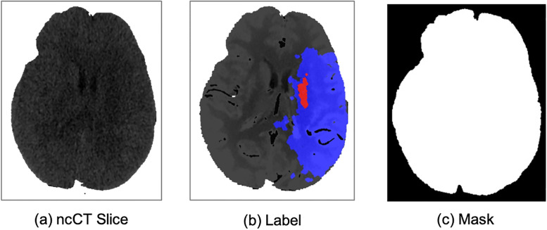
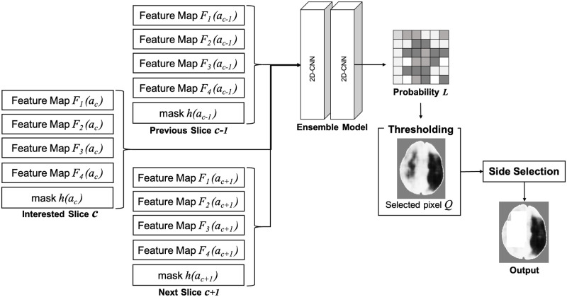
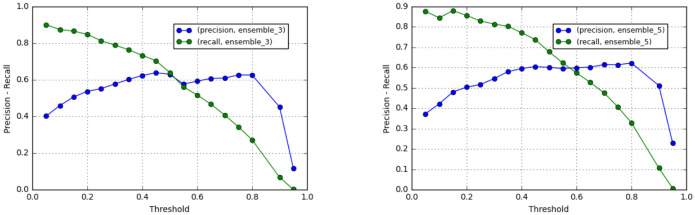
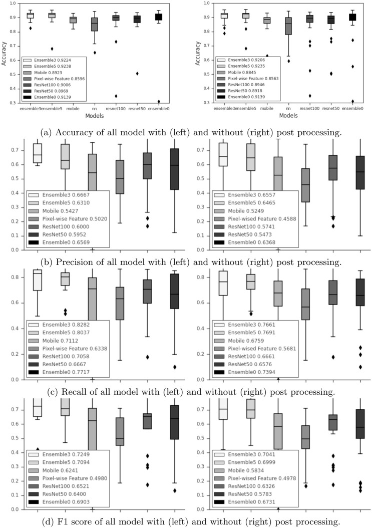
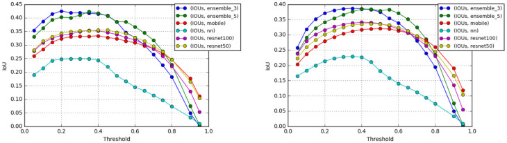
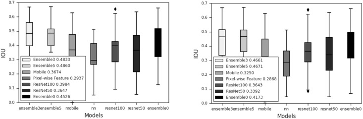
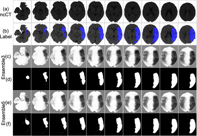
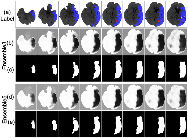
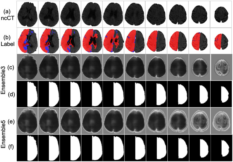
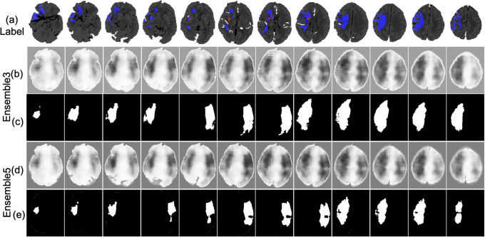
A non-contrast cranial computer tomography (ncCT) is often employed for the diagnosis of the early stage of the ischemic stroke. However, the number of false negatives is high. More accurate results are obtained by an MRI. However, the MRI is not available in every hospital. Moreover, even if it is available in the clinic for the routine tests, emergency often does not have it. Therefore, this paper proposes an end-to-end framework for detection and segmentation of the brain infarct on the ncCT. The computer tomography perfusion (CTp) is used as the ground truth. The proposed ensemble model employs three deep convolution neural networks (CNNs) to process three end-to-end feature maps and a hand-craft features characterized by specific contra-lateral features. To improve the accuracy of the detected infarct area, the spatial dependencies between neighboring slices are employed at the postprocessing step. The numerical experiments have been performed on 18 ncCT-CTp paired stroke cases (804 image-pairs). The leave-one-out approach is applied for evaluating the proposed method. The model achieves 91.16% accuracy, 65.15% precision, 77.44% recall, 69.97% F1 score, and 0.4536 IoU.
Copyright: © 2022 Kaothanthong et al. This is an open access article distributed under the terms of the Creative Commons Attribution License, which permits unrestricted use, distribution, and reproduction in any medium, provided the original author and source are credited.
Conflict of interest statement
The authors have declared that no competing interests exist.
Figures
Similar articles
-
Noncontrast Computed Tomography e-Stroke Infarct Volume Is Similar to RAPID Computed Tomography Perfusion in Estimating Postreperfusion Infarct Volumes.Stroke. 2021 Jan;52(2):634-641. doi: 10.1161/STROKEAHA.120.031651. Epub 2021 Jan 12. Stroke. 2021. PMID: 33430633
-
Automatic ischemic stroke lesion segmentation from computed tomography perfusion images by image synthesis and attention-based deep neural networks.Med Image Anal. 2020 Oct;65:101787. doi: 10.1016/j.media.2020.101787. Epub 2020 Jul 18. Med Image Anal. 2020. PMID: 32712524
-
Improvement of automatic ischemic stroke lesion segmentation in CT perfusion maps using a learned deep neural network.Comput Biol Med. 2021 Oct;137:104849. doi: 10.1016/j.compbiomed.2021.104849. Epub 2021 Sep 9. Comput Biol Med. 2021. PMID: 34530336
-
Diagnostic and prognostic utility of computed tomography perfusion imaging in posterior circulation acute ischemic stroke: A systematic review and meta-analysis.Eur J Neurol. 2021 Aug;28(8):2657-2668. doi: 10.1111/ene.14934. Epub 2021 Jun 12. Eur J Neurol. 2021. PMID: 34021664
-
An East Coast Perspective on Artificial Intelligence and Machine Learning: Part 2: Ischemic Stroke Imaging and Triage.Neuroimaging Clin N Am. 2020 Nov;30(4):467-478. doi: 10.1016/j.nic.2020.08.002. Neuroimaging Clin N Am. 2020. PMID: 33038997 Review.
Cited by
-
Role of artificial intelligence and machine learning in the diagnosis of cerebrovascular disease.Front Hum Neurosci. 2023 Sep 7;17:1254417. doi: 10.3389/fnhum.2023.1254417. eCollection 2023. Front Hum Neurosci. 2023. PMID: 37746051 Free PMC article. Review.
-
Artificial Intelligence in Optimizing the Functioning of Emergency Departments; a Systematic Review of Current Solutions.Arch Acad Emerg Med. 2024 Jan 27;12(1):e22. doi: 10.22037/aaem.v12i1.2110. eCollection 2024. Arch Acad Emerg Med. 2024. PMID: 38572221 Free PMC article. Review.
-
Cerebral ischemia detection using deep learning techniques.Health Inf Sci Syst. 2025 May 20;13(1):36. doi: 10.1007/s13755-025-00352-8. eCollection 2025 Dec. Health Inf Sci Syst. 2025. PMID: 40400660 Free PMC article.
References
-
- Zhao H, Shi J, Qi X, Wang X, Jia J. Pyramid Scene Parsing Network. In: 2017 IEEE Conference on Computer Vision and Pattern Recognition (CVPR); 2017. p. 6230–6239.
-
- Çiçek Ö, Abdulkadir A, Lienkamp SS, Brox T, Ronneberger O. 3D U-net: Learning dense volumetric segmentation from sparse annotation. In: Lecture Notes in Computer Science (including subseries Lecture Notes in Artificial Intelligence and Lecture Notes in Bioinformatics). vol. 9901 LNCS. Springer; Verlag; 2016. p. 424–432.
-
- Milletari F, Navab N, Ahmadi SA. V-Net: Fully convolutional neural networks for volumetric medical image segmentation. In: Proceedings—2016 4th International Conference on 3D Vision, 3DV 2016. Institute of Electrical and Electronics Engineers Inc.; 2016. p. 565–571.
Publication types
MeSH terms
LinkOut - more resources
Full Text Sources
Medical
