Randomized Controlled Trial
. 2022 Dec 1;17(12):e0278412.
doi: 10.1371/journal.pone.0278412. eCollection 2022.

Gamma frequency sensory stimulation in mild probable Alzheimer's dementia patients: Results of feasibility and pilot studies

Affiliations
Randomized Controlled Trial

Gamma frequency sensory stimulation in mild probable Alzheimer's dementia patients: Results of feasibility and pilot studies

Diane Chan et al. PLoS One. .

Abstract

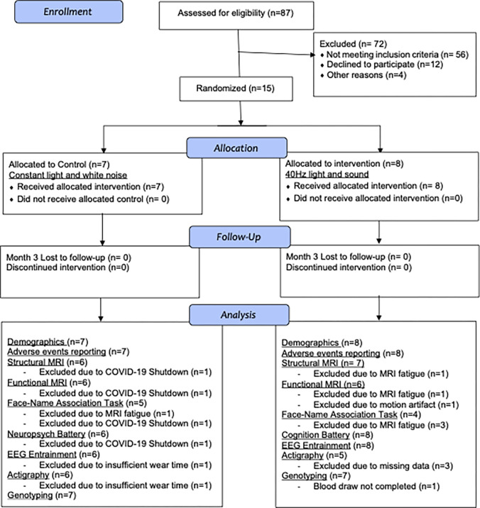
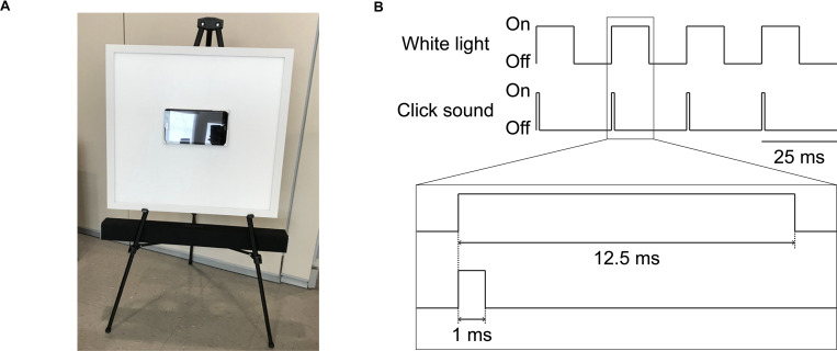
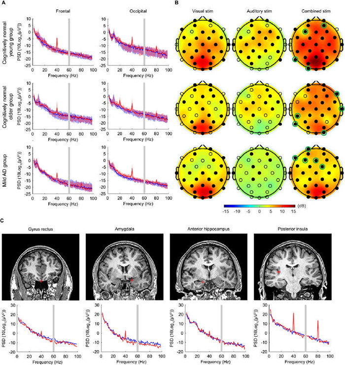
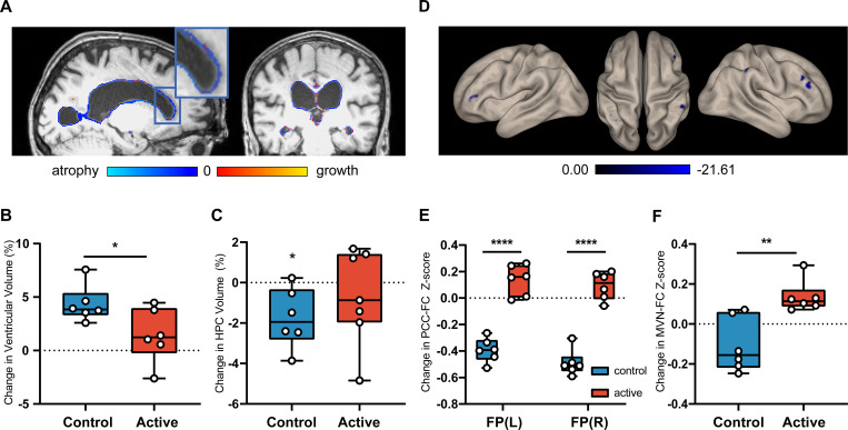
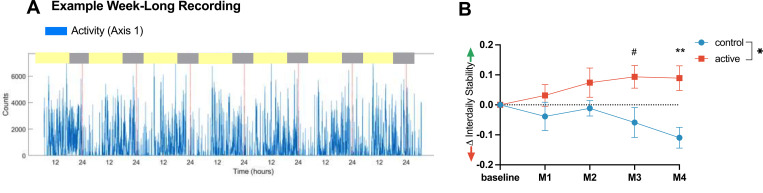
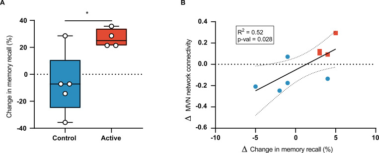
Non-invasive Gamma ENtrainment Using Sensory stimulation (GENUS) at 40Hz reduces Alzheimer's disease (AD) pathology such as amyloid and tau levels, prevents cerebral atrophy, and improves behavioral testing performance in mouse models of AD. Here, we report data from (1) a Phase 1 feasibility study (NCT04042922, ClinicalTrials.gov) in cognitively normal volunteers (n = 25), patients with mild AD dementia (n = 16), and patients with epilepsy who underwent intracranial electrode monitoring (n = 2) to assess safety and feasibility of a single brief GENUS session to induce entrainment and (2) a single-blinded, randomized, placebo-controlled Phase 2A pilot study (NCT04055376) in patients with mild probable AD dementia (n = 15) to assess safety, compliance, entrainment, and exploratory clinical outcomes after chronic daily 40Hz sensory stimulation for 3 months. Our Phase 1 study showed that 40Hz GENUS was safe and effectively induced entrainment in both cortical regions and other cortical and subcortical structures such as the hippocampus, amygdala, insula, and gyrus rectus. Our Phase 2A study demonstrated that chronic daily 40Hz light and sound GENUS was well-tolerated and that compliance was equally high in both the control and active groups, with participants equally inaccurate in guessing their group assignments prior to unblinding. Electroencephalography recordings show that our 40Hz GENUS device safely and effectively induced 40Hz entrainment in participants with mild AD dementia. After 3 months of daily stimulation, the group receiving 40Hz stimulation showed (i) lesser ventricular dilation and hippocampal atrophy, (ii) increased functional connectivity in the default mode network as well as with the medial visual network, (iii) better performance on the face-name association delayed recall test, and (iv) improved measures of daily activity rhythmicity compared to the control group. These results support further evaluation of GENUS in a pivotal clinical trial to evaluate its potential as a novel disease-modifying therapeutic for patients with AD.


Conflict of interest statement

LHT is a scientific co-founder, SAB member and Board of Director of Cognito Therapeutics. ESB is a scientific co-founder, SAB member of Cognito Therapeutics. EBK has consulted for the American Academy of Sleep Medicine Foundation, the Sleep Research Foundation, Yale University Press, the National Sleep Foundation, Sanofi Genzyme, and Circadian Therapeutics; her partner owns Chronsulting. BCD is a consultant for Acadia, Alector, Arkuda, Biogen, Denali, Lilly, Merck, Novartis, Takeda, and Wave Lifesciences, and receives royalties from Cambridge University Press, Elsevier, Oxford University Press. DC, HJS, BJ, NPM, DS, SDB, EK, VSFA, ADB, BU, PG, MH, GDW, AB, EJS, ANC, SA, SWG has nothing to disclose. This does not alter our adherence to PLOS ONE policies on sharing data and materials.

Figures

Fig 1
Fig 1. CONSORT flow diagram: Phase 2A study cohort.
Participant recruitment, allocation, follow-up, and analysis counts by activity.
Fig 2
Fig 2. GENUS device and 40Hz stimulation schematic.
(A) Light and sound stimulation device. The white board is a programmable LED light display panel that measures at 2 feet x 2 feet. Sitting at the center of the light panel is a tablet loaded with videos to keep the viewer’s attention on the device. The black bar below the light panel is the programmable soundbar audio system. The device is supported by the legs of a black easel and positioned so the tablet is eye level when the viewer is seated 5 feet away. (B) Schematic of the electrical signals that turn the white light and the click sound pulses on or off for concurrent light and sound stimulation at 40Hz.
Fig 3
Fig 3. Acute 40Hz combined visual and auditory stimulation entrains cortical and subcortical regions.
(A) Scalp EEG power spectral density (PSD) at the frontal (Fz, F3, F4, F7, F8) and the occipital (Oz, O1, O2) electrode sites, in cognitively normal young participants (n = 13; top row), cognitively normal older participants (n = 12; middle row), and patients with mild AD (n = 16; bottom row). Solid lines, group median; shaded areas, 95% confidence interval; blue, baseline; red, 40Hz GENUS light and sound stimulation (active condition). Gray bar indicates 60 Hz line noise. (B) Topographic maps showing the median change in 40Hz PSD from the baseline level with 40Hz visual alone, 40Hz auditory alone, and 40Hz combined stimulation, in cognitively normal young participants (n = 13; top row), cognitively normal older participants (n = 12; middle row), and patients with mild AD (n = 16; bottom row). Filled circles represent scalp electrodes at which the increase in 40Hz PSD from the baseline level was significant (p < 0.05, Wilcoxon’s sign rank test, Bonferroni corrected for 32 electrodes). Green circles indicate electrodes entrained with synchronized 40Hz GENUS light and sound stimulation but not by light or sound alone. (C) Example coronal MRI images before electrode implantation (top row) and intracranial EEG PSD (bottom row) from a single patient with medically intractable epilepsy (Patient 483), for depth electrode contacts placed in the gyrus rectus, amygdala, anterior hippocampus, and posterior insula. Red plus sign, approximate location of the depth electrode contact; blue, baseline; red, 40Hz GENUS light and sound stimulation (active condition). For the PSD between 58Hz and 62Hz, interpolated values are plotted because of the bandstop filtering around 60Hz.
Fig 4
Fig 4. Daily GENUS leads to group-level differences in structural and functional MRI outcomes at Month 3.
(A) Visualization of change in ventricular volume of an example control participant from baseline to Month 3 on sagittal and coronal T1-weighted structural MRI (B) Group-level analysis of percent change in ventricular volume between the control and active groups (n = 13, p = 0.024) (C) Group-level analysis of control vs active groups HPC volume compared to 0 (n = 13, control p = 0.034, active p = 0.4384) (D) Seed-to-Voxel analysis of PCC-FC in control group from baseline to Month 3 on resting state fMRI (See S3 Table) (E) Seed-to-voxel analysis of PCC-FC for between group comparisons from baseline to Month 3 (n = 12, p < 0.05 FEW-corrected) (F) Group-level analysis of changes in mean functional connectivity of the MVN from baseline to Month 3. (n = 12, p = 0.004). FP: Frontal Pole; MVN: Medial Visual Network; PCC-FC: Posterior Cingulate Cortex functional connectivity.
Fig 5
Fig 5. Changes in rest-activity patterns relative to baseline.
(A) Example 7-day activity recording of activity counts (blue) with red dotted lines every 24 hours from an individual. Inter-daily Stability (IS) quantifies the regularity of day-to-day rest-activity patterns. (B) Improved IS compared to baseline (means ± SEM) in active group (red) but not control group (blue), with significant differences between active and control groups at Month 4 (**p < 0.01) and a trend at Month 3 (#p < 0.1). Statistical analysis with a 2-Way ANOVA with Šídák’s multiple comparisons at each monthly timepoint. Mean of control = -0.055, mean of active = 0.072. Main effect of intervention p = 0.037, interaction of intervention x time, p = 0.025. Multiple comparison at Month 3, p = 0.051, Month 4, p = 0.007.
Fig 6
Fig 6. Chronic GENUS improves face-name association delayed recall test (FNA-DRT) results.
(A) Group-level analysis of changes in FNA-DRT from baseline to Month 3 (B) Correlation of change in FNA-DRT score with change in mean functional connectivity of the MVN.

References

    1. Canter RG, Penney J, Tsai LH. The road to restoring neural circuits for the treatment of Alzheimer’s disease. Nature. 2016. Nov;539(7628):187–96. doi: 10.1038/nature20412 - DOI - PubMed
    1. Uhlhaas PJ, Singer W. Neural Synchrony in Brain Disorders: Relevance for Cognitive Dysfunctions and Pathophysiology. Neuron. 2006. Oct;52(1):155–68. doi: 10.1016/j.neuron.2006.09.020 - DOI - PubMed
    1. Nimmrich V, Draguhn A, Axmacher N. Neuronal Network Oscillations in Neurodegenerative Diseases. Neuromol Med. 2015. Sep;17(3):270–84. doi: 10.1007/s12017-015-8355-9 - DOI - PubMed
    1. Herrmann CS, Demiralp T. Human EEG gamma oscillations in neuropsychiatric disorders. Clinical Neurophysiology. 2005. Dec;116(12):2719–33. doi: 10.1016/j.clinph.2005.07.007 - DOI - PubMed
    1. Palop JJ, Mucke L. Network abnormalities and interneuron dysfunction in Alzheimer disease. Nat Rev Neurosci. 2016. Dec;17(12):777–92. doi: 10.1038/nrn.2016.141 - DOI - PMC - PubMed

Publication types

Associated data