. 2023 Feb 1;29(3):560-570.
doi: 10.1158/1078-0432.CCR-22-1987.

Safety and Efficacy of MEDI0457 plus Durvalumab in Patients with Human Papillomavirus-Associated Recurrent/Metastatic Head and Neck Squamous Cell Carcinoma

Affiliations

Safety and Efficacy of MEDI0457 plus Durvalumab in Patients with Human Papillomavirus-Associated Recurrent/Metastatic Head and Neck Squamous Cell Carcinoma

Charu Aggarwal et al. Clin Cancer Res. .

Abstract

Purpose: Tumoral programmed cell death ligand-1 (PD-L1) expression is common in human papillomavirus (HPV)-associated head and neck squamous cell carcinoma (HNSCC). We assessed whether a DNA vaccine targeting HPV-16/18 E6/E7 with IL12 adjuvant (MEDI0457) combined with the PD-L1 inhibitor durvalumab could enhance HPV-specific T-cell response and improve outcomes in recurrent/metastatic HPV-16/18-associated HNSCC.

Patients and methods: In this phase Ib/IIa study, immunotherapy-naïve patients with ≥1 previous platinum-containing regimen (neoadjuvant/adjuvant therapy or for recurrent/metastatic disease) received MEDI0457 7 mg intramuscularly with electroporation on weeks 1, 3, 7, and 12, then every 8 weeks, plus durvalumab 1,500 mg intravenously on weeks 4, 8, and 12, then every 4 weeks, until confirmed progression and/or unacceptable toxicity. Coprimary objectives were safety and objective response rate (ORR; H0: ORR ≤ 15%); secondary objectives included 16-week disease control rate (DCR-16), overall survival (OS), and progression-free survival (PFS).

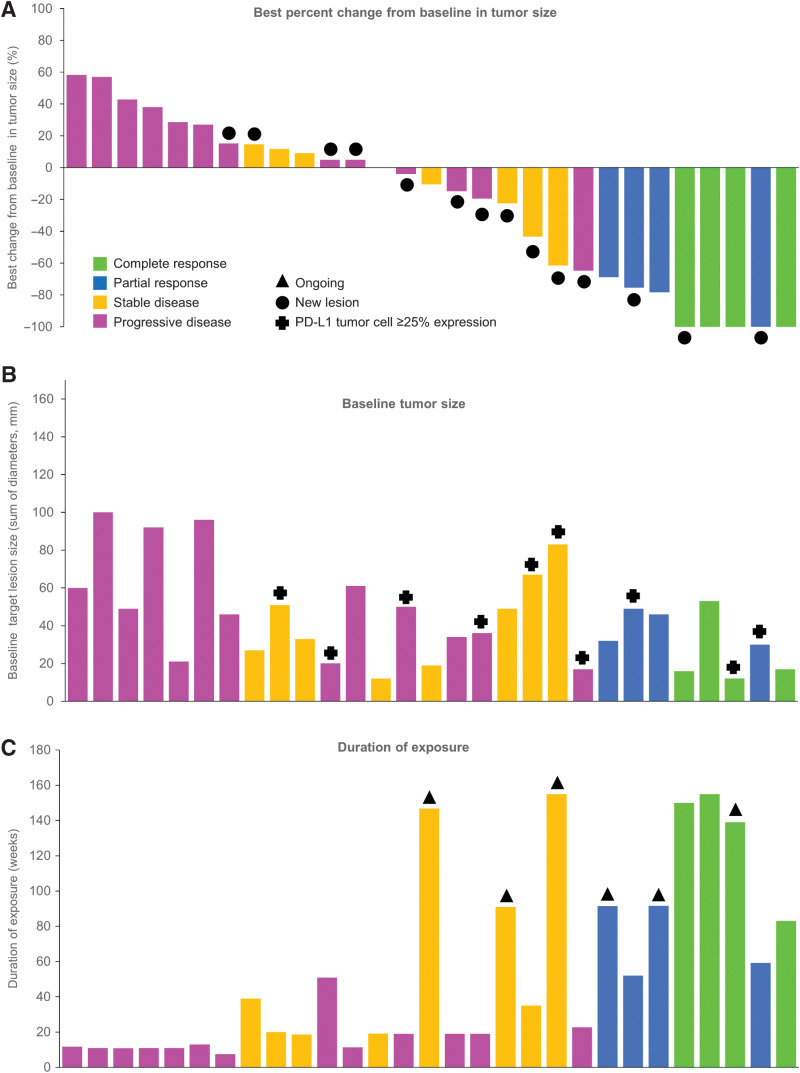
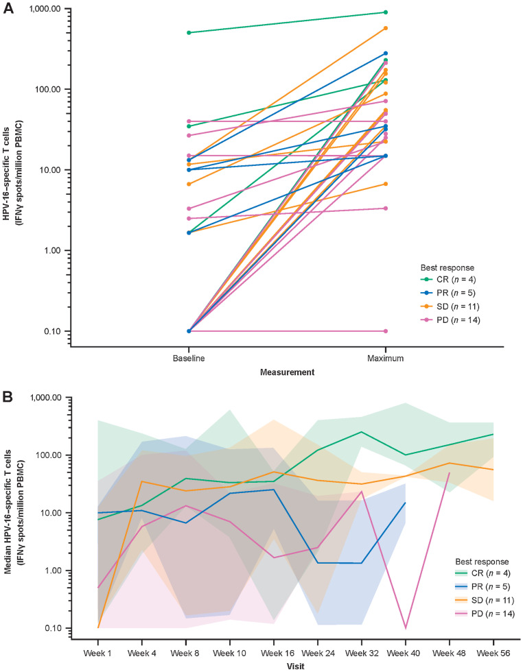
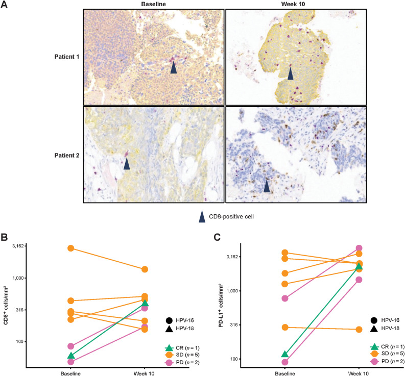
Results: Of 35 treated patients, 29 were response evaluable (confirmed HPV-associated disease; received both agents). ORR was 27.6% [95% confidence interval (CI), 12.7-47.2; four complete responses, four partial responses]; responses were independent of PD-L1 tumor-cell expression (≥25% vs. <25%). DCR-16 was 44.8% (95% CI, 26.5-64.3). Median PFS was 3.5 months (95% CI, 1.9-9.0); median OS was 29.2 months (15.2-not calculable). Twenty-eight (80.0%) patients had treatment-related adverse events [grade 3: 5 (14.3%); no grade 4/5], resulting in discontinuation in 2 (5.7%) patients. HPV-16/18-specific T cells increased on treatment; 4 of 8 evaluable patients had a >2-fold increase in tumor-infiltrating CD8+ T cells.

Conclusions: MEDI0457 plus durvalumab was well tolerated. While the primary efficacy endpoint was not reached, clinical benefit was encouraging.


Figures

Figure 1. Best percent change from baseline in tumor size and duration of exposure (response-evaluable population). A, Best change from baseline in tumor size, with bars color coded according to best response achieved; black filled circles indicate new lesions. B, Baseline tumor size; crosses indicate patients with PD-L1 tumor cell expression of ≥25%. C, Duration of exposure (any treatment); black triangles indicate patients ongoing on treatment at data cutoff. Data for individual patients are shown in the same order on the x-axis in each panel. PD-L1, programmed cell death ligand-1.
Figure 1.
Best percent change from baseline in tumor size and duration of exposure (response-evaluable population). A, Best change from baseline in tumor size, with bars color coded according to best response achieved; black filled circles indicate new lesions. B, Baseline tumor size; crosses indicate patients with PD-L1 tumor cell expression of ≥25%. C, Duration of exposure (any treatment); black triangles indicate patients ongoing on treatment at data cutoff. Data for individual patients are shown in the same order on the x-axis in each panel.
Figure 2. Peripheral HPV-16–specific T cells on IFNγ ELISpot assay of PMBCs. Baseline and maximum T-cell count by patient and response type (n = 34; A), and median T-cell count over time by response category (n = 34), plotted on a log10 y-axis (B). To facilitate visualization, T-cell counts of <0.1 were floored to 0.1. In A, each dot-to-dot line represents an individual patient, color coded according to best response. In B, the median lines are color coded according to best response, and the shaded areas represent the 95% CIs around the medians. Week 1 is the start of MEDI0457 dosing. Week 4 is the start of durvalumab dosing. CR, complete response; ELISpot, enzyme-linked immunospot; HPV, human papillomavirus; IFNγ, interferon gamma; PD, progressive disease; PBMC, peripheral blood mononuclear cell; PR, partial response, SD, stable disease.
Figure 2.
Peripheral HPV-16–specific T cells on IFNγ ELISpot assay of PMBCs. Baseline and maximum T-cell count by patient and response type (n = 34; A), and median T-cell count over time by response category (n = 34), plotted on a log10y-axis (B). To facilitate visualization, T-cell counts of <0.1 were floored to 0.1. In A, each dot-to-dot line represents an individual patient, color coded according to best response. In B, the median lines are color coded according to best response, and the shaded areas represent the 95% CIs around the medians. Week 1 is the start of MEDI0457 dosing. Week 4 is the start of durvalumab dosing. CR, complete response; ELISpot, enzyme-linked immunospot; HPV, human papillomavirus; IFNγ, interferon gamma; PBMC, peripheral blood mononuclear cell; PD, progressive disease; PR, partial response, SD, stable disease.
Figure 3. Intratumoral immunomodulation: CD8 IHC images from tumors of 2 patients showing increased numbers of CD8+ T cells at week 10 versus baseline—CD8+ T cells are stained purple and cytokeratin is stained yellow in the top two and the bottom left images, and CD8+ T cells are stained brown in the bottom right image (A); change in tumoral CD8+ T-cell count from baseline to week 10 (n = 8; B); and change in tumoral PD-L1+ cell count from baseline to week 10 (n = 8) by HPV status on IHC analysis of individual patients with paired tumor biopsies, with lines color coded according to best response (C).
Figure 3.
Intratumoral immunomodulation: CD8 IHC images from tumors of 2 patients showing increased numbers of CD8+ T cells at week 10 versus baseline—CD8+ T cells are stained purple and cytokeratin is stained yellow in the top two and the bottom left images, and CD8+ T cells are stained brown in the bottom right image (A); change in tumoral CD8+ T-cell count from baseline to week 10 (n = 8; B) and change in tumoral PD-L1+ cell count from baseline to week 10 (n = 8; C) by HPV status on IHC analysis of individual patients with paired tumor biopsies, with lines color coded according to best response.

Comment in

References

    1. Kobayashi K, Hisamatsu K, Suzui N, Hara A, Tomita H, Miyazaki T. A review of HPV-related head and neck cancer. J Clin Med 2018;7:241. - PMC - PubMed
    1. Marur S, D'Souza G, Westra WH, Forastiere AA. HPV-associated head and neck cancer: a virus-related cancer epidemic. Lancet Oncol 2010;11:781–9. - PMC - PubMed
    1. Kreimer AR, Clifford GM, Boyle P, Franceschi S. Human papillomavirus types in head and neck squamous cell carcinomas worldwide: a systematic review. Cancer Epidemiol Biomarkers Prev 2005;14:467–75. - PubMed
    1. Johnson DE, Burtness B, Leemans CR, Lui VWY, Bauman JE, Grandis JR. Head and neck squamous cell carcinoma. Nat Rev Dis Primers 2020;6:92. - PMC - PubMed
    1. Future IIS Group. Quadrivalent vaccine against human papillomavirus to prevent high-grade cervical lesions. N Engl J Med 2007;356:1915–27. - PubMed

Publication types

MeSH terms