. 2023 Mar 30;141(13):1524-1532.
doi: 10.1182/blood.2022017764.

The spectrum of GATA2 deficiency syndrome

Affiliations

The spectrum of GATA2 deficiency syndrome

Katherine R Calvo et al. Blood. .

Abstract

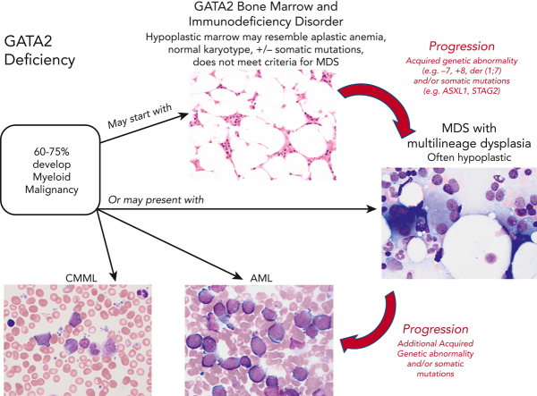
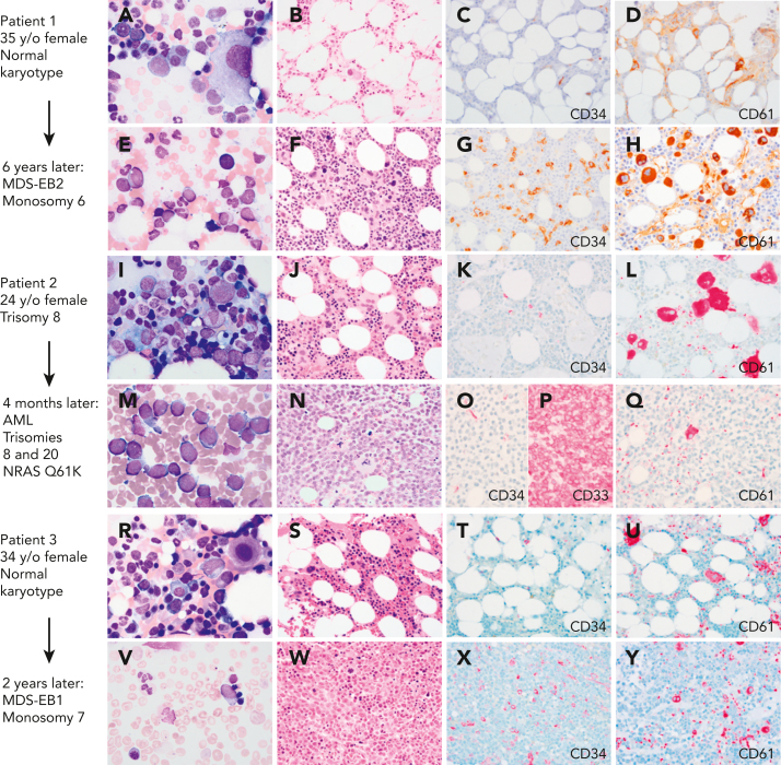
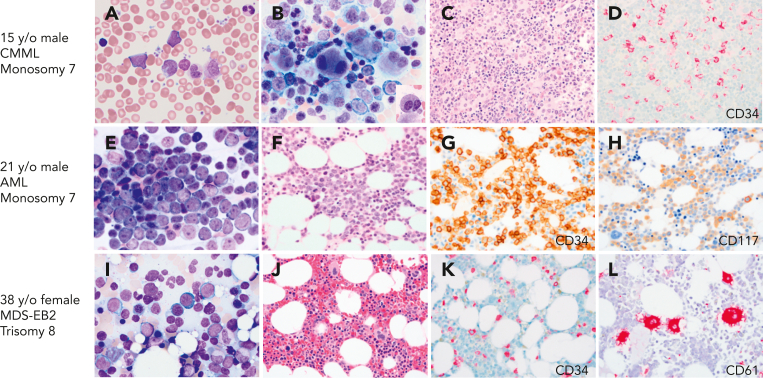
Inherited or de novo germ line heterozygous mutations in the gene encoding the transcription factor GATA2 lead to its deficiency. This results in a constellation of clinical features including nontuberculous mycobacterial, bacterial, fungal, and human papillomavirus infections, lymphedema, pulmonary alveolar proteinosis, and myelodysplasia. The onset, or even the presence, of disease is highly variable, even in kindreds with the identical mutation in GATA2. The clinical manifestations result from the loss of a multilineage progenitor that gives rise to B lymphocytes, monocytes, natural killer cells, and dendritic cells, leading to cytopenias of these lineages and subsequent infections. The bone marrow failure is typically characterized by hypocellularity. Dysplasia may either be absent or subtle but typically evolves into multilineage dysplasia with prominent dysmegakaryopoiesis, followed in some instances by progression to myeloid malignancies, specifically myelodysplastic syndrome, acute myelogenous leukemia, and chronic myelomonocytic leukemia. The latter 3 malignancies often occur in the setting of monosomy 7, trisomy 8, and acquired mutations in ASXL1 or in STAG2. Importantly, myeloid malignancy may represent the primary presentation of disease without recognition of other syndromic features. Allogeneic hematopoietic stem cell transplantation (HSCT) results in reversal of the phenotype. There remain important unanswered questions in GATA2 deficiency, including the following: (1) Why do some family members remain asymptomatic despite harboring deleterious mutations in GATA2? (2) What are the genetic changes that lead to myeloid progression? (3) What causes the apparent genetic anticipation? (4) What is the role of preemptive HSCT?


Conflict of interest statement

Conflict-of-interest disclosure: The authors declare no competing financial interests.

Figures

None
Graphical abstract
Figure 1.
Figure 1.
Myeloid progression in GATA2 deficiency. (A-D) A 35-year-old (35 y/o) female with neutropenia, normal hemoglobin and platelet count, and very low monocytes, B cells, and NK cells count. (A) BM aspirate showed dysplastic MKs with minimal morphologic dysplasia in other lineages and no increase in blasts. (B) The biopsy was hypocellular with trilineage hematopoiesis, (C) low number of CD34+ cells, (D) few atypical MKs highlighted by CD61, and normal cytogenetics. (E-F) Six years later, she presented with severe anemia with 16% circulating blasts. (E)The marrow aspirate showed increased scattered blasts. (F) The marrow biopsy was hypercellular with (G) 10% CD34+ blasts, (H) increased CD61+ dysplastic MKs and increased reticulin fibrosis (not shown) indicative of MDS with excess blasts. Cytogenetics showed presence of monosomy 6. A germ line GATA2 mutation was subsequently identified (T354M). (I-L) A 24-year-old female with a prior diagnosis of aplastic anemia in adolescence presented with moderate pancytopenia and very low monocytes, B cells, and NK cells count. (I) The marrow aspirate and (J) BM biopsy showed normocellular marrow with trilineage hematopoiesis, (K) no increase in CD34 positive blasts, and (L) moderate dysmegakaryopoiesis. Cytogenetics revealed trisomy 8. Germ line GATA2 mutation was identified (L375V). (M-P) Four months later she presented with a platelet count of 10 × 103/μL. (M) The BM aspirate showed increased blasts. (N) The marrow biopsy was markedly hypercellular with (O) sheets of blasts that were negative for CD34 and (P) positive for CD33, and (Q) markedly decreased dysplastic MKs. Flow cytometry analysis of the marrow aspirate (not shown) identified 85% monoblasts that expressed CD56, CD64, CD36, and CD123 with minimal expression of CD14 indicative of acute monoblastic leukemia. Cytogenetics showed new trisomy 20 plus trisomy 8 in 65% of metaphases. (R-U) A 34-year-old female with mild pancytopenia, low monocytes, B cells, and NK cells count. (R)The marrow aspirate showed trilineage hematopoiesis with a subset of mononuclear MKs. (S) The BM biopsy was hypocellular for age with trilineage hematopoiesis with (T) no increase in CD34+ blasts, (U) mild megakaryocytic atypia, and trisomy 8. (V-Y) Two years later she presented with circulating blasts. (V) The marrow aspirate was paucicellular with scattered blasts. (W) The BM biopsy was markedly hypercellular with (X) 8% CD34+ myeloblasts confirmed by flow cytometry, and (Y) dysplastic megakaryopoiesis with microMKs. Cytogenetics showed new monosomy 7. Germ line GATA2 mutation was identified (N371K). Marrow aspirates were stained with Wright-Giemsa stain (1000×). BM biopsies were stained with hematoxylin and eosin or immunohistochemistry (IHC) as indicated (500×). Images were taken using an Olympus BX41 microscope equipped with a DP74 camera using Olympus cellSens software. EB1, excess blasts.
Figure 2.
Figure 2.
BMs with CMML, AML, and MDS with excess blasts on initial presentation with GATA2 deficiency. (A-D) A 17-year-old healthy male with family history of GATA2 deficiency (c.1017+572C>T) and AML (mother), MDS (aunt), and CMML (grandfather), presented for his initial evaluation. White blood cell counts of 15.6 × 103/μL. (A) Peripheral blood smear with leukocytosis and circulating blasts (6%), monocytosis (1.5 × 103/μL), and left-shifted granulocytes. (B) BM aspirate showing marked megakaryocytic dysplasia with dysplastic myelopoiesis (inset). (C) BM core biopsy was markedly hypercellular. (D) CD34 IHC on the marrow biopsy showed increased blasts. Cytogenetic analysis revealed monosomy 7, and myeloid next-generation sequencing analysis detected ASXL1, SETBP1, and U2AF1 mutations. Findings indicative of CMML. (E-H) A 21-year-old male presented with fatigue and pancytopenia with no history of infections or family history of myeloid malignancy. (E) BM aspirate with sheets of blasts confirmed by flow cytometry as myeloblasts. (F) BM core biopsy was hypocellular for age. (G) CD34 IHC on the marrow biopsy showed marked increase in blasts >50%. (H) CD117 IHC highlighting the blasts. Monosomy 7 detected on cytogenetic analysis. The patient was diagnosed with AML and subsequently found to have a germ line mutation in GATA2 (T354M). (I-L) A 39-year-old female who was a healthy relative of a proband with GATA2 deficiency (R398W), was recently found to be positive for the mutation and presented for evaluation. Peripheral blood counts were normal to slightly decreased. (I) BM aspirate with increased blasts (∼10%). (J) BM core biopsy was normocellular. (K) CD34 IHC showing increased blasts (∼10%). (L) CD61 IHC staining MKs with a few small forms. The patient was diagnosed with MDS with excess blasts. All peripheral blood and BM aspirates were stained with Wright-Giemsa stain at 1000×. Core biopsies were stained with H&E or IHC as indicated. Images were taken using an Olympus BX41 microscope equipped with a DP74 camera using Olympus cellSens software.

Comment in

References

    1. Vinh DC, Patel SY, Uzel G, et al. Autosomal dominant and sporadic monocytopenia with susceptibility to mycobacteria, fungi, papillomaviruses, and myelodysplasia. Blood. 2010;115(8):1519–1529. - PMC - PubMed
    1. Bigley V, Haniffa M, Doulatov S, et al. The human syndrome of dendritic cell, monocyte, B and NK lymphoid deficiency. J Exp Med. 2011;208(2):227–234. - PMC - PubMed
    1. Mansour S, Connell F, Steward C, et al. Emberger syndrome—primary lymphedema with myelodysplasia: report of seven new cases. Am J Med Genet. 2010;152A(9):2287–2296. - PubMed
    1. Hahn CN, Chong CE, Carmichael CL, et al. Heritable GATA2 mutations associated with familial myelodysplastic syndrome and acute myeloid leukemia. Nat Genet. 2011;43(10):1012–1017. - PMC - PubMed
    1. Hsu AP, Sampaio EP, Khan J, et al. Mutations in GATA2 are associated with the autosomal dominant and sporadic monocytopenia and mycobacterial infection (MonoMAC) syndrome. Blood. 2011;118(10):2653–2655. - PMC - PubMed

Publication types

MeSH terms

Substances