Review
. 2023 Jan;44(1):2-6.
doi: 10.3174/ajnr.A7721. Epub 2022 Dec 1.

The Monro-Kellie Doctrine: A Review and Call for Revision

Affiliations
Review

The Monro-Kellie Doctrine: A Review and Call for Revision

J C Benson et al. AJNR Am J Neuroradiol. 2023 Jan.

Abstract

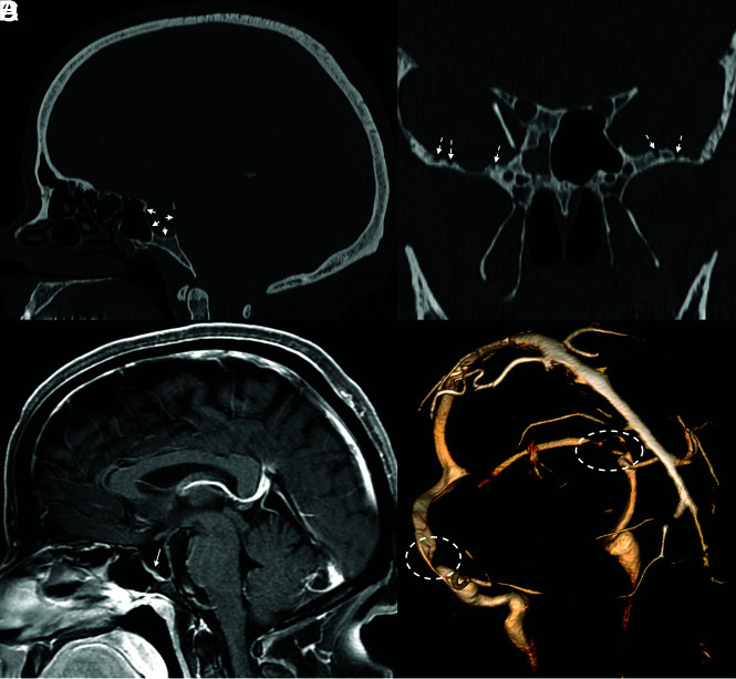
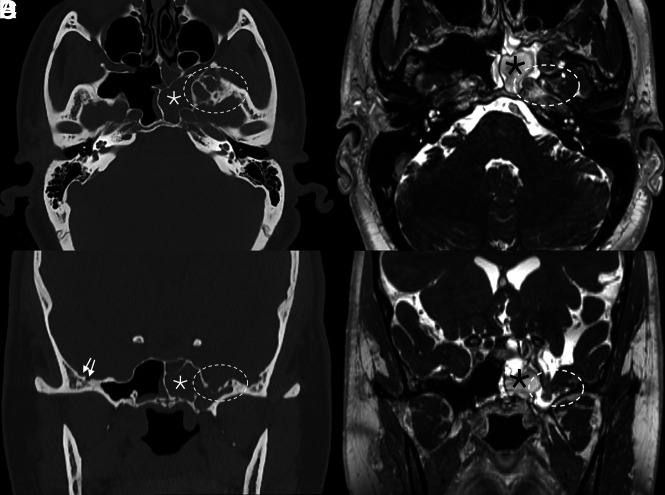
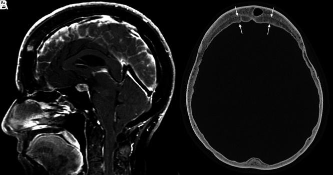
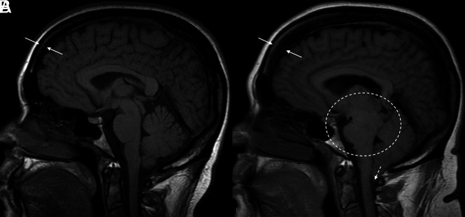
The Monro-Kellie doctrine is a well-accepted principle of intracranial hemodynamics. It has undergone few consequential revisions since it was established. Its principle is straightforward: The combined volume of neuronal tissue, blood, and CSF is constant. To maintain homeostatic intracranial pressure, any increase or decrease in one of these elements leads to a reciprocal and opposite change in the others. The Monro-Kellie doctrine assumes a rigid, unadaptable calvaria. Recent studies have disproven this assumption. The skull expands and grows in response to pathologic changes in intracranial pressure. In this review, we outline what is known about calvarial changes in the setting of pressure dysregulation and suggest a revision to the Monro-Kellie doctrine that includes an adaptable skull as a fourth component.


Figures

FIG 1.
FIG 1.
Sella expansion and skull base pitting in IIH. Sagittal CT image (A) in a 38-year-old woman with pseudotumor cerebri demonstrates marked expansion of the osseous walls of the sella (short solid arrows), with frank dehiscence posteriorly. Corresponding sagittal MR image (B) shows flattening of the pituitary tissue along the floor of the sella (long solid arrow). Prominent pitting is also noted along the anterior margins of both middle cranial fossae (dashed arrows, C). 3D reconstruction image of an MRV (D) demonstrates smooth tapering of the bilateral transverse sinuses (dashed ovals), compatible with IIH.
FIG 2.
FIG 2.
Skull pitting, meningoceles, and CSF leak in a patient who presented with rhinorrhea. Axial and coronal CT images (A and B) demonstrate substantial pitting along the anteromedial aspect of the left middle cranial fossa (dashed ovals, A and B), with complete opacification of the left sphenoid sinus (asterisks), concerning for a CSF leak. Milder pitting is noted on the right (solid arrows). Corresponding MR imaging (C and D) confirms a meningocele protruding into the left sphenoid sinus (dashed ovals, C and D). MRV (not shown) noted smooth tapered stenoses involving both transverse sinuses, and the patient was ultimately diagnosed with pseudotumor cerebri.
FIG 3.
FIG 3.
Example of layered calvarial thickening in a patient with SIH. Sagittal MR images (A) demonstrate classic findings of SIH, including pituitary enlargement, brain sag, and dural venous sinus engorgement. Axial CT image (B) in the same patient shows thickening of the bifrontal calvaria with characteristic layering related to preferential growth along the inner table of the skull (between straight arrows).
FIG 4.
FIG 4.
Hyperostosis related to a CSF leak. Sagittal T1-weight images before (A) and after (B) the development and diagnosis of SIH, with 16 years between examinations. Multiple classic findings of SIH are seen, including brain sag (dashed oval) and herniation of the cerebellar tonsils through the foramen magnum (dashed arrow). The patient also developed substantial frontal-predominant calvarial hyperostosis (between solid arrows), particularly along the inner table of the skull.

References

    1. Mokri B. The Monro-Kellie hypothesis: applications in CSF volume depletion. Neurology 2001;56:1746–48 10.1212/wnl.56.12.1746 - DOI - PubMed
    1. Rosenberg KI, Banik R. Pseudotumor cerebri syndrome associated with giant arachnoid granulation. J Neuroophthalmol 2013;33:417–19 10.1097/WNO.0b013e3182a5943b - DOI - PubMed
    1. Babcock JC, Johnson DR, Benson JC, et al. . Diffuse calvarial hyperostosis and spontaneous intracranial hypotension: a case-control study. AJNR Am J Neuroradiol 2022;43:978–83 10.3174/ajnr.A7557 - DOI - PMC - PubMed
    1. Wu OC, Manjila S, Malakooti N, et al. . The remarkable medical lineage of the Monro family: contributions of Alexander primus, secundus, and tertius. J Neurosurg 2012;116:1337–46 10.3171/2012.2.JNS111366 - DOI - PubMed
    1. Wilson MH. Monro-Kellie 2.0: The dynamic vascular and venous pathophysiological components of intracranial pressure. J Cereb Blood Flow Metab 2016;36:1338–50 10.1177/0271678X16648711 - DOI - PMC - PubMed

MeSH terms

LinkOut - more resources