. 2022 Nov;8(2):e002672.
doi: 10.1136/rmdopen-2022-002672.

Dysregulated long non-coding RNA in Sjögren's disease impacts both interferon and adaptive immune responses

Affiliations

Dysregulated long non-coding RNA in Sjögren's disease impacts both interferon and adaptive immune responses

Michelle L Joachims et al. RMD Open. 2022 Nov.

Abstract

Objective: Sjögren's disease (SjD) is an autoimmune disease characterised by inflammatory destruction of exocrine glands. Patients with autoantibodies to Ro/SSA (SjDRo+) exhibit more severe disease. Long non-coding RNAs (lncRNAs) are a functionally diverse class of non-protein-coding RNAs whose role in autoimmune disease pathology has not been well characterised.

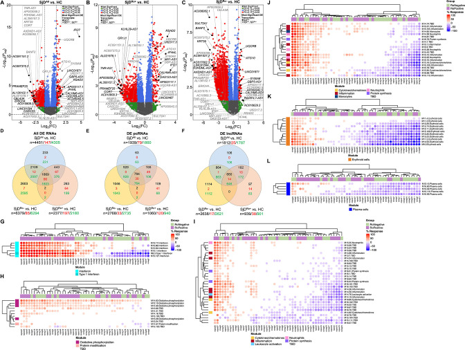
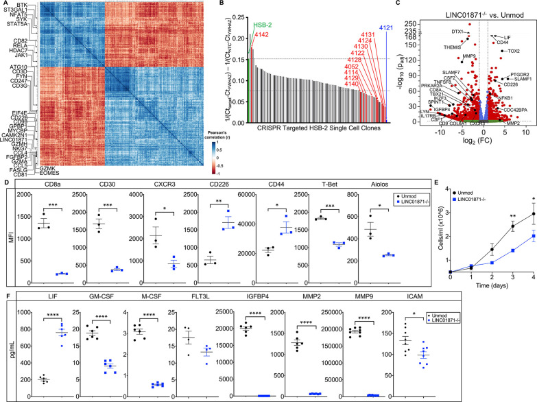
Methods: Whole blood RNA-sequencing (RNA-seq) was performed on SjD cases (n=23 Ro/SSA negative (SjDRo-); n=27 Ro/SSA positive (SjDRo+) and healthy controls (HCs; n=27). Bioinformatics and pathway analyses of differentially expressed (DE) transcripts (log2 fold change ≥2 or ≤0.5; padj<0.05) were used to predict lncRNA function. LINC01871 was characterised by RNA-seq analyses of HSB-2 cells with CRISPR-targeted LINC01871 deletion (LINC01871-/ -) and in vitro stimulation assays.

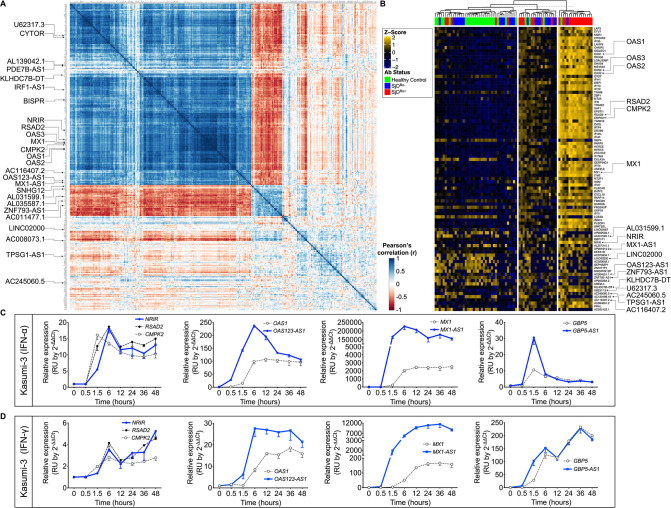
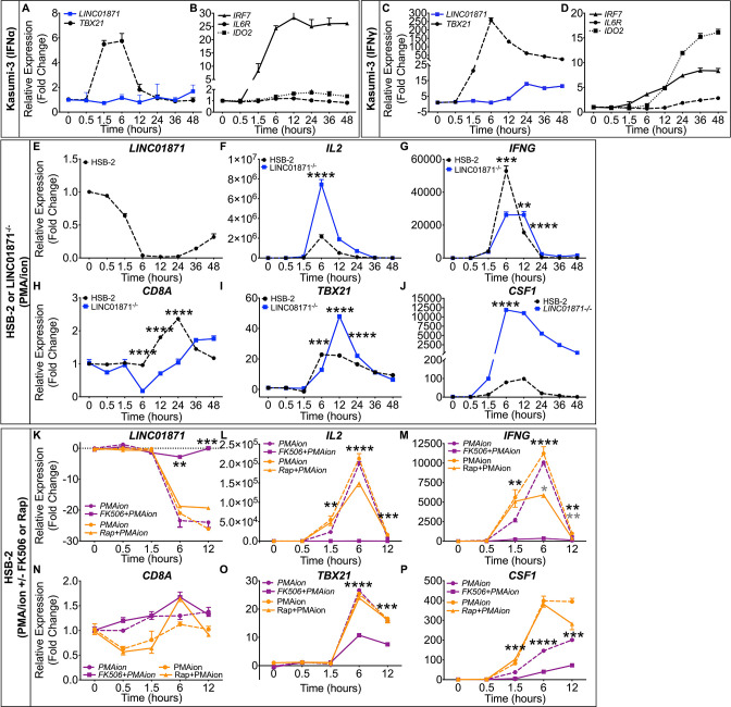
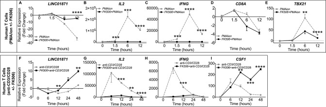
Results: Whole blood RNA-seq revealed autoantibody-specific transcription profiles and disproportionate downregulation of DE transcripts in SjD cases relative to HCs. Sixteen DE lncRNAs exhibited correlated expression with the interferon (IFN)-regulated gene, RSAD2, in SjDRo+ (r≥0.65 or ≤-0.6); four antisense lncRNAs exhibited IFN-regulated expression in immune cell lines. LINC01871 was upregulated in all SjD cases. RNA-seq and pathway analyses of LINC01871-/ - cells implicated roles in cytotoxic function, differentiation and IFNγ induction. LINC01871 was induced by IFNγ in a myeloid cell line and regulated by calcineurin/NFAT pathway and T cell receptor (TCR) signalling in primary human T cells.

Conclusion: LINC01871 influences expression of many immune cell genes and growth factors, is IFNγ inducible, and regulated by calcineurin signalling and TCR ligand engagement. Altered LINC01871 expression may influence the dysregulated T cell inflammatory pathways implicated in SjD.

Trial registration: ClinicalTrials.gov NCT02327884.

Keywords: Autoantibodies; Autoimmune Diseases; Autoimmunity; Polymorphism, Genetic; Sjogren's Syndrome.


Conflict of interest statement

Competing interests: KS is a current employee of Janssen. ADF and CJL have an active collaborative research agreement with Janssen. All other authors have reported that they have no competing interests to report.

Figures

Figure 1
Figure 1
Protein-coding (pc)RNAs and long non-coding (lnc)RNAs are differentially expressed in the whole blood of SjD cases compared with healthy controls (HCs). (A–C) Differentially expressed (DE) transcripts from whole blood RNA-sequencing analysis of (A) all SjD cases (SjDAll; n=50), (B) anti-Ro positive SjD cases (SjDRo+; n=27) or (C) anti-Ro negative SjD cases (SjDRo−; n=23) compared with HCs (n=27). Y-axis shows the −log10 of the FDR-adjusted p value (padj); x-axis shows the log2 of the fold change (FC). Black dots indicate the top three upregulated and downregulated pcRNAs and lncRNAs in the analysis. Grey dots indicate the top three upregulated and downregulated pcRNAs and lncRNAs in the other analyses. (D–F) Distribution of (D) all DE transcripts, (E) DE pcRNAs or (F) DE lncRNAs across the three analyses. Black text indicates total DE transcripts; red text indicates upregulated transcripts; green text indicates downregulated transcripts. (G–L) Hierarchical clustering of annotated module aggregates across individual SjDRo+ (purple) or SjDRo− (green) cases. Displayed individual modules had DE of >20% of constitutive transcripts in at least one case. Colour gradient indicates the proportion of DE transcripts ranging from 100% increased (red) to 100% decreased (blue), respective to HCs. FDR, false discovery rate; SjD, Sjögren’s disease.
Figure 2
Figure 2
Differentially expressed (DE) interferon (IFN)-responsive lncRNAs were coordinately modulated with associated pcRNAs and correlated with antibody status of SjD cases. (A) RSAD2 correlation analysis of the normalised RNA-seq data from the SjDRo+-only primary expression matrix (r≥0.65 or ≤−0.6; p<0.05). Type I IFN-responsive pcRNAs and other pcRNAs and lncRNAs of interest are indicated. (B) Individual-level expression of transcripts that were correlated with RSAD2 expression and DE in any of the whole blood RNA-seq analyses. Z-scores were computed using scale function in R, after calculating fold change of expression relative to healthy controls. K-means clustering (K=3) segregated cases by high, moderate and low IFN status, and are shown relative to patient antibody status. pcRNAs and lncRNAs of interest are indicated. (C, D) RT-qPCR analysis of indicated DE transcripts in Kasumi-3 cells stimulated with (C) universal type I IFN (IFNα; 150 U/mL) or (D) IFNγ (2800 U/mL) from 0 to 48 hours. Target lncRNAs are shown in blue and respective pcRNAs in grey (n>3). lncRNAs, long non-coding RNAs; pcRNAs, protein-coding RNAs; RNA-seq, RNA sequencing; RT-qPCR, quantitative reverse transcription PCR; SjD, Sjögren’s disease; SjDRo+, anti-Ro positive SjD; SjDRo−, anti-Ro negative SjD.
Figure 3
Figure 3
Loss of LINC01871 disrupts basal expression of genes involved in immune cell regulation. (A) LINC01871 correlation analysis of normalised RNA-seq data from the SjDRo−-only primary expression matrix (r≥0.7 or ≤−0.6; p<0.05). Transcripts implicated in immune function or SjD pathology are indicated. (B) Quantitative PCR screen of LINC01871 expression in HSB-2 single-cell clones after CRISPR-targeted deletion of LINC01871. (C) Differentially expressed transcripts from the RNA-seq analysis of HSB-2 clone 4121 (hereafter LINC01871−/) relative to HSB-2 parental cell line. Y-axis shows the −log10 of the FDR-adjusted p value (padj); x-axis shows the log2 of the fold change (FC). Black dots indicate independently replicated transcripts of interest. (D) Levels of indicated surface or intracellular proteins, reported as mean fluorescence intensity (MFI), in HSB-2 parental cells (black) and LINC01871−/ cells (blue); n=3; unpaired t-test where *p<0.05, **p<0.01 or ***p<0.001. (E) Growth curve analysis of HSB-2 parental cells (black) and LINC01871−/ cells (blue) from 0 to 96 hours; n=3; unpaired t-test where *p<0.05 or **p<0.01. (F) Concentration of indicated secreted protein in supernatant collected from HSB-2 parental cells (black) and LINC01871−/ cells (blue) at 96 hours; n>6; unpaired t-test where *p<0.05 or ****p<0.0001. FDR, false discovery rate; RNA-seq, RNA sequencing; SjD, Sjögren’s disease; SjDRo−, anti-Ro negative SjD.
Figure 4
Figure 4
LINC01871 is modulated by interferon (IFN)γ and calcineurin/NFAT signalling in immune cell lines. (A−D) RT-qPCR analysis of LINC01871 (A and C), TBX21 (A and C), IRF7 (B and D), IL6R (B and D) and IDO2 (B and D) in Kasumi-3 cells stimulated with either (A, B) universal type I IFN (IFNα; 150 U/mL) or (C, D) IFNγ (2800 U/mL) from 0 to 48 hours. Expression is reported as fold change relative to unstimulated controls. (E−J) RT-qPCR analysis of LINC01871 (E), IL2 (F), IFNG (G), CD8A (H), TBX21 (I) and CSF1 (J) in LINC01871−/− cells (blue) and HSB-2 parental (black) cells stimulated from 0 to 48 hours with PMA/I (100 ng/mL; 1000 ng/mL). Expression is reported as fold change relative to unstimulated controls; n=3; unpaired t-test where **p<0.01, ***p<0.001 or ****p<0.0001. (K−P) RT-qPCR analysis of LINC01871 (K), IL2 (L), IFNG (M), CD8A (N), TBX21 (O) and CSF1 (P) in HSB-2 parental cells stimulated from 0 to 12 hours with PMA/I (100 ng/mL; 1000 ng/mL) with and without FK506 (2.5 µM; purple) or rapamycin (Rap; 2.5 µM; orange). Expression is reported as fold change relative to unstimulated inhibitor controls; n=3; paired t-test where *p<0.05, **p<0.01, ***p<0.001 or ****p<0.0001. Black * indicates FK506 with PMA/I relative to PMA/I alone and grey * indicates Rap with PMA/I relative to PMA/I alone. PMA/I, phorbol 12-myristate 13-acetate and ionomycin; RT-qPCR, quantitative reverse transcription PCR.
Figure 5
Figure 5
LINC01871 is modulated by IFNγ and calcineurin/NFAT signalling in purified human T cells. (A–E) Negatively selected human donor T cells were treated with DMSO or 2.5 µM FK506 overnight, then stimulated with PMA/I (100 ng/mL; 1000 ng/mL) for 12 hours. Expression of LINC01871 (A), IL2 (B), IFNG (C), CD8A (D) and TBX21 (E) was assessed by RT-qPCR and reported as fold change relative to DMSO-treated cells; n=4; paired t-test where *p<0.05, **p<0.01, ***p<0.001 or ****p<0.0001. Grey lines indicate PMA/I. Black lines indicate PMA/I with FK506 treatment. (F–I) Negatively selected human donor T cells were treated with either DMSO or 2.5 µM FK506 overnight, then stimulated with anti-CD3/CD28 for 48 hours. Expression of LINC01871 (F), IL2 (G), IFNG (H) and CSF1 (I) was assessed by RT-qPCR and reported as fold change relative to DMSO-treated cells at each time point; n=4; paired t-test where *p<0.05, **p<0.01, ***p<0.001 or ****p<0.0001. Grey lines indicate anti-CD3/CD28. Black lines indicate anti-CD3/CD28 with FK506 treatment. IFN, interferon; PMA/I, phorbol 12-myristate 13-acetate and ionomycin; RT-qPCR, quantitative reverse transcription PCR.

Comment in

Similar articles

Cited by

  • Multiomic single-cell sequencing defines tissue-specific responses in Stevens-Johnson Syndrome and Toxic epidermal necrolysis.
    Gibson A, Ram R, Gangula R, Li Y, Mukherjee E, Palubinsky AM, Campbell CN, Thorne M, Konvinse KC, Choshi P, Deshpande P, Pedretti S, O'Neil RT, Wanjalla CN, Kalams SA, Gaudieri S, Lehloenya RJ, Bailin SS, Chopra A; Jason A Trubiano on behalf of the AUS-SCAR study group; Jonny G Peter on behalf of the AFRI-SCAR and IMARI-Africa study group; Mallal SA, Phillips EJ. Gibson A, et al. bioRxiv [Preprint]. 2024 Feb 18:2023.11.26.568771. doi: 10.1101/2023.11.26.568771. bioRxiv. 2024. Update in: Nat Commun. 2024 Oct 8;15(1):8722. doi: 10.1038/s41467-024-52990-3. PMID: 38405793 Free PMC article. Updated. Preprint.
  • Variants in the DDX6-CXCR5 autoimmune disease risk locus influence the regulatory network in immune cells and salivary gland.
    Wiley MM, Khatri B, Joachims ML, Tessneer KL, Stolarczyk AM, Rasmussen A, Anaya JM, Aqrawi LA, Bae SC, Baecklund E, Björk A, Brun JG, Bucher SM, Dand N, Eloranta ML, Engelke F, Forsblad-d'Elia H, Fugmann C, Glenn SB, Gong C, Gottenberg JE, Hammenfors D, Imgenberg-Kreuz J, Jensen JL, Johnsen SJA, Jonsson MV, Kelly JA, Khanam S, Kim K, Kvarnström M, Mandl T, Martín J, Morris DL, Nocturne G, Norheim KB, Olsson P, Palm Ø, Pers JO, Rhodus NL, Sjöwall C, Skarstein K, Taylor KE, Tombleson P, Thorlacius GE, Venuturupalli S, Vital EM, Wallace DJ, Grundahl KM, Radfar L, Brennan MT, James JA, Scofield RH, Gaffney PM, Criswell LA, Jonsson R, Appel S, Eriksson P, Bowman SJ, Omdal R, Rönnblom L, Warner BM, Rischmueller M, Witte T, Farris AD, Mariette X, Shiboski CH; Sjögren’s International Collaborative Clinical Alliance (SICCA); Wahren-Herlenius M, Alarcón-Riquelme ME; PRECISESADS Clinical Consortium; Ng WF; UK Primary Sjögren’s Syndrome Registry; Sivils KL, Guthridge JM, Adrianto I, Vyse TJ, Tsao BP, Nordmark G, Lessard CJ. Wiley MM, et al. bioRxiv [Preprint]. 2023 Oct 6:2023.10.05.561076. doi: 10.1101/2023.10.05.561076. bioRxiv. 2023. Update in: Ann Rheum Dis. 2025 May 29:S0003-4967(25)00949-5. doi: 10.1016/j.ard.2025.04.023. PMID: 39071447 Free PMC article. Updated. Preprint.
  • Role of ncRNAs in the Pathogenesis of Sjögren's Syndrome.
    Al-Haidose A, Hassan S, Elhassan M, Ahmed E, Al-Riashi A, Alharbi YM, Ghunaim M, Alhejaili T, Abdallah AM. Al-Haidose A, et al. Biomedicines. 2024 Jul 11;12(7):1540. doi: 10.3390/biomedicines12071540. Biomedicines. 2024. PMID: 39062113 Free PMC article. Review.
  • [Sex-specific aspects in rheumatology].
    Albrecht K, Ohrndorf S, Strangfeld A. Albrecht K, et al. Z Rheumatol. 2024 Nov;83(9):749-759. doi: 10.1007/s00393-024-01527-6. Epub 2024 Jun 13. Z Rheumatol. 2024. PMID: 38869669 German.
  • Multiomic single-cell sequencing defines tissue-specific responses in Stevens-Johnson syndrome and toxic epidermal necrolysis.
    Gibson A, Ram R, Gangula R, Li Y, Mukherjee E, Palubinsky AM, Campbell CN, Thorne M, Konvinse KC, Choshi P, Deshpande P, Pedretti S, Fear MW, Wood FM, O'Neil RT, Wanjalla CN, Kalams SA, Gaudieri S, Lehloenya RJ, Bailin SS, Chopra A, Trubiano JA; AUS-SCAR Consortium; Peter JG; AFRiSCAR Consortium; Mallal SA, Phillips EJ. Gibson A, et al. Nat Commun. 2024 Oct 8;15(1):8722. doi: 10.1038/s41467-024-52990-3. Nat Commun. 2024. PMID: 39379371 Free PMC article.

References

    1. Qin B, Wang J, Yang Z, et al. . Epidemiology of primary Sjögren's syndrome: a systematic review and meta-analysis. Ann Rheum Dis 2015;74:1983–9. 10.1136/annrheumdis-2014-205375 - DOI - PubMed
    1. Helmick CG, Felson DT, Lawrence RC, et al. . Estimates of the prevalence of arthritis and other rheumatic conditions in the United States. Part I. Arthritis Rheum 2008;58:15–25. 10.1002/art.23177 - DOI - PubMed
    1. Gøransson LG, Haldorsen K, Brun JG, et al. . The point prevalence of clinically relevant primary Sjögren's syndrome in two Norwegian counties. Scand J Rheumatol 2011;40:221–4. 10.3109/03009742.2010.536164 - DOI - PubMed
    1. Goules AV, Kapsogeorgou EK, Tzioufas AG. Insight into pathogenesis of Sjögren's syndrome: dissection on autoimmune infiltrates and epithelial cells. Clin Immunol 2017;182:30–40. 10.1016/j.clim.2017.03.007 - DOI - PubMed
    1. Mariette X, Criswell LA. Primary Sjögren's syndrome. N Engl J Med 2018;378:931–9. 10.1056/NEJMcp1702514 - DOI - PubMed

Publication types

Associated data