. 2023 Feb;49(1):e12865.
doi: 10.1111/nan.12865.

Abundant copathologies of polyglucosan bodies, frontotemporal lobar degeneration with TDP-43 inclusions and ageing-related tau astrogliopathy in a family with a GBE1 mutation

Affiliations

Abundant copathologies of polyglucosan bodies, frontotemporal lobar degeneration with TDP-43 inclusions and ageing-related tau astrogliopathy in a family with a GBE1 mutation

Maiko T Uemura et al. Neuropathol Appl Neurobiol. 2023 Feb.

Abstract

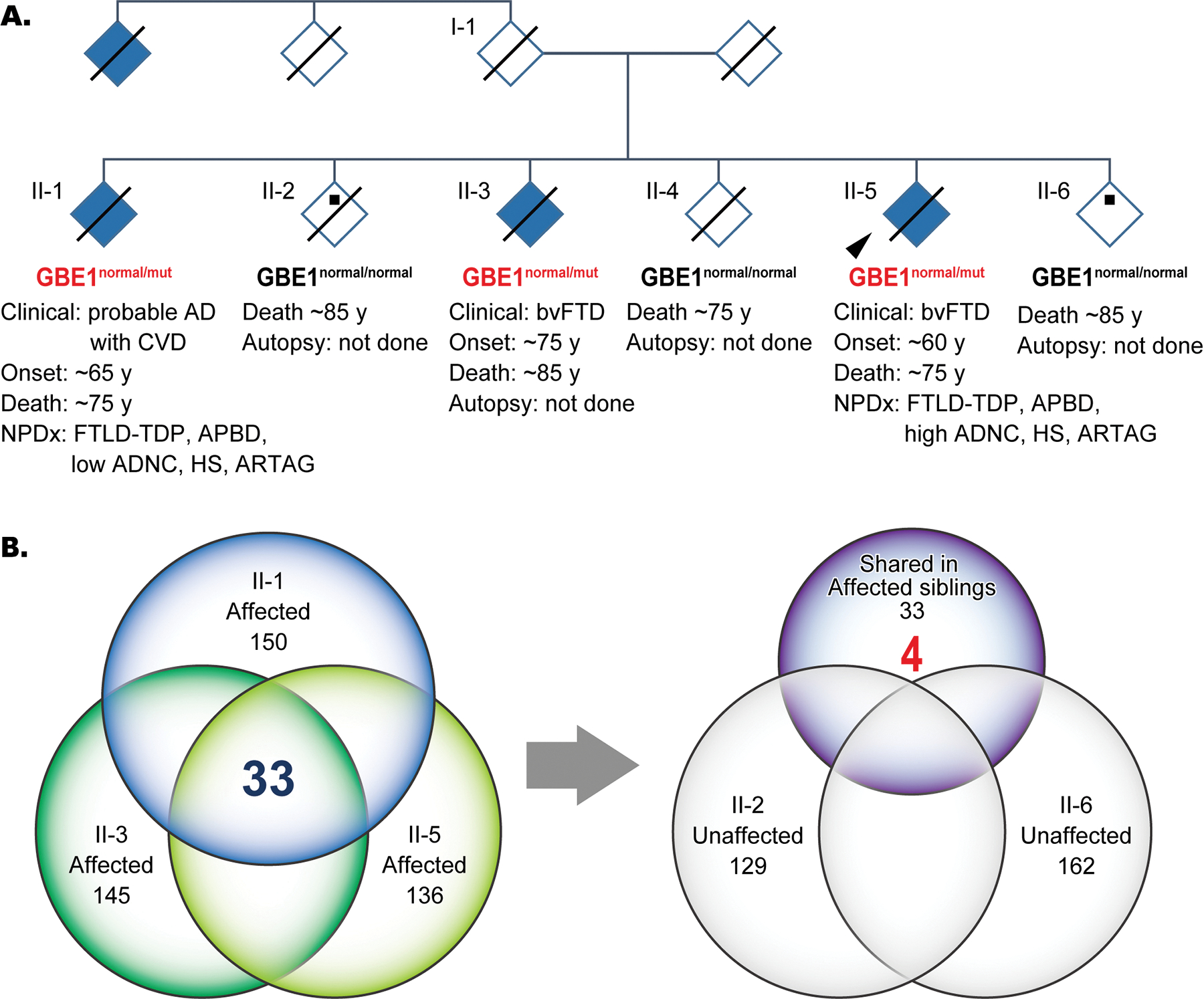
Aims: Adult polyglucosan body disease (APBD) is a progressive neurogenetic disorder caused by 1,4-alpha-glucan branching enzyme 1 (GBE1) mutation with an accumulation of polyglucosan bodies (PBs) in the central and peripheral nervous systems as a pathological hallmark. Here, we report two siblings in a family with a GBE1 mutation with prominent frontotemporal lobar degeneration with TAR DNA-binding protein 43 (FTLD-TDP) and ageing-related tau astrogliopathy (ARTAG) copathologies with PBs in the central nervous system.

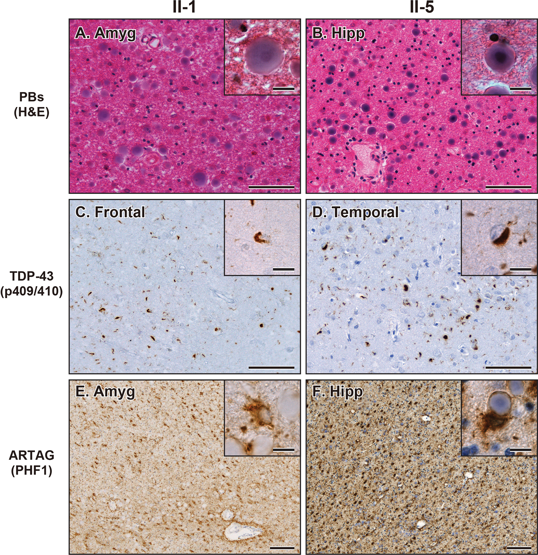
Methods: Whole-genome sequencing (WGS) followed by Sanger sequencing (SS) was performed on three affected and two unaffected siblings in a pedigree diagnosed with familial frontotemporal dementia. Out of the affected siblings, autopsies were conducted on two cases, and brain samples were used for biochemical and histological analyses. Brain sections were stained with haematoxylin and eosin and immunostained with antibodies against ubiquitin, tau, amyloid β, α-synuclein, TDP-43 and fused in sarcoma (FUS).

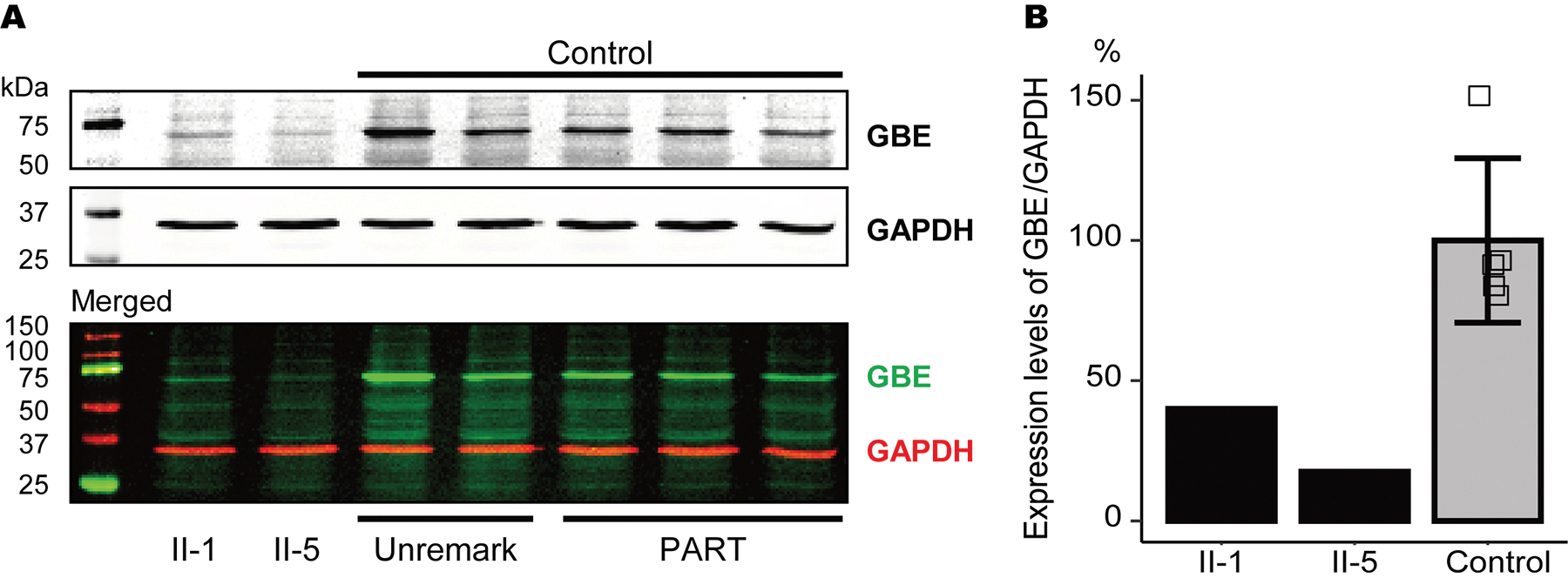
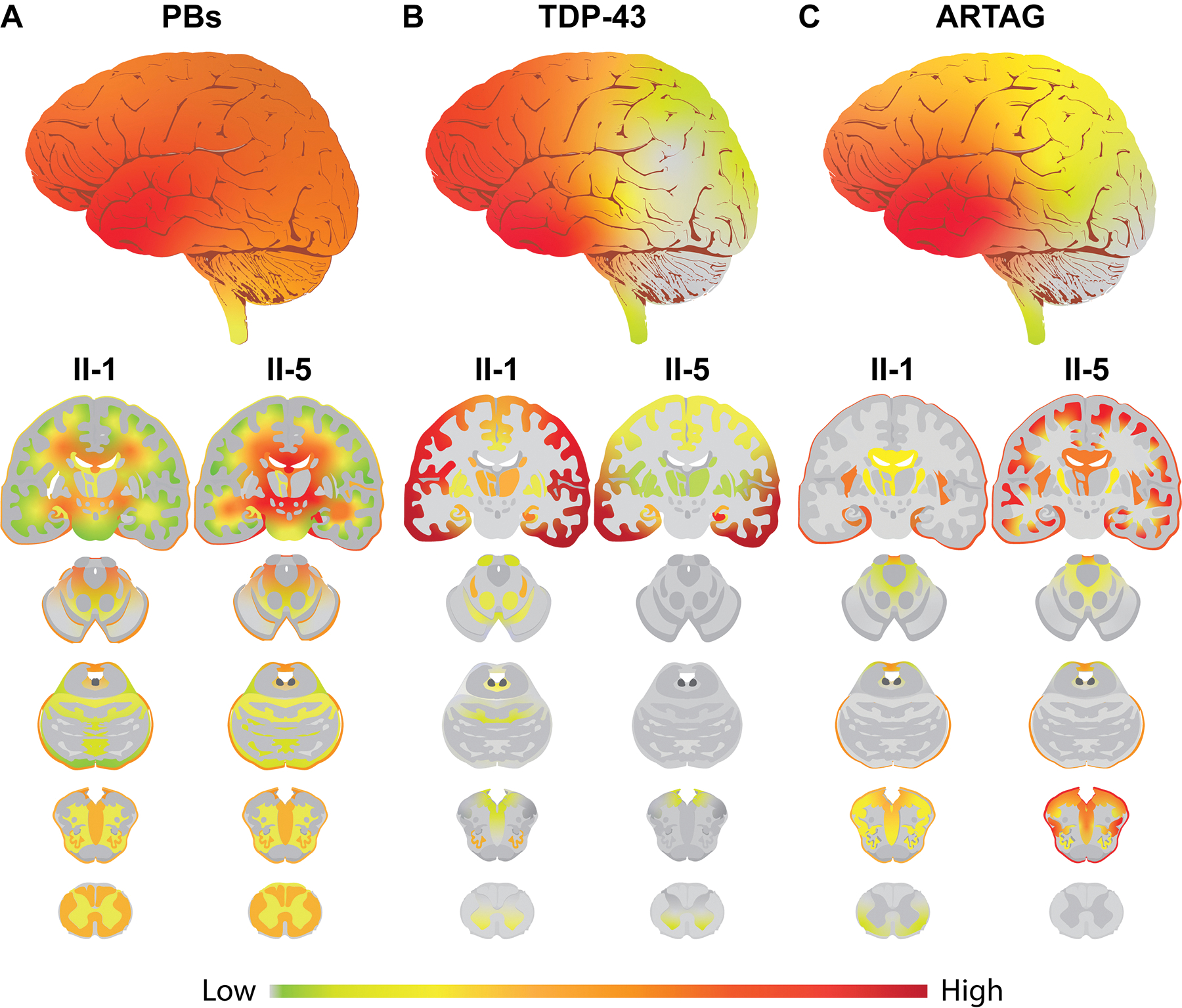
Results: A novel single nucleotide deletion in GBE1, c.1280delG, was identified, which is predicted to result in a reading frameshift, p.Gly427Glufs*9. This variant segregated with disease in the family, is absent from population databases and is predicted to cause loss of function, a known genetic mechanism for APBD. The affected siblings showed a greater than 50% decrease in GBE protein levels. Immunohistochemical analysis revealed widespread FTLD-TDP (type A) and ARTAG pathologies as well as PBs in the brains of two affected siblings for whom an autopsy was performed.

Conclusions: This is the first report of a family with several individuals with a FTD clinical phenotype and underlying copathologies of APBD, FTLD-TDP and ARTAG with a segregating GBE1 loss-of-function mutation in affected siblings. The finding of copathologies of APBD and FTLD-TDP suggests these processes may share a disease mechanism resulting from this GBE1 mutation.

Keywords: ARTAG; FTLD-TDP; GBE1; adult polyglucosan body disease; corpora amylacea.


Figures

Figure 1.
Figure 1.. Pedigree with FTD and Genetic analysis
(A) Pedigree diagnosed with FTD. An autopsy was performed on two affected siblings (II-1 and -5). Arrowhead shows proband. Black dots and solid lines indicate cases with available DNA and those deceased, respectively. Whole genome sequencing was performed with samples from 3 affected siblings (II-1, -3, -5) and two unaffected siblings (II-2 and -6). The heterozygous status of the GBE1 mutation is shown in red. (B) Venn diagrams of numbers of rare variants identified from whole genome sequencing among the affected and unaffected siblings. Thirty-three variants that were shared among the three affected siblings were filtered against the variants identified in unaffected siblings, which resulted in 4 variants. Abbreviations: ADNC, Alzheimer’s disease neuropathologic change; APBD, adult polyglucosan body disease; ARTAG, ageing-related tau astrogliopathy; bvFTD, behavioural variant of frontotemporal dementia; CVD, cerebrovascular disease; FTLD-TDP, frontotemporal lobar degeneration with TDP-43 inclusions; mut, mutant; PDD, Parkinson’s disease dementia
Figure 2.
Figure 2.. Western blot analysis for GBE1 protein expression levels.
(A) Representative images of western blot analysis using GBE and GAPDH antibodies. (B) Relative expression levels of GBE/GAPDH in II-1, II-5, and control cases. The GBE protein expression levels were reduced in affected sibling cases compared with control cases. Vertical bars represent mean ± SD. Abbreviations: PART, primary age-related tauopathy; Unremark, unremarkable adult brain.
Figure 3.
Figure 3.. Neuropathological findings in the cases with GBE1 mutation.
(A, B) Representative images of H&E staining showing PBs in the amygdala in case II-1 (A) and hippocampus in case II-5 (B). Bars represent 50 μm and 10 μm (insets). (C, D) Representative images of TDP-43 (p409/410) staining showing NCI and DN in the frontal cortex in case II-1 (C) and temporal cortex in case II-5 (D). Bars represent 50 μm and 10 μm (insets). (E, F) Representative images of PHF1 staining showing ARTAG in the amygdala in case II-1 (E) and hippocampus in case II-5 (F). Insets show PBs adjacent to ARTAG. Bars represent 200 μm and 10 μm (insets). Abbreviations: Amyg, amygdala; Hipp, hippocampus; Temp, temporal cortex.
Figure 4.
Figure 4.. Distribution of PBs, TDP-43 NCI and DN, and ARTAG in the CNS with GBE1 mutation.
(A) Heatmap of PB pathology. The upper whole brain image represents the average of two cases with interpolated smoothing. Lower sagittal and coronal images represent PB distribution in II-1 and II-5. PBs are observed in the subpial matter and the deep white matter of frontal, temporal, parietal, and occipital lobes, and the cerebellum as well as some parts of the brainstem and the spinal cord. II-5 shows more abundant PB pathology than II-1. (B) Heatmap of TDP-43 pathology. The upper whole brain image represents the average of two cases with interpolated smoothing. Lower sagittal and coronal images represent TDP-43 distribution in II-1 and II-5. TDP-43 NCI and DN are observed in the neocortical, allocortical, and subcortical grey matter and partially in the grey matter of the brainstem and spinal cord. The TDP-43 pathologies are especially abundant in the frontotemporal cortices. II-1 shows more broadly distributed pathology than II-5. (C) Heatmap of ARTAG pathology. The upper whole brain image represents the average of two cases with interpolated smoothing. Lower sagittal and coronal images represent ARTAG distribution in II-1 and II-5. ARTAG is abundantly observed in the subpial matter and the white matter of frontotemporal lobes, amygdala, and hippocampus and partially observed in the subpial matter and the white matter of the brainstem and spinal cord. II-5 shows more abundant and broadly distributed ARTAG than II-1. A gradient smoothing method was used for the boundaries between each assessed legion.

Similar articles

Cited by

References

    1. Mochel F, Schiffmann R, Steenweg ME, Akman HO, Wallace M, Sedel F, Laforêt P, Levy R, Powers MJ, Demeret S, Maisonobe T, Froissart R, Nobrega B, Fogel BL, Natowicz MR, Lubetzki C, Durr A, Brice A, Rosenmann H, Barash V, Kakhlon O, Gomori MJ, Knaap MSvd, Lossos A. Adult polyglucosan body disease: Natural History and Key Magnetic Resonance Imaging Findings. Annals of neurology 2012; 72: 433–41 - PMC - PubMed
    1. Akman HO, Kakhlon O, Coku J, Peverelli L, Rosenmann H, Rozenstein-Tsalkovich L, Turnbull J, Meiner V, Chama L, Lerer I, Shpitzen S, Leitersdorf E, Paradas C, Wallace M, Schiffmann R, DiMauro S, Lossos A, Minassian BA. Deep Intronic GBE1 Mutation in Manifesting Heterozygous Patients With Adult Polyglucosan Body Disease. Jama Neurol 2015; 72: 441–5 - PubMed
    1. Lossos A, Meiner Z, Barash V, Soffer D, Schlesinger I, Abramsky O, Argov Z, Shpitzen S, Meiner V. Adult polyglucosan body disease in Ashkenazi Jewish patients carrying the Tyr329Ser mutation in the glycogen-branching enzyme gene. Annals of neurology 1998; 44: 867–72 - PubMed
    1. Harigaya Y, Matsukawa T, Fujita Y, Mizushima K, Ishiura H, Mitsui J, Morishita S, Shoji M, Ikeda Y, Tsuji S. Novel GBE1 mutation in a Japanese family with adult polyglucosan body disease. Neurology Genetics 2017; 3 (2): e138. - PMC - PubMed
    1. Akman OH, Kurt Y, Wallace M, Schiffmann R, DiMauro S. Compound heterozygosis of a splice site and the common Ashkenazi Jewish mutation in GBE1 causes adult onset polyglucosan body disease. (P2.026). Neurology 2015; 84: P 2.026

MeSH terms

Supplementary concepts

LinkOut - more resources