. 2022 Dec 1;12(1):20758.
doi: 10.1038/s41598-022-24870-7.

Modulation of EEG theta by naturalistic social content is not altered in infants with family history of autism

Collaborators, Affiliations

Modulation of EEG theta by naturalistic social content is not altered in infants with family history of autism

Rianne Haartsen et al. Sci Rep. .

Abstract

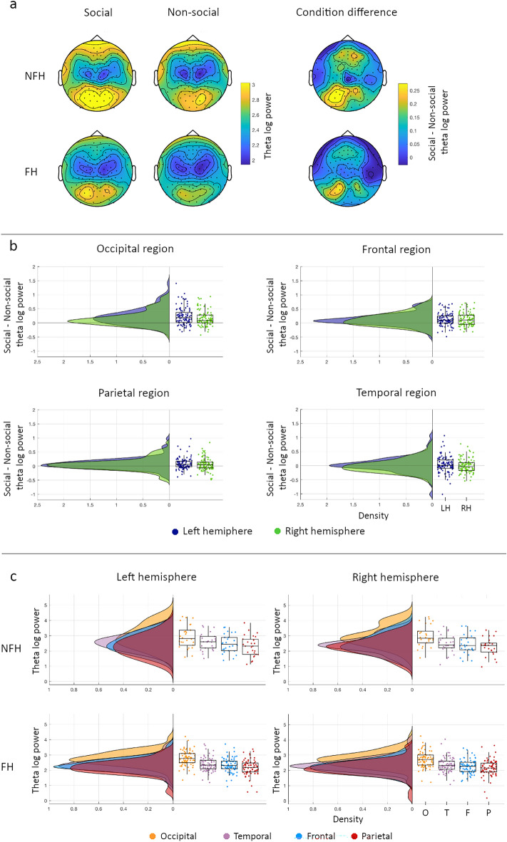
Theta oscillations (spectral power and connectivity) are sensitive to the social content of an experience in typically developing infants, providing a possible marker of early social brain development. Autism is a neurodevelopmental condition affecting early social behaviour, but links to underlying social brain function remain unclear. We explored whether modulations of theta spectral power and connectivity by naturalistic social content in infancy are related to family history for autism. Fourteen-month-old infants with (family history; FH; N = 75) and without (no family history; NFH; N = 26) a first-degree relative with autism watched social and non-social videos during EEG recording. We calculated theta (4-5 Hz) spectral power and connectivity modulations (social-non-social) and associated them with outcomes at 36 months. We replicated previous findings of increased theta power and connectivity during social compared to non-social videos. Theta modulations with social content were similar between groups, for both power and connectivity. Together, these findings suggest that neural responses to naturalistic social stimuli may not be strongly altered in 14-month-old infants with family history of autism.


Conflict of interest statement

The authors declare no competing interests.

Figures

Figure 1
Figure 1
Paradigm and attrition rates. (a) Screenshot of one of the toy dynamic videos. (b) Top view of the layout of the EEG high-density net. Electrodes on the outer rim were excluded since these were bad during the recording (red diamonds). Spectral theta power was calculated for frontal (blue), parietal (orange), temporal (green), and occipital (purple) regions (after). (c) Attrition rates for the samples included in the analyses.
Figure 2
Figure 2
Theta power modulations by social context. (a) Topoplots for the social and non-social condition (left and middle) and the difference (social–non-social) between conditions (right) for the NFH (top) and FH (bottom) group. (b) Density and boxplots for condition differences in power for the left (LH in navy) and right (RH in green) hemisphere in for each region (occipital in top left, frontal in top right, parietal in bottom left, and temporal in bottom right plot). Each dot represents 1 infant. Positive values indicate higher power for social than the non-social condition. (c) Density and boxplots for power within each region (occipital (O) in yellow, Temporal (T) in purple, Frontal (F) in blue, and Parietal (P) in red) within the left hemisphere (left) and the right hemisphere (right) for the NFH (top) and the FH (bottom) group. Each dot represents 1 infant.
Figure 3
Figure 3
Theta connectivity modulations by social context. (a) Density and boxplots for global connectivity for the social (blue) and non-social (orange) condition in the NFH group (top) and FH group (bottom). Each dot represents 1 infant. (b) Network of edges and nodes showing elevated connectivity during the social versus non-social condition. (c) Density and boxplots for connectivity within the social network for the social (blue) and non-social (orange) condition in the NFH group (top) and FH group (bottom). Each dot represents 1 infant.

References

    1. Grossmann T, Johnson MH. The development of the social brain in human infancy. Eur. J. Neurosci. 2007;25:909–919. doi: 10.1111/j.1460-9568.2007.05379.x. - DOI - PubMed
    1. Jones EJH, Gliga T, Bedford R, Charman T, Johnson MH. Developmental pathways to autism: A review of prospective studies of infants at risk. Neurosci. Biobehav. Rev. 2014;39:1–33. doi: 10.1016/j.neubiorev.2013.12.001. - DOI - PMC - PubMed
    1. American Psychological Association . Diagnostic and statistical manual of mental disorders. American Psychiatric Publishing; 2013.
    1. De La Torre-Ubieta L, Won H, Stein JL, Geschwind DH. Advancing the understanding of autism disease mechanisms through genetics. Nat. Med. 2016;22:345–361. doi: 10.1038/nm.4071. - DOI - PMC - PubMed
    1. Gao W, et al. A review on neuroimaging studies of genetic and environmental influences on early brain development. Neuroimage. 2019;185:802–812. doi: 10.1016/j.neuroimage.2018.04.032. - DOI - PMC - PubMed

Publication types