. 2022 Dec 1;12(1):20791.
doi: 10.1038/s41598-022-19710-7.

Analysis of genome-wide knockout mouse database identifies candidate ciliopathy genes

Collaborators, Affiliations

Analysis of genome-wide knockout mouse database identifies candidate ciliopathy genes

Kendall Higgins et al. Sci Rep. .

Abstract

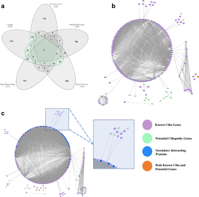
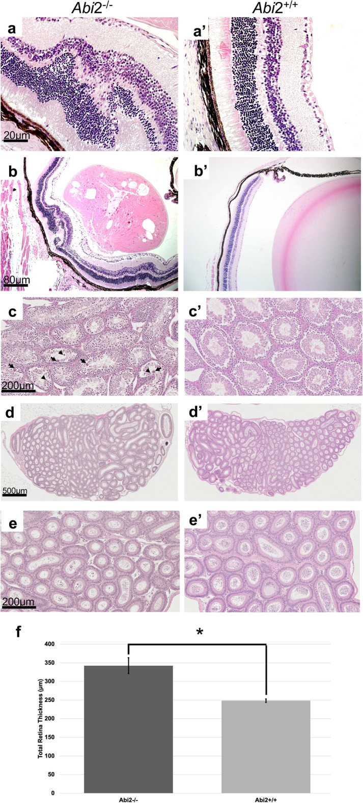
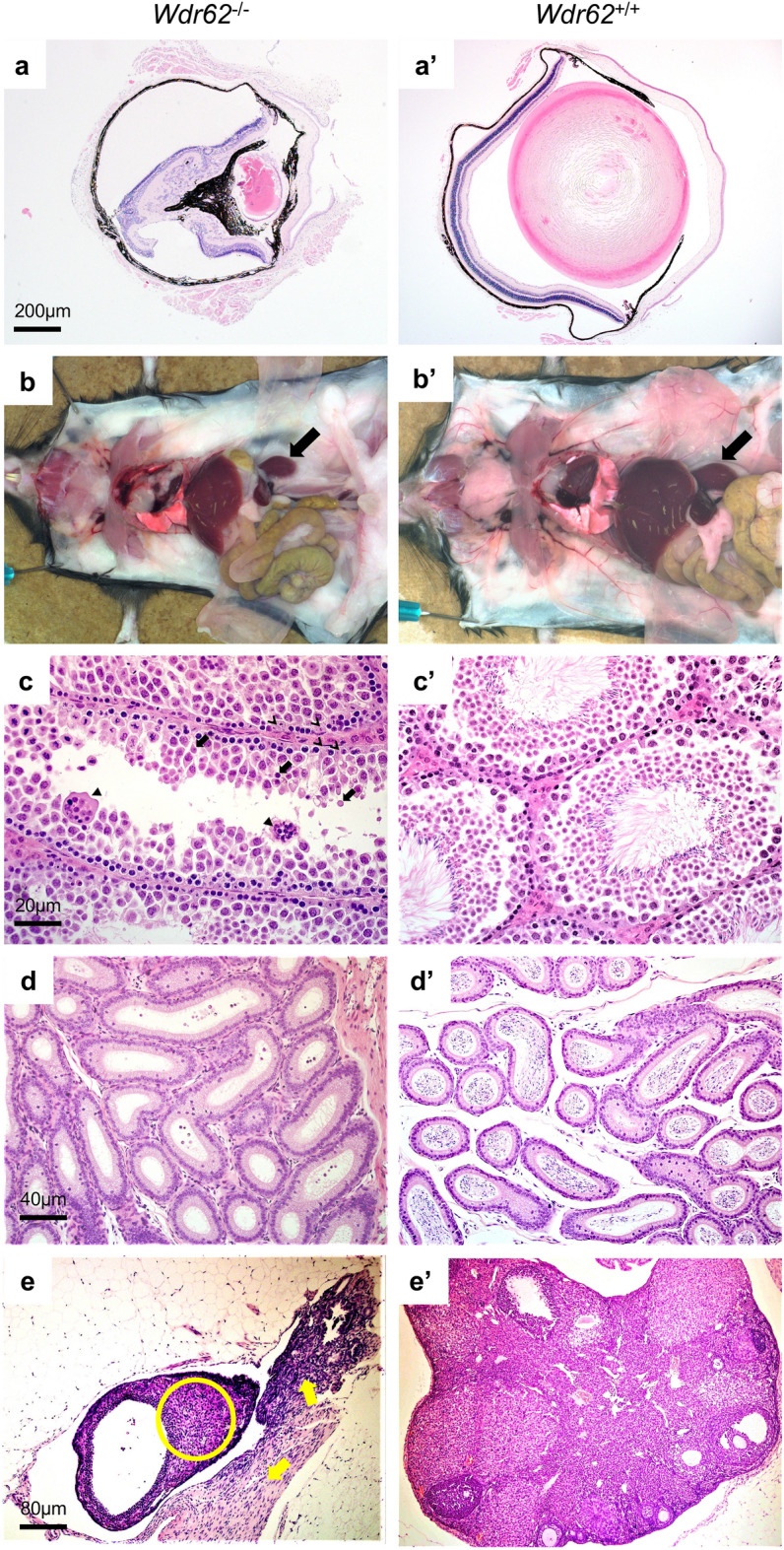
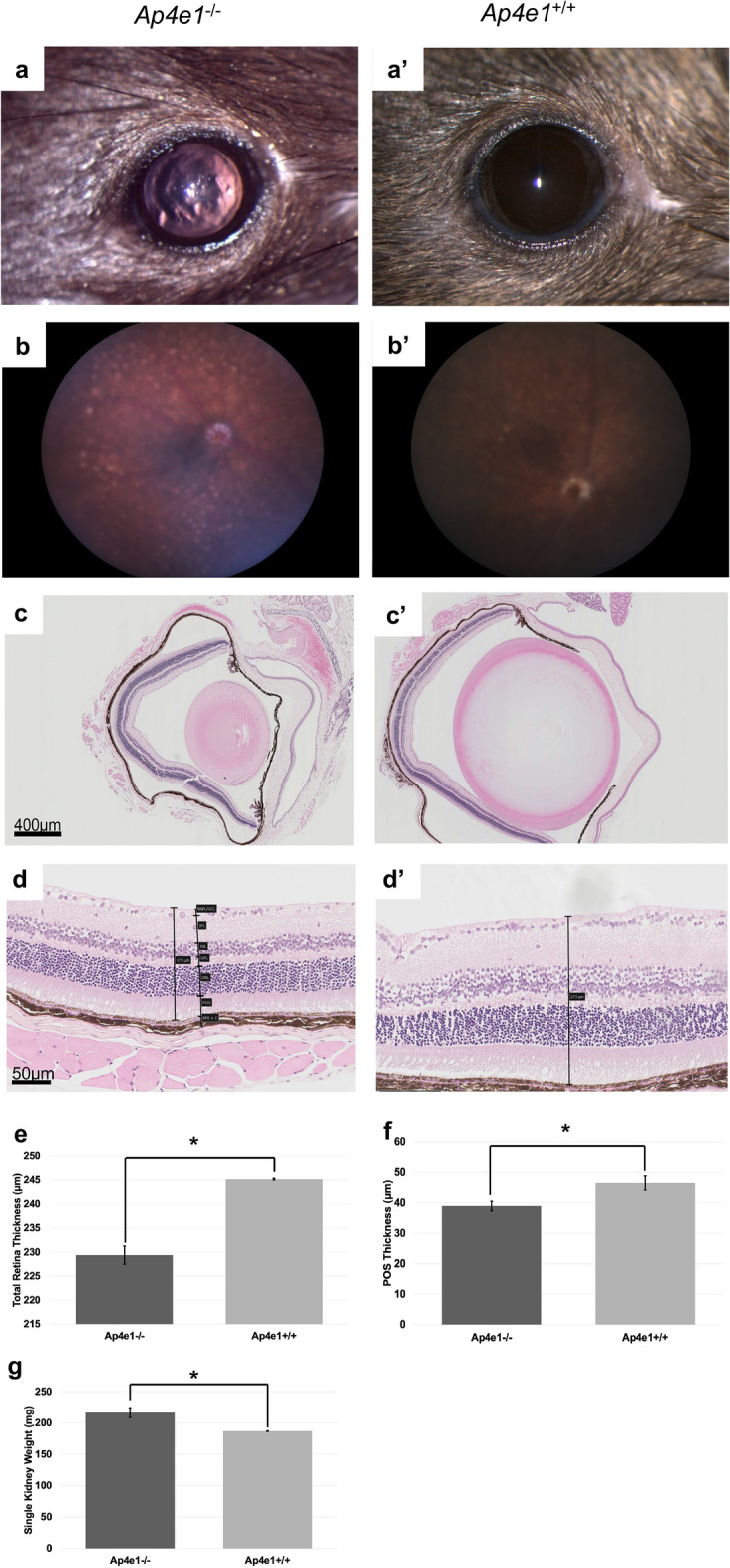
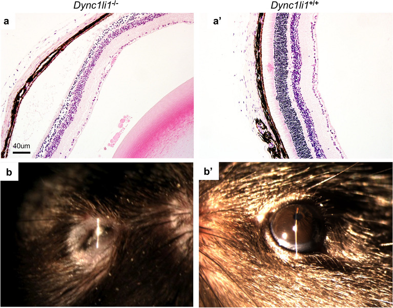
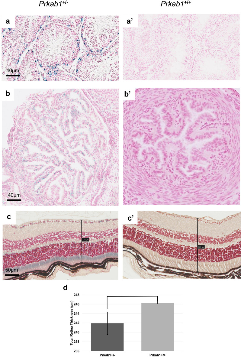
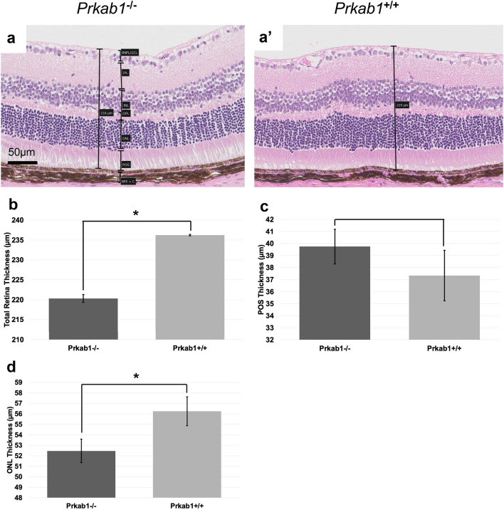
We searched a database of single-gene knockout (KO) mice produced by the International Mouse Phenotyping Consortium (IMPC) to identify candidate ciliopathy genes. We first screened for phenotypes in mouse lines with both ocular and renal or reproductive trait abnormalities. The STRING protein interaction tool was used to identify interactions between known cilia gene products and those encoded by the genes in individual knockout mouse strains in order to generate a list of "candidate ciliopathy genes." From this list, 32 genes encoded proteins predicted to interact with known ciliopathy proteins. Of these, 25 had no previously described roles in ciliary pathobiology. Histological and morphological evidence of phenotypes found in ciliopathies in knockout mouse lines are presented as examples (genes Abi2, Wdr62, Ap4e1, Dync1li1, and Prkab1). Phenotyping data and descriptions generated on IMPC mouse line are useful for mechanistic studies, target discovery, rare disease diagnosis, and preclinical therapeutic development trials. Here we demonstrate the effective use of the IMPC phenotype data to uncover genes with no previous role in ciliary biology, which may be clinically relevant for identification of novel disease genes implicated in ciliopathies.


Conflict of interest statement

The authors declare no competing interests.

Figures

Figure 1
Figure 1
Identification of potential ciliopathy genes and protein network interactions of potential ciliopathy genes and known ciliopathy proteins. (a) Venn diagram showing intersection of IMPC genes associated with various phenotype subgroups, those highlighted in green outline have ocular and one or more other concomitant renal or reproductive trait abnormality and are subsequently considered “Potential ciliopathy genes” (b) STRING interaction map of known ciliopathy genes with potential ciliopathy genes. Proteins are shown as nodes on the map where green is indicative of a potential ciliopathy gene, purple shown known or gold standard ciliopathy genes, orange are those that were identified to be potential ciliopathy genes and are previously known as ciliopathy genes. Each line represents a STRING interaction score of greater than or equal to .9 based on 5 pieces of evidence: “experiments,” “databases,” “co-expression,” “neighborhood,” “gene fusion,” and “co-occurrence.” 24 of the 140 potential ciliopathy genes are shown to be directly interacting with known ciliopathy genes. (c) Incorporation of additional proteins or nodes into the network where blue nodes are genes not found in either gold standard or potential ciliopathy genes. Highlighted is an interacting oculorenal gene WDR62 which is predicted to interact with known ciliopathy genes through an additional protein CEP63.
Figure 2
Figure 2
Abi2−/− mice had eye and male reproductive tract abnormalities consistent with ciliopathy compared to controls. (a) Marked, chronic, multifocal localized retinal dysplasia characterized by multiple clusters of external nuclear structures within the outer plexiform layer. (b) Cortical cataract is present with marked, chronic, focally extensive swollen and disrupted lens fibers with abnormally retained nuclei (balloon cells) with capsular thickening and wrinkling in the cortical region of the lens. (c) 20 × magnification histopathology showed testicular degeneration, marked, chronic, bilateral, multifocal vacuolation of the seminiferous tubule epithelia with primary and secondary spermatocyte and spermatid hypocellularity, with very few spermatozoa. Apoptotic bodies (arrowheads) and multinucleated giant cells (arrows) were frequent. (d) On magnification (5 ×), the epididymis showed marked hypospermia. Epididymal ducts in all segments of the epididymis (caput, corpus, cauda) contained cell and protein debris in the lumen with few mature spermatozoa. (e) Magnification (20 ×) of (d). (f) Quantitative measurement of average total retina thickness, where Abi2+/+ n = 12, average = 342.35 µm, SE = 4.88 and Abi2−/− n = 9, average = 342.35 µm, SE = 21.35 p-value = 0.0190. All error bars represent standard error of the mean, * indicates p-value < .05 result of student’s two-tailed t-test.
Figure 3
Figure 3
Wdr62−/− mice have ocular, renal, and reproductive organ abnormalities consistent with ciliopathy. (a) Microphthalmia with undeveloped lens and residual lens vesicle remnant in the vitreous cavity. The ocular contents are small, malformed, and disorganized. (b) Small kidneys at necropsy (arrow), but histologically normal in appearance (data not shown). (c) Testis with seminiferous tubule degeneration characterized by defective spermatogenesis and aspermia with increased apoptosis of meiosis I spermatocytes (arrow), and multinucleated syncytia (solid arrowhead). There are numerous spermatogonia and spermatocytes, but rare spermatids and mature spermatozoa. Sertoli cells are prominent (line arrowhead) and seminiferous tubules contain degenerated cells with irregular dense nuclei. (d) Epididymal ducts containing scattered cell debris and no spermatozoa. (e) Ovaries are small, hypoplastic with no detectable folliculogenesis. The ovarian bursa is hyperplastic, dominated by fibrous connective tissue stroma with spindle shaped cells (solid yellow arrows) and rare immature solid tubular structures (yellow circles) representing mesonephric remnants in the ovary. Germinal epithelium, oocytes, and follicles are absent.
Figure 4
Figure 4
Ap4e1−/− mice have ocular abnormalities in the retina, cornea, lens, and have small kidneys. (a) Corneal epithelial irregularity on gross examination of the eye, focally extensive corneal epithelial squamous hyperplasia. (b) Extensive retinal hypopigmentary lesions on color fundus photography. (c) Abnormal lens morphology. (d) Decreased total retinal thickness. Retina layers labeled where RNFL/GCL, retinal nerve fiber layer/ganglion cell layer; IPL, inner plexiform layer; INL, inner nuclear layer; OPL, outer plexiform layer; ONL, outer nuclear layer; POS, photoreceptor outer segments; RPE, retinal pigmented epithelium; C, choroid. (e) Quantitative measurement of average total retina thickness, where Ap4e1+/+ n = 3861, average = 245.20 µm, SE = 0.164 and Ap4e1−/− n = 45, average = 229.39 µm, SE = 1.89, p-value = 7.78 × 10–11. (f) Quantitative measurement of POS layer thickness. Where Ap4e1+/+ n = 8, average = 46.54 µm, SE = 2.35 and Ap4e1−/− n = 16, average = 38.99 µm, SE = 1.58, p-value = 0.019 . (g) Quantitative measurement of single kidney weight where Ap4e1+/+ n = 3629, 187.18 mg, SE = 0.581 and Ap4e1−/− n = 29, 217.54 mg, SE = 7.85, p-value = 0.00085. All error bars represent standard error of the mean, * indicates p-value < .05 result of student's two-tailed t-test.
Figure 5
Figure 5
Dync1li1−/− mice have ocular abnormalities in the retina and microphthalmia. (a) Extensive retinal hypoplasia, atrophy, and cellular disorganization across all layers of the retina. In addition, the retina has decreased total thickness compared to control. (b) Microphthalmia evident on slit lap examination compared to control.
Figure 6
Figure 6
Prkab1+/− LacZ expression under Prkab1 promoter is found in multiple reproductive tissues and photoreceptor outer segments of the retina. (a) lacZ reporter shows expression in the spermatogonia layer of the testis. (b) lacZ reporter expression under the control of the Prkab1 promoter in the oviduct shows faint scattered expression. (c) lacZ reporter expression under the control of the Prkab1 promoter shows strong signal in the wavy and disorganized photoreceptor inner and outer segments. (d) Quantitative measurement of average total retina thickness, where Prkab1+/+ n = 3698, average = 246.25 µm, SE = .148 and Prkab1+/- n = 28, average = 241.93, SE 2.40, p-value = 0.084. No significant difference was found in total retinal thickness based on student’s two-tailed t-test. All error bars represent standard error of the mean.
Figure 7
Figure 7
Prkab1−/− mice have decreased total retinal thickness and reduced thickness of the ONL. (a) Prkab1−/− mice retinas show decreased total thickness with evidence of reduced thickness in the ONL. (b) Quantitative measurement of average total retina thickness, where Prkab1+/+ n = 6567, average = 236.21 µm, SE = 0.19 and Prkab1−/− n = 85, average = 220.29 µm, SE = .998. p = 1.078 × 10–27 (c) Quantitative measurement of POS layer thickness where Prkab1+/+ n = 9, average = 37.33 µm, SE = 2.09 and Prkab1−/− n = 14, average = 39.74 µm, SE = 1.43 p-value = .357 (d) Quantitative measurement of ONL thickness where Prkab1+/+ n = 11, average = 56.24 µm, SE = 1.37 and Prkab1−/− n = 14, average = 52.57 µm, SE 1.12 p-value = .039. All error bars represent standard error of the mean, * indicates p-value < .05 result of student’s two-tailed t-test. Retinal layers abbreviated as above.

References

    1. Waters AM, Beales PL. Ciliopathies: An expanding disease spectrum. Pediatr. Nephrol. 2011;26:1039–1056. doi: 10.1007/s00467-010-1731-7. - DOI - PMC - PubMed
    1. Bartoloni L, et al. Mutations in the DNAH11 (axonemal heavy chain dynein type 11) gene cause one form of situs inversus totalis and most likely primary ciliary dyskinesia. Proc. Natl. Acad. Sci. U.S.A. 2002;99:10282–10286. doi: 10.1073/pnas.152337699. - DOI - PMC - PubMed
    1. Ichioka K, Kohei N, Okubo K, Nishiyama H, Terai A. Obstructive azoospermia associated with chronic sinopulmonary infection and situs inversus totalis. Urology. 2006;68:204.e5. doi: 10.1016/j.urology.2006.01.072. - DOI - PubMed
    1. Inaba K, Mizuno K. Sperm dysfunction and ciliopathy. Reprod. Med. Biol. 2015;15:77–94. doi: 10.1007/s12522-015-0225-5. - DOI - PMC - PubMed
    1. Rosenfeld CS. Male reproductive tract cilia beat to a different drummer. Proc. Natl. Acad. Sci. U. S. A. 2019;116:3361–3363. doi: 10.1073/pnas.1900112116. - DOI - PMC - PubMed

Publication types