Review
. 2023 Mar;23(3):173-188.
doi: 10.1038/s41568-022-00531-9. Epub 2022 Dec 1.

Improving head and neck cancer therapies by immunomodulation of the tumour microenvironment

Affiliations
Review

Improving head and neck cancer therapies by immunomodulation of the tumour microenvironment

Ayana T Ruffin et al. Nat Rev Cancer. 2023 Mar.

Abstract

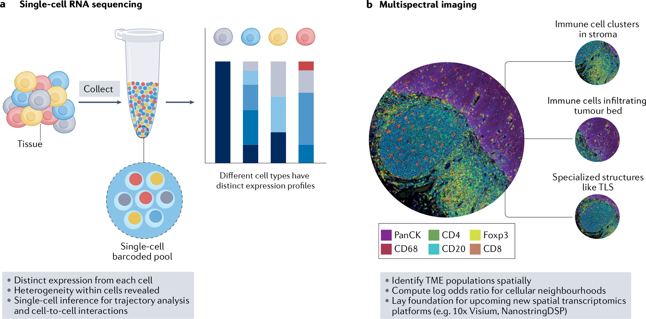
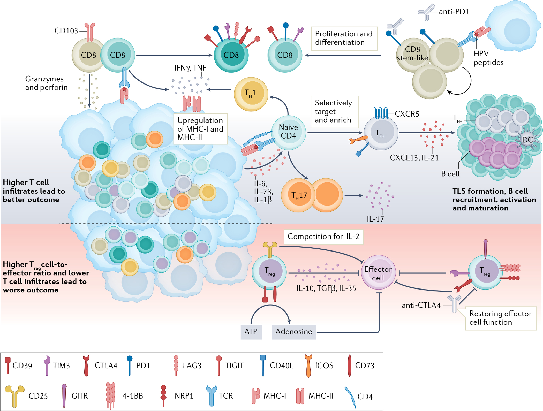
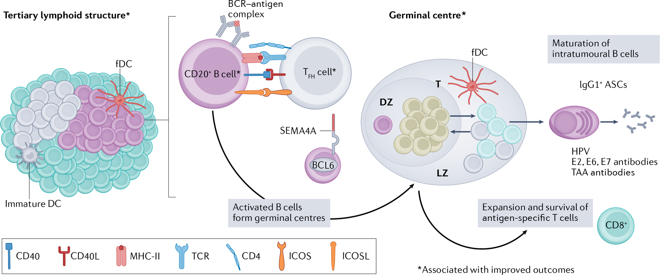
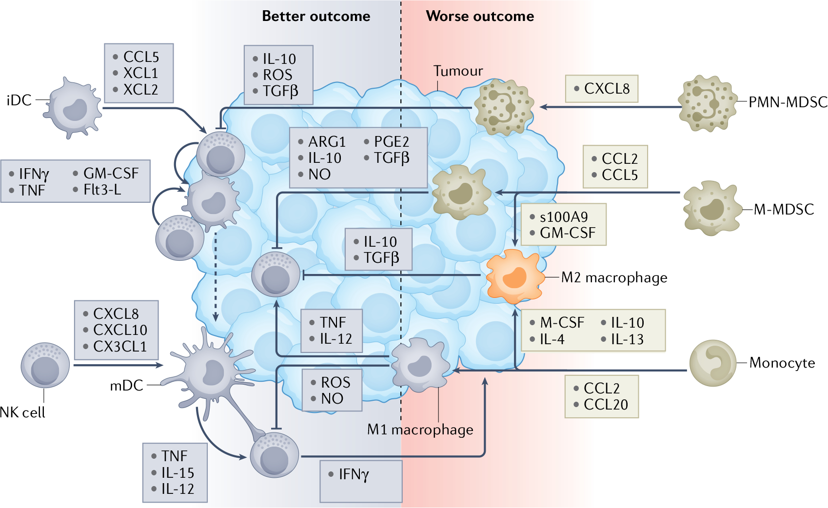
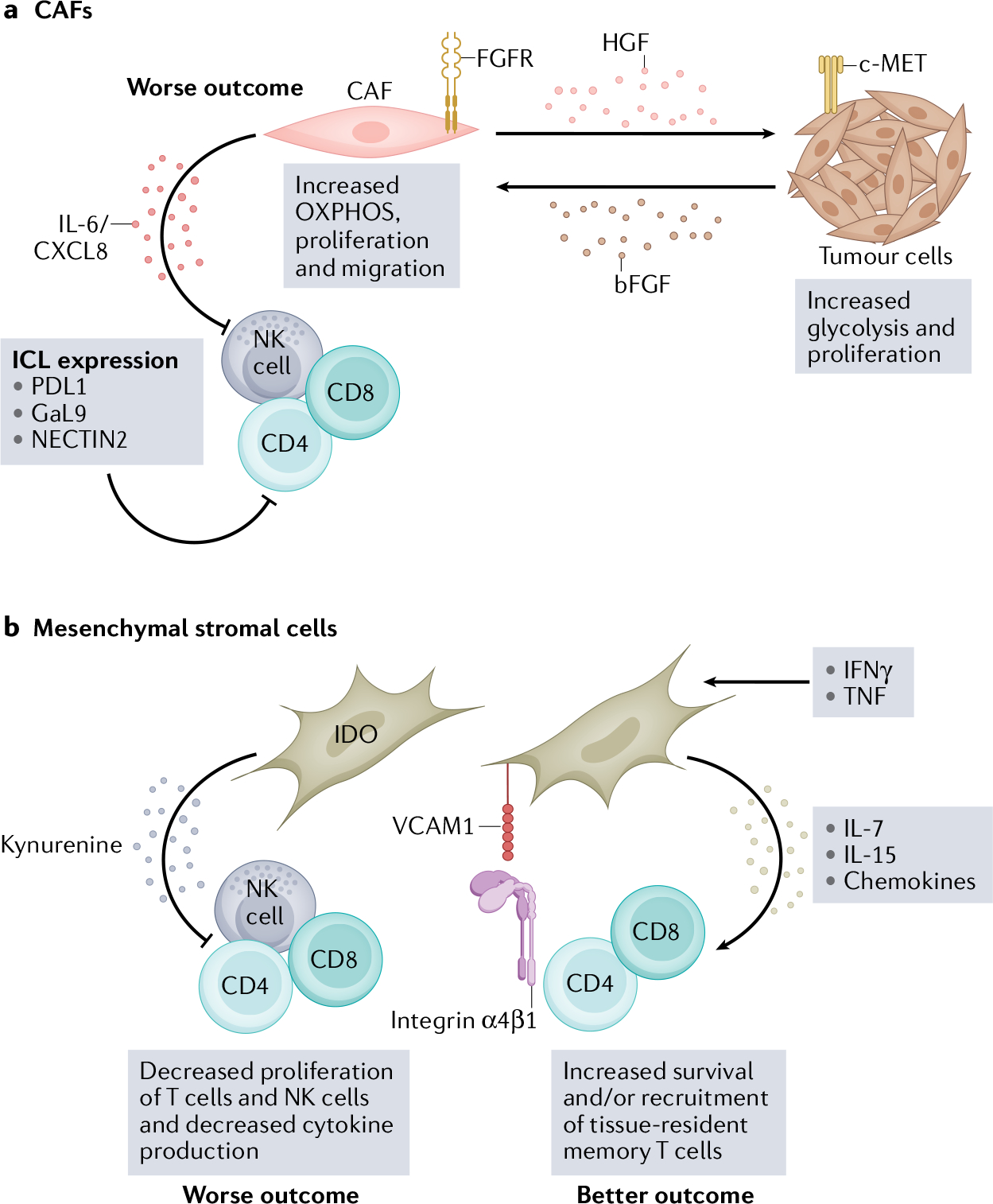
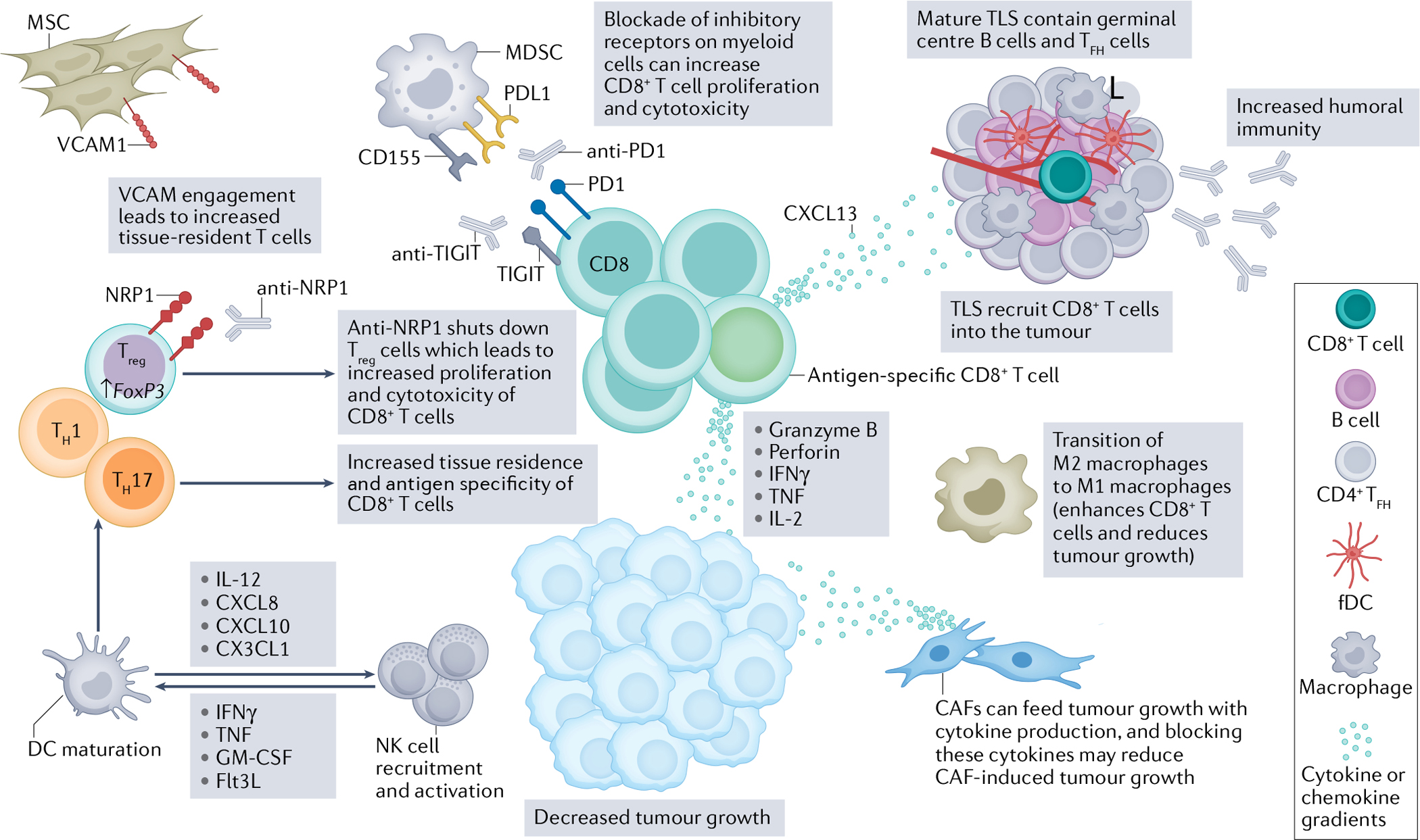
Targeted immunotherapy has improved patient survival in head and neck squamous cell carcinoma (HNSCC), but less than 20% of patients produce a durable response to these treatments. Thus, new immunotherapies that consider all key players of the complex HNSCC tumour microenvironment (TME) are necessary to further enhance tumour-specific T cell responses in patients. HNSCC is an ideal tumour type in which to evaluate immune and non-immune cell differences because of two distinct TME aetiologies (human papillomavirus (HPV)-positive and HPV-negative disease), multiple anatomic sites for tumour growth, and clear distinctions between patients with locally advanced disease and those with recurrent and/or metastatic disease. Recent technological and scientific advancements have provided a more complete picture of all cellular constituents within this complex TME and have evaluated the interplay of both immune and non-immune cells within HNSCC. Here, we include a comprehensive analysis of the complete ecosystem of the HNSCC TME, performed utilizing data-rich resources such as The Cancer Genome Atlas, and cutting-edge techniques, such as single-cell RNA sequencing, high-dimensional flow cytometry and spatial multispectral imaging, to generate improved treatment strategies for this diverse disease.


Conflict of interest statement

Competing interests

L.V. is a co-inventor of a methodology licensed to INmune Bio, Inc., where soluble TNF sequestration using DN-TNF can be used to prevent or treat malignancies. D.P.Z. declares competing interests with Blueprint Medicines (advisory board), Macrogenics (consulting), Prelude Therapeutics (advisory board), and Merck (advisory board) and research support (institutional) from Merck, BMS, AstraZeneca, GlaxoSmithKline, Aduro, Astellas, Macrogenics, Lilly, Bicara, Checkmate Pharma, and Novasenta. R.L.F. declares competing interests with Aduro Biotech, Inc. (consulting), AstraZeneca/MedImmune (clinical trial, research funding), Bristol-Myers Squibb (advisory board, clinical trial, research funding), EMD Serono (advisory board), MacroGenics Inc. (advisory board), Merck (advisory board, clinical trial), Novasenta (consulting, stock, research funding), Numab Therapeutics AG (advisory board), Pfizer (advisory board), Sanofi (consultant), Tesaro (research funding) and Zymeworks Inc. (consultant). T.C.B. declares competing interests with Walking Fish Therapeutics (Scientific Advisory Board). The other authors declare no competing interests.

Figures

Fig. 1 |
Fig. 1 |. Novel technologies have begun to elucidate the clear complexity of the HNSCC TME.
a, Single-cell RNA sequencing enables distinct expression from each cell and reveals heterogeneity within immune and non-immune cell subsets. Furthermore, bioinformatic analyses can provide single-cell inference for trajectory analysis and cell-to-cell interactions. b, Multispectral imaging is paramount to identifying tumour microenvironment (TME) populations spatially and analysing cellular interactions within and between tumour and stroma. Bioinformatic analyses can also determine cellular neighbourhoods within the head and neck squamous cell carcinoma (HNSCC) TME, and these imaging technologies have laid the foundation for new and upcoming spatial transcriptomic platforms that will increase our spatial knowledge of the HNSCC TME. TLS, tertiary lymphoid structure.
Fig. 2 |
Fig. 2 |. New T cell targets in the HNSCC TME.
T cells are important in antitumour immunity, and human papillomavirus (HPV)-positive tumours generally show higher T cell infiltration and are associated with better outcomes than HPV-negative tumours. CD8+ T cells can directly lyse target cells by releasing granzymes and perforin in an antigen-directed manner. Chronic engagement with tumour antigens leads to a dysfunctional state characterized by high expression of immune-checkpoint receptors (PD1, T cell immunoglobulin and mucin domain 3 (TIM3), lymphocyte-activation protein 3 (LAG3) and CTLA4). An HPV16-specific CD8+PD1+ T cell population in HPV-positive tumours contains a stem-like CD8+ T cell subset. These cells are capable of proliferating and differentiating into effector cells upon HPV peptide stimulation and represent a population of future therapeutic targets. CD4+ T lymphocytes recognize major histocompatibility complex (MHC) class II antigens and differentiate into different subtypes upon antigen stimulation. A higher number of T helper 1 (TH1) and TH17 cells are found in HPV-positive tumours than in HPV-negative tumours,. TH1 cells release IFNγ and TNF and promote MHC class I and II upregulation on cancer cells, facilitating tumour elimination,. IL-17-releasing TH17 cells are induced by IL-6, IL-23 and IL-1β produced by primary tumour cells,. CXCR5+PD1+ICOS+CD40L+ T follicular helper (TFH) cells produce CXCL13 and IL-21, recruiting B cells into the tumour microenvironment (TME),. TFH cells are essential for B cell activation, maturation and tertiary lymphoid structure (TLS) formation in tumours, and their presence is associated with improved outcomes. T regulatory (Treg) cells have an immunosuppressive role in the TME. Treg cells can suppress effector cells through the release of IL-10, TGFβ and IL-35. High levels of CD25 on Treg cells compete with effector cells for local IL-2, so that effector cells might not have enough IL-2 to survive and function. Treg cells also express CD39 and CD73, which convert extracellular ATP to adenosine and impair effector T cell function,. Co-expression of 4–1BB, GITR and neuropilin 1 (NRP1) on intratumoural Treg cells demonstrated an enhanced suppression function,,. Immune-checkpoint receptors, such as TIM3 and CTLA4, are reportedly expressed by Treg cells in head and neck squamous cell carcinoma (HNSCC). Both are highly expressed by intratumoural Treg cells and able to suppress effector cell functions,,,. Therapeutically targeting Treg cells might help rejuvenate effector cell function. DC, dendritic cell; ICOS, inducible T cell co-stimulator; TCR, T cell receptor; TIGIT, T cell immunoreceptor with immunoglobulin and ITIM domains.
Fig. 3 |
Fig. 3 |. Increasing cellular interactions with tertiary lymphoid structures in the HNSCC TME for maximal humoral and cellular immunity.
Tertiary lymphoid structures (TLS) form within the tumour or surrounding the tumour stroma in patients with head and neck squamous cell carcinoma (HNSCC),,. TLS vary in size and cell composition in patients, but an increased presence of TLS correlates with improved survival and reduced risk of recurrence in both human papillomavirus (HPV)-positive and HPV-negative HNSCC. Within TLS, B cells co-localize with T follicular helper (TFH) cells and receive activation signals via CD40, inducible T cell co-stimulator ligand (ICOSL) and major histocompatibility complex class II (MHC-II) engagement,,. Activation of B cells via TFH cells leads to germinal centre formation, which is regulated by transcription factor B cell lymphoma 6 (BCL6) and marked by surface expression of semaphorin 4A (SEMA4A) on germinal centre B cells,. Intratumoural germinal centre B cells in HPV-positive HNSCC have three distinct cell states: dark zone (DZ), light zone (LZ) and transitional (T), marked by unique patterns of gene expression that are important for the selection and maturation of B cells. Formation of germinal centres within TLS is an independent predictor of superior progression-free survival in HNSCC. As a result of successful germinal centre formation in HPV-positive HNSCC, germinal centres produce terminally differentiated somatic hypermutated antibody-secreting cells (ASCs) that have undergone immunoglobulin class-switching to an IgG1 isotype. IgG1+ ASCs in patients with HPV-positive disease recognize HPV viral proteins E6, E7 and E2 (ref.). Antibodies directed at tumour-associated antigens (TAAs) have also been detected in the serum of patients with HPV-positive and HPV-negative HNSCC. Activated B cells might also provide co-stimulatory signals to CD8+ T cells, resulting in their expansion and survival in HNSCC tumours,. Co-localization of B cells with CD8+ T cells is associated with favourable outcomes in HNSCC,. Together, these features provide a new targetable axis for future immunotherapies, which should be directed at enhancing TLS formation, maturation of B cells and increasing B–T cell interactions within the TME of HNSCC. BCR, B cell receptor; fDC, follicular dendritic cell; ICOS, inducible T cell co-stimulator; TCR, T cell receptor.
Fig. 4 |
Fig. 4 |. Innate cell interactions generate inflammatory signals that drive outcomes in patients with HNSCC.
Innate immunity, an evolutionary defence against pathogens or tissue damage, plays a central role in antitumour defence as it dictates the robustness and function of tumour immune infiltrates. Human papillomavirus (HPV)-positive lesions generally have a greater level of innate immune infiltrates than HPV-negative lesions, which generally correlates with a better clinical outcome. Natural killer (NK) cell–dendritic cell (DC) crosstalk plays a pivotal role in antitumour immunity,. Enhanced NK cell and DC infiltrates correlate with better patient survival,. Tumour-infiltrating NK cells can recruit immature DC (iDCs) into the tumour microenvironment (TME) by releasing CCL5, XCL1 and XCL2 (ref.). Once inside the tumour, iDCs can take up cell debris from killed tumour cells, which is the first step required for effective antigen presentation. As antigen-loaded DCs start to mature, they can engage and crosstalk with NK cells with their long dendrites. NK cells enhance DC maturation and polarization through release of IFNγ, TNF, granulocyte–macrophage colony-stimulating factor (GM-CSF) and Flt3-L,. Maturing DCs (mDCs) in turn enhance NK cell activation through IL-12 secretion as well as cell-to-cell contact mediated by transmembrane TNF and trans-presented IL-15 (ref.). Furthermore, they release a number of chemokines, including CXCL8, CXCL10 and CX3CL1, that enhance the number of NK cells infiltrating the tumour,. This cascade of events initiates and potentiates tumour-specific adaptive T cell responses that are required for effective tumour elimination. The myeloid compartment is highly diverse and consists of various cell subsets that exhibit a broad range of immune functions. Myeloid cells commonly infiltrate the tumours and associated stroma and their presence influences outcomes in patients with head and neck squamous cell carcinoma (HNSCC). Patients with worse clinical outcome generally display higher numbers of tumour-infiltrating macrophages and myeloid-derived suppressor cells (MDSCs),,,,. Monocytes, monocytic MDSCs (M-MDSCs) and polymorphonuclear MDSCs (PMN-MDSCs) are recruited into a tumour by specific chemokines,. Once in the tumour, monocytes can differentiate into antitumour M1 macrophages and immunosuppressive M2 macrophages depending on the inflammatory signals they receive within the TME. M-MDSCs can maintain their immature myeloid profile within the TME or can further differentiate into M2 macrophages under the influence of endogenous S100A9 and exogenous GM-CSF,–. M2 macrophages, M-MDSCs and PMN-MDSCs all have a variety of mechanisms by which they can suppress NK cell and DC activation and skew their polarization towards an immunoregulatory phenotype. M1 macrophages, which can activate NK cells by IL-12 and TNF production, can also inhibit NK cell function by reactive oxygen species (ROS) and nitric oxide (NO) release,.
Fig. 5 |
Fig. 5 |. The stromal microenvironment is functionally important for the HNSCC TME.
The head and neck squamous cell carcinoma (HNSCC) tumour microenvironment (TME) consists of a diverse stromal cell compartment — which includes cancer-associated fibroblasts (CAFs) and mesenchymal stromal cells (MSCs) — that interacts with neighbouring tumour cells and infiltrating immune cells,. These interactions primarily promote tumour growth, progression and metastases but, under some conditions, can provide a supportive environment for immune cells and recruit immune cells to the HNSCC TME,. Combining immune-checkpoint therapy with therapies that target the stromal compartment might improve patient outcomes in HNSCC. a, CAFs and tumour cells have a metabolic relationship mediated by hepatocyte growth factor (HGF) and basic fibroblast growth factor basic (bFGF), which increases oxidative phosphorylation (OXPHOS) in CAFs and glycolysis in tumour cells. Increased oxidative phosphorylation in CAFs leads to IL-6 and CXCL8 production, which suppress immune cell function,. CAFs also express several immune-checkpoint ligands (ICL), including PDL1, galectin 9 (GaL9) and nectin cell adhesion molecule 2 (NECTIN2), which interact with corresponding inhibitory receptors on natural killer (NK) cells and T cells. b, MSCs can also suppress immune cell function in vitro via indoleamine-pyrrole 2,3-dioxygenase (IDO), which metabolizes tryptophan into kynurenine and is cytotoxic to T cells and NK cells,,. However, some in vitro studies suggest that MSCs can support tissue-resident memory T cells via IL-7 and IL-15 production in the presence of IFNγ and TNF. VCAM1, vascular cell adhesion protein 1.
Fig. 6 |
Fig. 6 |. Immune and non-immune therapeutic targets in the HNSCC TME.
The head and neck squamous cell carcinoma (HNSCC) tumour microenvironment (TME) hosts a complex interplay of immune and non-immune cells that can amplify tumour killing. Augmenting aspects of antitumour immunity while suppressing pro-tumour immunity will be key in next-generation therapeutics. Although the CD8+ T cell has been the centre of immunotherapeutic strategies for several years, several other axes can be targeted for improved antitumour CD8+ T cell responses. First, blockade of the PD1–PDL1 pathway on myeloid cells, in particular myeloid-derived suppressor cells (MDSCs), has been an important consideration for improved intratumoural CD8+ T cell function. Additionally, other inhibitory receptors, like T cell immunoreceptor with immunoglobulin and ITIM domains (TIGIT), can be blocked on myeloid cells to improve responses to anti-PD1 immunotherapy. Further, reinvigoration of these antigen-specific CD8+ T cells can result in increased production of key chemokines, such as CXCL13, that can trigger tertiary lymphoid structure (TLS) development in the TME. Amplifying mature, germinal centre-containing TLS will lead to increased recruitment of CD8+ T cells and increased antitumour humoral immunity. Further, directly enhancing germinal centre B cells and T follicular helper (TFH) cells could be paramount for increased antitumour immunity as they correlate with increased prognosis,. TFH cells also secrete CXCL13, which helps with further recruitment of B cells and CD8+ T cells into the TME and TLS. Evidence exists to indicate that the transition of M2 macrophages to M1 macrophages will further enhance antitumour immunity. Furthermore, dendritic cell (DC) maturation can be fuelled by natural killer (NK) cells via secretion of IFNγ, TNF, granulocyte–macrophage colony-stimulating factor (GM-CSF) and Flt3L,, which in turn will also help to activate and recruit more NK cells via IL-12, CXCL8, CXCL10 and CX3CL1 (refs.,,). DC maturation is also instrumental in educating productive T helper 1 (TH1) and TH17 responses, which ultimately lead to increased tissue residence and antigen specificity of CD8+ T cells. While T regulatory (Treg) cells have dampened CD8+ T cells in the TME, new therapeutics, such as neuropilin 1 (NRP1) blockade, can specifically reduce immunosuppression without any effects on homeostatic balance in patients,. Finally, non-immune cells, such as cancer associated fibroblasts (CAFs) and mesenchymal stromal cells (MSCs), have a dichotomy of function in the TME. CAFs can feed tumour growth via cytokine production, and the metabolic crosstalk between CAFs and tumour cells could be a targetable axis to suppress tumour growth. MSCs can be advantageous in recruiting tissue-resident T cells to the TME in a vascular cell adhesion protein 1 (VCAM1)-dependent manner. Ultimately, next-generation immunotherapies for HNSCC will be multi-faceted to improve patient response and treatment durability. fDC, follicular dendritic cell.

References

    1. Ferlay J et al. Estimating the global cancer incidence and mortality in 2018: GLOBOCAN sources and methods. Int. J. Cancer 144, 1941–1953 (2019). - PubMed
    1. Sung H et al. Global cancer statistics 2020: GLOBOCAN estimates of incidence and mortality worldwide for 36 cancers in 185 countries. CA Cancer J. Clin. 71, 209–249 (2021). - PubMed
    1. Johnson DE et al. Head and neck squamous cell carcinoma. Nat. Rev. Dis. Prim. 6, 92 (2020). - PMC - PubMed
    1. HHS.gov. Smoking Cessation: A Report of the Surgeon General — Key Findings https://www.hhs.gov/surgeongeneral/reports-and-publications/tobacco/2020... (2020).
    1. Bray F et al. Global cancer statistics 2018: GLOBOCAN estimates of incidence and mortality worldwide for 36 cancers in 185 countries. CA Cancer J. Clin. 68, 394–424 (2018). - PubMed

Publication types

MeSH terms

Substances