. 2023 Feb;30(2):397-406.
doi: 10.1038/s41418-022-01091-z. Epub 2022 Dec 1.

DCC/netrin-1 regulates cell death in oligodendrocytes after brain injury

Affiliations

DCC/netrin-1 regulates cell death in oligodendrocytes after brain injury

Madelen M Díaz et al. Cell Death Differ. 2023 Feb.

Abstract

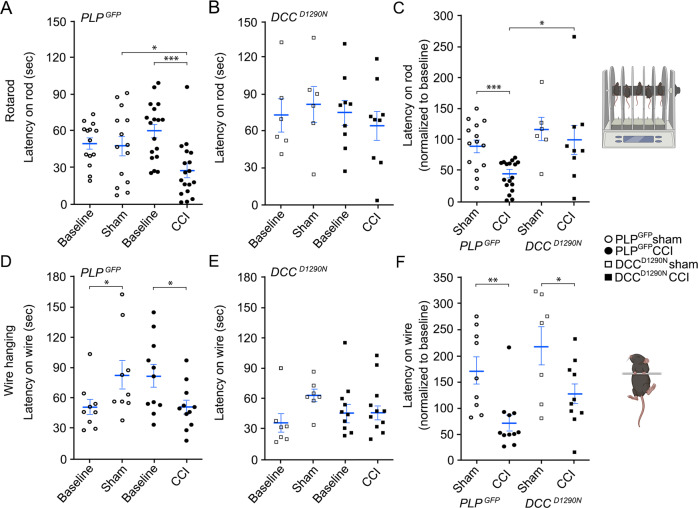
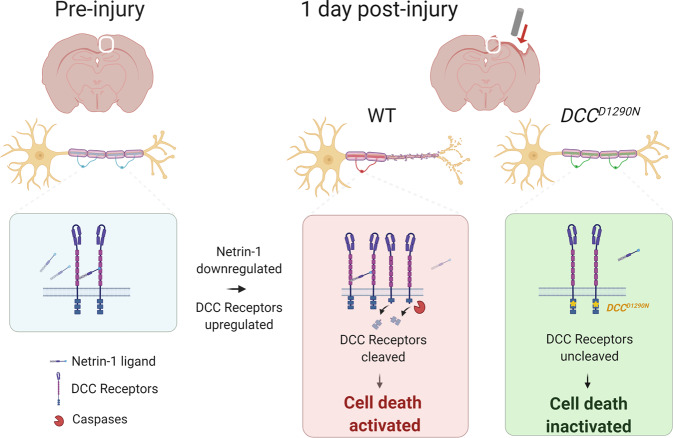
Hallmark pathological features of brain trauma are axonal degeneration and demyelination because myelin-producing oligodendrocytes (OLs) are particularly vulnerable to injury-induced death signals. To reveal mechanisms responsible for this OL loss, we examined a novel class of "death receptors" called dependence receptors (DepRs). DepRs initiate pro-death signals in the absence of their respective ligand(s), yet little is known about their role after injury. Here, we investigated whether the deleted in colorectal cancer (DCC) DepR contributes to OL loss after brain injury. We found that administration of its netrin-1 ligand is sufficient to block OL cell death. We also show that upon acute injury, DCC is upregulated while netrin-1 is downregulated in perilesional tissues. Moreover, after genetically silencing pro-death activity using DCCD1290N mutant mice, we observed greater OL survival, greater myelin integrity, and improved motor function. Our findings uncover a novel role for the netrin-1/DCC pathway in regulating OL loss in the traumatically injured brain.


Conflict of interest statement

The authors declare no competing interests.

Figures

Fig. 1
Fig. 1. DCC receptors are expressed by oligodendrocytes (OLs) and are dependent on netrin-1 for survival.
A–F Representative images showing DCC (B, E) is expressed in both PDGFRα-labeled oligodendrocyte progenitor cells (OPCs) (A) and myelin basic protein (MBP)-positive mature OLs (D) in vitro. Blue channel indicates DAPI signal. Scale bar represents 10 μm. G Netrin-1 dose-dependent (4, 40, 400 ng/mL) administration protects cultured mature OLs from 20 nM Staurosporine-induced cell death (n = 9–20). H,I The percentage of DCC receptors in total OLs (H) and mean fluorescent intensity (MFI) per OL (I) are increased in FACS cortical O1+ OLs at 1-day post-CCI injury (dpi) (n = 6) as compared to sham controls (n = 5). J Western blot analysis shows increased netrin-1 expression at only 3 dpi (n = 3–5). K Fold change of netrin-1 mRNA transcripts from sham, 1 dpi, and 3 dpi WT cortices (n = 3–5). LR Representative coronal images of αNetrin-1 (magenta) co-labeling with αNeuN + neurons (green) in WT sham (L, NP) and 1 dpi (M, Q, R) mouse tissues. L αNetrin-1 has widespread expression in the cortex, white matter tracts, and subcortical regions. Arrow denotes the apex of the CC, which designates our definition of the medial cortex (mCtx) from other lateral cortical tissues. M αNetrin-1 labeling is not detectable in regions of the ventral CC (arrowheads) and perilesional tissues (white outline, QR) at 1 dpi. Scale bars are 500 µm (LM), or 50 µm (NR). Values represent mean ± SEM. One-way ANOVA with Bonferroni’s multiple comparison test or Welsh’s unpaired, two-tailed t-test. *p < 0.05, **p < 0.01, ***p < 0.001. Ctx: cortex, CC: corpus callosum, H: hippocampus.
Fig. 2
Fig. 2. DCCD1290N mice have reduced OL cell death after CCI injury.
A–C Representative coronal images of a control sham brain (A), and a CCI-injured brain from a PLPGFP mice (B) and DCCD1290N:PLPGFP mice (C) at 1 dpi. The injury epicenter is noted by the loss of tissue and GFP-fluorescence. Blue channel indicates DAPI signal. Scale bar represents 500 μm. D–F Unbiased relative counts of GFP-labeled OLs show significant deficits within all accessed cortical tissues (D) and differentially between the perilesional cortex (E) and medial cortex (mCtx, F) from PLPGFP mice and DCCD1290N:PLPGFP mice at 1 dpi (n = 4–5). Values represent mean ± SEM. Welsh’s unpaired, two-tailed t-test. *p < 0.05. **p < 0.01.
Fig. 3
Fig. 3. DCCD1290N mice have improved cortical sparing and myelination after CCI injury.
A–D Representative coronal images showing Black Gold II myelin stain of sham controls (A,C) and 7 dpi (B,D) from PLPGFP (A, B) and DCCD1290N:PLPGFP mice (C, D). E Quantification of cortical spared tissue (mm3) in sham and 7 dpi from PLPGFP mice and DCCD1290N:PLPGFP mice (n = 5–13). Magnified Black Gold II myelin stain of the CC of sham controls (F, H) and 7 dpi (G, I) from PLPGFP (F, G) and DCCD1290N:PLPGFP mice (H, I). J Quantification of Black Gold II intensity within the CC of sham and 7 dpi PLPGFP mice and DCCD1290N:PLPGFP mice. Values represent mean ± SEM. Two-way ANOVA with Tukey’s post hoc comparison. *** p < 0.001.
Fig. 4
Fig. 4. DCCD1290N mice have improved locomotor function after CCI injury.
A,B Latency on the rotating rod for of sham (open) and CCI injured (closed) PLPGFP (A) and DCCD1290N:PLPGFP (B) mice as compared to baseline. The latency to fall (seconds) was measured prior to surgery and at 6 dpi (n = 6–18). C Percent normalized time on the rod at 6 dpi relative to the baseline of each animal. D,E Wire hanging test of sham (open) and CCI injured (closed) PLPGFP (D) and DCCD1290N:PLPGFP (E) mice as compared to baseline. The latency to fall (seconds) was measured prior to surgery and at 6 dpi (n = 7–11). F Percent normalized time on the wire at 6 dpi relative to the baseline of each animal. Values represent mean ± SEM. Repeated two-way ANOVA with Sidak’s multiple comparison test, and Welsh’s unpaired, two-tailed t-test. *p < 0.05.**p < 0.01. ***p < 0.001.
Fig. 5
Fig. 5. Schematic model of Netrin-1/DCC receptor function in the injured brain.
After cortical injury, DCC receptors are acutely upregulated while netrin-1 is downregulated in perilesional tissues, leading to a receptor-ligand imbalance. The DCC receptors without netrin-1 binding will undergo intracellular cleavage at the conserved aspartic acid residue at 1290, which leads to significant OL cell death. However, the knock-in DCCD1290N point mutation blocks dependence receptor-mediated apoptosis, leading to greater cell survival, myelin integrity, and motor behavior after brain injury.

References

    1. Dewan MC, Rattani A, Gupta S, Baticulon RE, Hung YC, Punchak M, et al. Estimating the global incidence of traumatic brain injury. J Neurosurg. 2019;130:1080–97. - PubMed
    1. Daugherty J, Waltzman D, Sarmiento K, Xu L. Traumatic Brain Injury-Related Deaths by Race/Ethnicity, Sex, Intent, and Mechanism of Injury - United States, 2000-2017. MMWR Morb Mortal Wkly Rep. 2019;68:1050–6. doi: 10.15585/mmwr.mm6846a2. - DOI - PMC - PubMed
    1. Vaishnavi S, Rao V, Fann JR. Neuropsychiatric problems after traumatic brain injury: unraveling the silent epidemic. Psychosomatics. 2009;50:198–205. doi: 10.1176/appi.psy.50.3.198. - DOI - PubMed
    1. McDonald S, Genova H. The effect of severe traumatic brain injury on social cognition, emotion regulation, and mood. Handb Clin Neurol. 2021;183:235–60. doi: 10.1016/B978-0-12-822290-4.00011-6. - DOI - PubMed
    1. Shah EJ, Gurdziel K, Ruden DM. Mammalian Models of Traumatic Brain Injury and a Place for Drosophila in TBI Research. Front Neurosci. 2019;13:409. doi: 10.3389/fnins.2019.00409. - DOI - PMC - PubMed

Publication types