. 2022 Dec;28(12):2555-2562.
doi: 10.1038/s41591-022-02074-w. Epub 2022 Dec 1.

Differential roles of Aβ42/40, p-tau231 and p-tau217 for Alzheimer's trial selection and disease monitoring

Affiliations

Differential roles of Aβ42/40, p-tau231 and p-tau217 for Alzheimer's trial selection and disease monitoring

Nicholas J Ashton et al. Nat Med. 2022 Dec.

Abstract

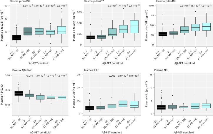
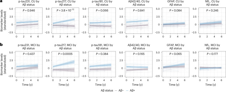
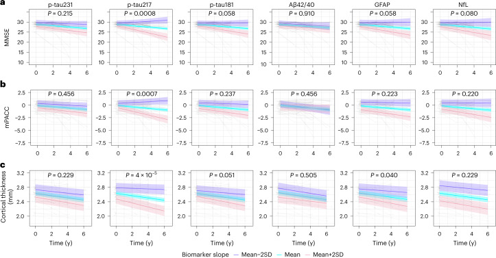
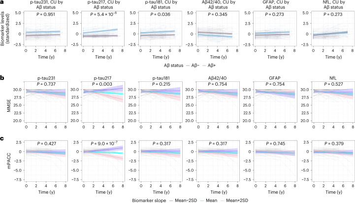
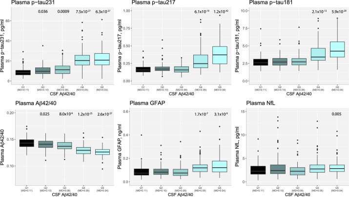
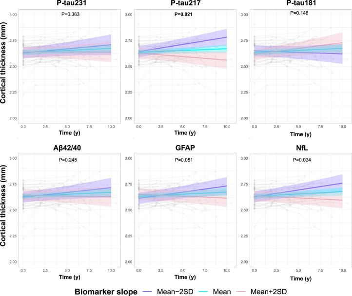
Blood biomarkers indicative of Alzheimer's disease (AD) pathology are altered in both preclinical and symptomatic stages of the disease. Distinctive biomarkers may be optimal for the identification of AD pathology or monitoring of disease progression. Blood biomarkers that correlate with changes in cognition and atrophy during the course of the disease could be used in clinical trials to identify successful interventions and thereby accelerate the development of efficient therapies. When disease-modifying treatments become approved for use, efficient blood-based biomarkers might also inform on treatment implementation and management in clinical practice. In the BioFINDER-1 cohort, plasma phosphorylated (p)-tau231 and amyloid-β42/40 ratio were more changed at lower thresholds of amyloid pathology. Longitudinally, however, only p-tau217 demonstrated marked amyloid-dependent changes over 4-6 years in both preclinical and symptomatic stages of the disease, with no such changes observed in p-tau231, p-tau181, amyloid-β42/40, glial acidic fibrillary protein or neurofilament light. Only longitudinal increases of p-tau217 were also associated with clinical deterioration and brain atrophy in preclinical AD. The selective longitudinal increase of p-tau217 and its associations with cognitive decline and atrophy was confirmed in an independent cohort (Wisconsin Registry for Alzheimer's Prevention). These findings support the differential association of plasma biomarkers with disease development and strongly highlight p-tau217 as a surrogate marker of disease progression in preclinical and prodromal AD, with impact for the development of new disease-modifying treatments.


Conflict of interest statement

N.J.A. has given lectures in symposia sponsored by Eli Lilly. S.J. has no disclosures. N.M.-C. has no disclosures. A.P.B. has no disclosures. O.S. has no disclosures. W.S.B. has no disclosures. T.K.K. has no disclosures. F.G.-O. has no disclosures. G.D.M. has no disclosures. F.J.M. has no disclosures. E.M.J. has no disclosures. R.L.K. has no disclosures. K.C. has no disclosures. T.J.B. has no disclosures. Y.L. has no disclosures. E.V. is a cofounder of ADx Neurosciences. S.P. has served on scientific advisory boards and/or given lectures in symposia sponsored by Biogen, Eli Lilly, Geras Solutions and Roche. E.S. has no disclosures. R.J.B. has equity ownership interest in C2N Diagnostics and may receive income based on technology licensed or optioned by Washington University to C2N Diagnostics, receives income from C2N Diagnostics for serving on the scientific advisory board and has received honoraria as a speaker/consultant/advisory board member from Amgen, Eisai and Roche. H.Z. has served at scientific advisory boards and/or as a consultant for Abbvie, Alector, Eisai, Denali, Roche Diagnostics, Wave, Samumed, Siemens Healthineers, Pinteon Therapeutics, Nervgen, AZTherapies, CogRx and Red Abbey Labs, has given lectures in symposia sponsored by Cellectricon, Fujirebio, Alzecure and Biogen, and is a cofounder of Brain Biomarker Solutions in Gothenburg AB (BBS), which is a part of the GU Ventures Incubator Program (outside submitted work). S.C.J. has within the past 3 years served at scientific advisory boards and/or as a consultant for Roche Diagnostics, Prothena and Eisai and has received research support from Cerveau Technologies. K.B. has served as a consultant, at advisory boards, or at data monitoring committees for Abcam, Axon, BioArctic, Biogen, JOMDD/Shimadzu. Julius Clinical, Lilly, MagQu, Novartis, Ono Pharma, Pharmatrophix, Prothena, Roche Diagnostics and Siemens Healthineers, and is a cofounder of Brain Biomarker Solutions in Gothenburg AB (BBS), which is a part of the GU Ventures Incubator Program, outside the work presented in this paper. O.H. has acquired research support (for the institution) from ADx, AVID Radiopharmaceuticals, Biogen, Eli Lilly, Eisai, Fujirebio, GE Healthcare, Pfizer and Roche. In the past 2 years, he has received consultancy/speaker fees from Amylyx, Alzpath, BioArctic, Biogen, Cerveau, Fujirebio, Genentech, Novartis, Roche and Siemens.

Figures

Fig. 1
Fig. 1. Associations between plasma biomarkers and Aβ-PET in BioFINDER-1 (cohort 1).
Log10-transformed plasma biomarker levels were compared between the centiloid (CL) groups, <12 (MD −2.6; n = 139; reference group), Q1 (range 12.0–35.9; MD 17.9; n = 27), Q2 (range 35.9–71.7; MD 50.1; n = 24), Q3 (range 71.7–95.3; MD 80.6; n = 25) and Q4 (>95.3; MD 114.1; n = 25) using univariate general linear models adjusting for age. Untransformed data are presented in the boxplots to aid interpretation of biomarker values across different comparisons. One NfL outlier is not shown but was included in the statistical analysis. Boxes show interquartile range, the horizontal lines are medians and the whiskers were plotted using the Tukey method. Two-sided P values were corrected for multiple comparisons using Benjamini–Hochberg FDR; uncorrected and corrected P values are shown in Extended Data Table 6.
Fig. 2
Fig. 2. Longitudinal plasma biomarker changes in BioFINDER-1 (cohort 2).
a,b, Longitudinal plasma biomarker changes stratified by β-amyloid status (negative, purple; positive, blue) in CU (a) and MCI (b). The x axis shows time from first plasma biomarker sample. Shaded areas represent 95% confidence intervals of the regression lines plotted from linear mixed effects models with the interaction between time and Aβ status as well as baseline Aβ status as independent variables and adjusting for age and sex. All p-tau biomarkers and Aβ42/40 were significantly changed in Aβ+ individuals at baseline in both CU and MCI (P < 0.001). Two-sided P values were corrected for multiple comparisons using Benjamini–Hochberg FDR; corrected and uncorrected P values are shown in Table 1 and Supplementary Table 1. Several outliers (p-tau231, n = 1; p-tau217, n = 9; p-tau181, n = 5; GFAP, n = 4; NfL, n = 4) are not shown but these data were included in the statistical analysis.
Fig. 3
Fig. 3. Associations of longitudinal plasma biomarkers with longitudinal cognitive decline and brain atrophy in BioFINDER-1 (cohort 2).
ac, The association between longitudinal plasma biomarkers and MMSE (a), mPACC (b) and cortical thickness of the typical AD signature regions (c) in Aβ positive cognitively unimpaired participants. The x axis shows time from first plasma biomarker samples. The model trajectories, shown as the mean slope and the mean ± 2 SD with 95% CI (shaded area), were plotted from linear mixed effects models with the interaction between time and standardized plasma biomarker slopes (derived from subject-level linear regression models) as an independent variable adjusting for age and sex; associations with cognition were also adjusted for years of education. Two-sided P values were corrected for multiple comparisons using Benjamini–Hochberg FDR; corrected and uncorrected P values are shown in Table 2 and Supplementary Table 2.
Fig. 4
Fig. 4. Longitudinal plasma biomarker changes and their association with cognitive decline in WRAP (cohort 3).
a, Longitudinal plasma biomarker change stratified by Aβ status (negative, purple; positive, blue) in CU participants. The average regression lines with 95% CI (shaded area) were plotted from linear mixed effects models with the interaction between time and Aβ status as well as baseline Aβ status independent variables and adjusting for age and sex. All p-tau biomarkers, Aβ42/40 (P < 0.001) and GFAP (P = 0.005) were significantly changed in Aβ+ individuals at baseline. Several outliers (p-tau217, n = 5; p-tau181, n = 2; GFAP, n = 1; NfL, n = 3) are not shown in (a) but these data were included in the statistical analysis. b,c, The association between longitudinal plasma biomarkers and MMSE (b) and mPACC (c) in Aβ-positive cognitively unimpaired participants. The model trajectories, shown as the mean slope and the mean ± 2 SD with 95% CI (shaded area), were plotted from linear mixed effects models with the interaction between time and standardized plasma biomarker slopes (derived from subject-level linear regression models) as an independent variable adjusting for age, sex and years of education. Two-sided P values were corrected for multiple comparisons using Benjamini–Hochberg FDR; corrected and uncorrected P values are shown in Tables 1 and 2 and Supplementary Tables 1 and 2. One outlier with MMSE value of 17 is not shown in b but was included in the statistical analysis The x axes in ac show time from first plasma biomarker samples.
Extended Data Fig. 1
Extended Data Fig. 1. Associations between plasma biomarkers and CSF Aβ42/40 in BioFINDER-1 (cohort 1).
Log10-transformed plasma biomarker levels were compared between the CSF Aβ42/40 quintile groups, Q1 ( > 0.102; median [MD], 0.108; n = 115, reference group), Q2 (range, 0.089-0.102; MD, 0.097; n = 115), Q3 (range 0.064-0.089; MD, 0.079; n = 115), Q4 (range, 0.042-0.064; MD, 0.051; n = 115) and Q5 (range, <0.042; MD, 0.035; n = 115) using univariate general linear models adjusting for age. Untransformed data are presented in the boxplots to aid interpretation of biomarker values across different comparisons. Outliers (p-tau217, n = 1; p-tau181, n = 2; GFAP, n = 1; NfL, n = 2) are not shown in the boxplots but were included in the statistical analysis. Boxes show interquartile range, the horizontal lines are medians, and the whiskers were plotted using Tukey method. Two-sided p-values were corrected for multiple comparisons using Benjamini–Hochberg false discovery rate; uncorrected and corrected p-values are shown in Extended Data Table 6.
Extended Data Fig. 2
Extended Data Fig. 2. Associations between longitudinal plasma biomarkers and brain atrophy in WRAP (cohort 3).
The association between longitudinal plasma biomarkers and cortical thickness of the typical AD signature regions in Aβ positive cognitively unimpaired participants. The x-axis show time from first plasma biomarker samples. The model trajectories, shown as the mean slope and the mean±2 SD with 95% CI (shaded area), were plotted from linear mixed effects models with the interaction between time and standardized plasma biomarker slopes (derived from subject-level linear regression models) as the independent variable adjusting for age and sex. Two-sided p-values were corrected for multiple comparisons using Benjamini–Hochberg false discovery rate; corrected and uncorrected p-values are shown in Table 2 and Supplementary Table 2.

References

    1. Hansson O. Biomarkers for neurodegenerative diseases. Nat. Med. 2021;27:954–963. doi: 10.1038/s41591-021-01382-x. - DOI - PubMed
    1. Hansson O. et al. The Alzheimer’s Association appropriate use recommendations for blood biomarkers in Alzheimer’s disease. Alzheimers Dement.10.1002/alz.12756 (2022). - PMC - PubMed
    1. Schindler SE, et al. High-precision plasma beta-amyloid 42/40 predicts current and future brain amyloidosis. Neurology. 2019;93:e1647–e1659. doi: 10.1212/WNL.0000000000008081. - DOI - PMC - PubMed
    1. Janelidze S, et al. Head-to-head comparison of 8 plasma amyloid-beta 42/40 assays in Alzheimer disease. JAMA Neurol. 2021;78:1375–1382. doi: 10.1001/jamaneurol.2021.3180. - DOI - PMC - PubMed
    1. Ashton NJ, et al. Plasma p-tau231: a new biomarker for incipient Alzheimer’s disease pathology. Acta Neuropathol. 2021;141:709–724. doi: 10.1007/s00401-021-02275-6. - DOI - PMC - PubMed

Publication types