

The roles of sclerostin and irisin on bone and muscle of orchiectomized rats
- PMID: 36456918
- PMCID: PMC9716692
- DOI: 10.1186/s12891-022-05982-7
The roles of sclerostin and irisin on bone and muscle of orchiectomized rats
Abstract
Background: The reduction in androgen level gives rise to a decrease in bone mineral density (BMD) and muscle strength, but the exact mechanisms are unclear. We investigated the roles of novel cytokines of sclerostin and irisin on bone and muscle of orchiectomized rats.
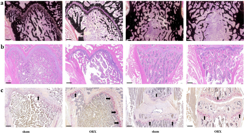
Methods: Twenty 3-month-old male rats were randomized to receive sham or orchiectomy (ORX) operation. Rats were euthanized after 8 weeks of surgery, and serum levels of sclerostin and irisin were measured by enzyme-linked immunosorbent assay at baseline and execution. Grip strength was measured by a grip strength tester at baseline and before execution. BMD and bone microarchitecture were measured by microcomputed tomography. The samples of bone and muscle were harvested at execution. Bone biomechanics were measured by three-point bending tests and vertebral body indentation tests. Bone and muscle histological features were analyzed by hematoxylin and eosin stain, Von Kossa's stain and tartrate resistant acid phosphatase stain. Simple linear regression analyses were used to analyze the relationships between serum levels of sclerostin, irisin and grip strength and BMD of ORX rats.
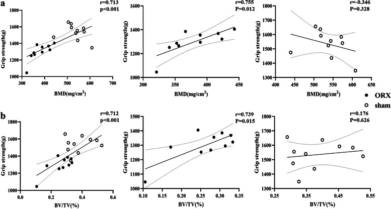
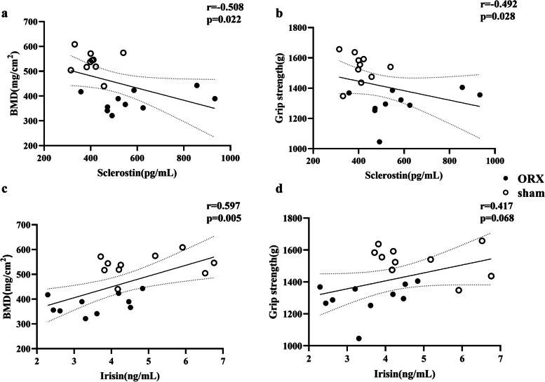
Results: Serum sclerostin level increased from 279 ± 44 pg/mL to 586 ± 57 pg/mL since baseline to 8 weeks after ORX (P = 0.002), which was significantly higher than that in sham rats (406 ± 20 pg/mL at execution) (P = 0.012). Serum irisin level decreased from 4.12 ± 0.20 ng/mL to 3.55 ± 0.29 ng/mL since baseline to 8 weeks of ORX (P = 0.048), which was significantly lower than sham rats (4.84 ± 0.37 pg/mL at execution) (P = 0.013). Trabecular BMD, parameters of bone microarchitecture, bone strength, grip strength and the myofibers size of soleus muscles were significantly lower in ORX rats than in sham group. Grip strength was positively correlated with femoral trabecular BMD (r = 0.713, P < 0.001) and bone volume/total volume (r = 0.712, P < 0.001) in all rats. The serum sclerostin level was negatively correlated to femoral trabecular BMD (r = -0.508, P = 0.022) and grip strength (r = -0.492, P = 0.028). Serum irisin level was positively correlated with femoral trabecular BMD (r = 0.597, P = 0.005), but no obvious correlation was found between irisin level and muscle strength in all rats.
Conclusions: Reduced BMD, impaired bone microarchitecture, weak strength of bone and muscle, and thin myofibers were induced by androgen deficiency of ORX rats. Serum sclerostin and irisin levels were significantly changed after ORX, which might be closely correlated with the occurrence of osteoporosis and sarcopenia in ORX rats.
Keywords: Irisin; Orchiectomy; Osteoporosis; Sarcopenia; Sclerostin.
© 2022. The Author(s).
Conflict of interest statement
No potential conflict of interest was reported by the authors.
Figures


References
MeSH terms
Substances
Grants and funding
LinkOut - more resources
Full Text Sources
