

Osteochondral Allograft Transplant for Combined Medial and Lateral Patellar Cartilage Lesions: The Osteochondral Wide Lesion (OWL) Technique
- PMID: 36457384
- PMCID: PMC9705722
- DOI: 10.1016/j.eats.2022.07.012
Osteochondral Allograft Transplant for Combined Medial and Lateral Patellar Cartilage Lesions: The Osteochondral Wide Lesion (OWL) Technique
Abstract
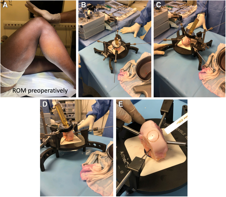
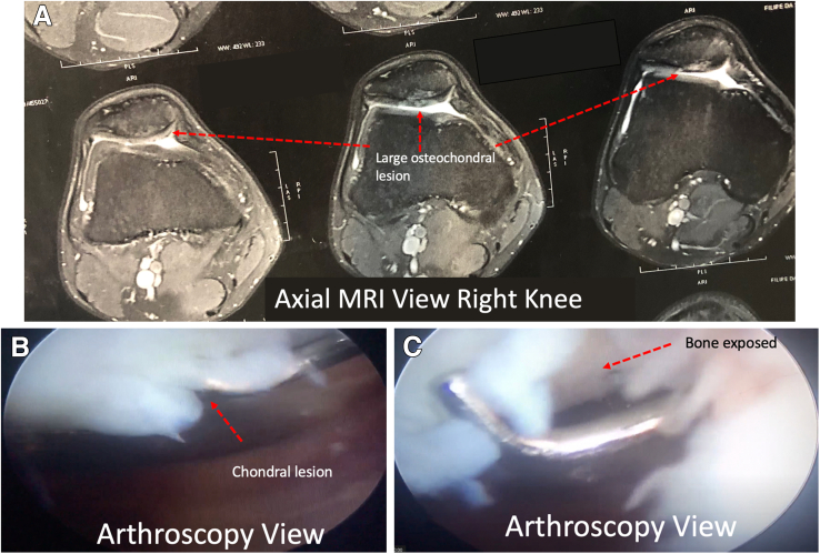
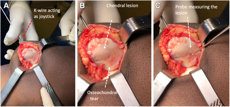
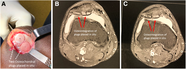
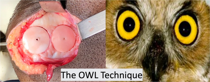
Symptomatic articular cartilage injuries are often seen in young active patients and athletes. Magnetic resonance imaging screening examinations have frequently identified such lesions in athletic patients. Patellofemoral chondral defects were previously identified as the most common knee cartilage lesion in high-level athletes. Chondral defects measuring 2 cm2 or greater and complex cartilage defects involving bone loss are ideally replaced with fresh osteochondral allograft. We describe a technique indicated for patients with symptomatic and recurrent anterior knee pain associated with osteochondral patellar defects including the lateral and medial patellar facets. Patients who have undergone previous interventions, including membrane techniques, microfracture, or autologous chondral transplantation, without clinical benefit are also eligible to undergo osteochondral allograft transplantation for combined medial and lateral patellar cartilage lesions, that is, the osteochondral wide lesion (OWL) technique.
© 2022 The Authors.
Figures


Similar articles
-
Combined Trochlear, Patellar, Medial and Lateral Condyle Fresh Osteochondral Allograft Transplantation: A Case Report.Video J Sports Med. 2023 Nov 6;3(6):26350254231193031. doi: 10.1177/26350254231193031. eCollection 2023 Nov-Dec. Video J Sports Med. 2023. PMID: 40309088 Free PMC article.
-
Surgical Treatment for Osteochondral Lesions of the Talus.Arthroscopy. 2021 Dec;37(12):3393-3396. doi: 10.1016/j.arthro.2021.10.002. Arthroscopy. 2021. PMID: 34863377
-
Combined Particulated Juvenile Cartilage Allograft Transplantation and Medial Patellofemoral Ligament Reconstruction for Symptomatic Chondral Defects in the Setting of Recurrent Patellar Instability.Arthrosc Tech. 2016 Oct 10;5(5):e1149-e1154. doi: 10.1016/j.eats.2016.06.008. eCollection 2016 Oct. Arthrosc Tech. 2016. PMID: 28224070 Free PMC article.
-
Chondral and osteochondral injuries associated with acute patellar dislocation.Arthroscopy. 2003 Sep;19(7):717-21. doi: 10.1016/s0749-8063(03)00401-8. Arthroscopy. 2003. PMID: 12966379 Review.
-
Osteochondral Autologous Transplantation for Treating Patellar High-Grade Chondral Defects: A Systematic Review.Orthop J Sports Med. 2019 Oct 17;7(10):2325967119876618. doi: 10.1177/2325967119876618. eCollection 2019 Oct. Orthop J Sports Med. 2019. PMID: 31667196 Free PMC article. Review.
References
-
- Carnes J., Stannus O., Cicuttini F., Ding C., Jones G. Knee cartilage defects in a sample of older adults: Natural history, clinical significance and factors influencing change over 2.9 years. Osteoarthritis Cartilage. 2012;20:1541–1547. - PubMed
-
- Major N.M., Helms C.A. MR imaging of the knee: Findings in asymptomatic collegiate basketball players. AJR Am J Roentgenol. 2002;179:641–644. - PubMed
-
- Kaplan L.D., Schurhoff M.R., Selesnick H., Thorpe M., Uribe J.W. Magnetic resonance imaging of the knee in asymptomatic professional basketball players. Arthroscopy. 2005;21:557–561. - PubMed
-
- Widuchowski W., Widuchowski J., Trzaska T. Articular cartilage defects: Study of 25,124 knee arthroscopies. Knee. 2007;14:177–182. - PubMed
-
- Flanigan D.C., Harris J.D., Trinh T.Q., Siston R.A., Brophy R.H. Prevalence of chondral defects in athletes’ knees: A systematic review. Med Sci Sports Exerc. 2010;42:1795–1801. - PubMed
LinkOut - more resources
Full Text Sources
