. 2022 Oct 20;11(11):e1937-e1944.
doi: 10.1016/j.eats.2022.07.008. eCollection 2022 Nov.

Repair of a Horizontal Cleavage Tear of the Lateral Meniscus with Circumferential Compression Stitches and Marrow Venting Augmentation

Affiliations

Repair of a Horizontal Cleavage Tear of the Lateral Meniscus with Circumferential Compression Stitches and Marrow Venting Augmentation

Enzo S Mameri et al. Arthrosc Tech. .

Abstract

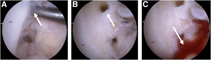
Horizontal cleavage tears (HCTs) are challenging meniscal tear patterns, as they split the meniscus into inferior and superior leaflets, while also involving the central, less vascular portions of the meniscus. Circumferential compression sutures using an all-inside self-retrieving suture passing device like the Novostitch Pro (Smith & Nephew, Andover, MA) have demonstrated the ability to create stable repair constructs with uniform compression across both leaflets in the setting of HCTs. Additionally, biological augmentation of meniscal repairs using a marrow venting procedure (MVP) has demonstrated superior clinical outcomes relative to isolated meniscal repairs. Thus, the purpose of this technical note is to outline our procedure for implementing circumferential compression sutures and biologic augmentation using an MVP for repairing an HCT of the lateral meniscus.


Figures

Fig 1
Fig 1
Tear of the lateral meniscus of the right knee. (A) Magnetic resonance imaging T2-coronal cut revealing a horizontal cleavage tear in the body of the lateral meniscus (white arrow) with associated meniscal parameniscal cyst (black arrow). (B) Initial arthroscopic visualization of the complex meniscal tear, with a small incomplete radial tear and an incomplete vertical tear on the upper surface of the meniscus. (C) Horizontal cleavage tear. (D) Tear site preparation with arthroscopic rasp (Smith & Nephew, Andover, MA), while viewing from the standard anterolateral portal and using the standard anteromedial portal as a working portal for instrumentation. For this lateral sided injury, the patient is placed in a figure-of-four position, paired with a slightly higher anteromedial portal in order to provide safe access to the posterior horn and midportion of the lateral meniscus.
Fig 2
Fig 2
Repair of a lateral meniscus tear of the right knee. The arthroscopic camera is inserted from the standard anterolateral portal, and the standard anteromedial portal is used as a working portal for instrumentation. For this lateral sided injury, the patient is placed in a figure-of-four position, in order to provide access to the posterior horn and midportion of the lateral meniscus. (A) The self-passing all-inside suture device is preloaded with 2-0 high-strength, nonabsorbable suture (Novostitch Pro; Smith & Nephew, Andover, MA) and is introduced into the joint with the lower jaw in the retracted position and the upper jaw in the horizontal, low-profile configuration. This is done for atraumatic insertion through the working anteromedial portal. The front, orange trigger is squeezed during introduction to maintain the upper jaw in this position. Upon accessing the site of the horizontal cleavage tear, the front trigger is released enough to elevate and place the upper jaw of the device on the femoral surface of the meniscus. The lower jaw is then protracted by activating the thumb lever on the side of the device, placing it between the tibial articular surface and the lower surface of the meniscus. (B) The suture is passed by pressing the posterior trigger of the all-inside suture device. The suture is passed using a minimally traumatic needle, through the meniscal tissue, and into the upper jaw, where it is self-captured. At the time of suture passage, the entire device should be gently driven toward the periphery of the meniscus as to prevent the meniscal tissue from extruding from the jaws of the device. (C) An arthroscopic knot is tied, followed by additional alternated half-stitches to complete the suture construct. Suture limbs are cut with a dedicated cutter. (D) Sequential stitching of the meniscus along the entire tear is performed until probing demonstrates adequate construct stability. Using a probe, knots should then be pushed toward the joint periphery to avoid knot placement in the meniscofemoral interface and prevent possible meniscal and/or cartilage injury.
Fig 3
Fig 3
Bone marrow-venting procedure of the right knee. The arthroscopic camera is inserted from the standard anterolateral portal, and the standard anteromedial portal is used as a working portal for instrumentation. (A) Anterolateral portal view of microfracture awl position on the lateral aspect of the intercondylar notch. (B) Anteromedial portal view of three perforations, which are anterior to the femoral origin of the anterior cruciate ligament (ACL) and allow for bone marrow elements to leak into the joint space. This marrow venting procedure provides a biologically enriched environment that promotes meniscal healing; (C) leakage of bone marrow products must be visually confirmed to ensure adequate marrow venting. If no leakage is initially observed, irrigation of the joint may be temporarily stopped to diminish the intra-articular pressure and confirm successful venting. LFC, lateral femoral condyle.

References

    1. Sallé de Chou E., Pujol N., Rochcongar G., et al. Analysis of short and long-term results of horizontal meniscal tears in young adults. Orthop Traumatol Surg Res. 2015;101:S317–S322. - PubMed
    1. Mordecai S.C., Al-Hadithy N., Ware H.E., Gupte C.M. Treatment of meniscal tears: An evidence based approach. World J Orthop. 2014;5:233–241. - PMC - PubMed
    1. Kurzweil P.R. Treatment of horizontal cleavage tears—Resection to repair. Oper Tech Sports Med. 2018;26:271–278.
    1. Woodmass J.M., Johnson J.D., Wu I.T., Saris D.B.F., Stuart M.J., Krych A.J. Horizontal cleavage meniscus tear treated with all-inside circumferential compression stitches. Arthrosc Tech. 2017;6:e1329–e1333. - PMC - PubMed
    1. Chahla J., Papalamprou A., Chan V., et al. Assessing the resident progenitor cell population and the vascularity of the adult human meniscus. Arthroscopy. 2021;37:252–265. - PMC - PubMed

LinkOut - more resources