Review
. 2022 Nov 28;7(1):bpac026.
doi: 10.1093/biomethods/bpac026. eCollection 2022.

HAT-field: a cheap, robust and quantitative Point-of-care serological test for Covid-19

Affiliations
Review

HAT-field: a cheap, robust and quantitative Point-of-care serological test for Covid-19

Etienne Joly et al. Biol Methods Protoc. .

Abstract

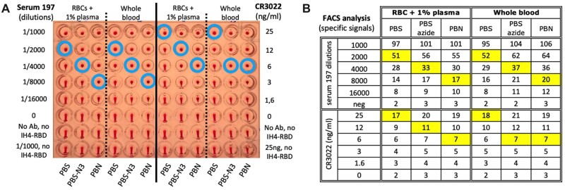
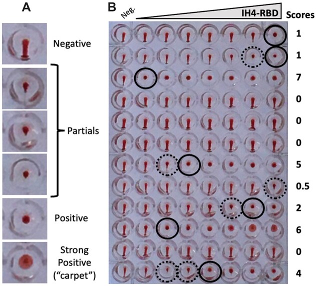
The haemagglutination test (HAT)-field protocol described here is an optimization of the recently published HAT, for the detection of antibodies directed against the receptor binding domain (RBD) of the SARS-Cov-2 virus. HAT and HAT-field are both based on haemagglutination triggered by a single reagent, the IH4-RBD recombinant protein. A sample of IH4-RBD sufficient for several thousand tests or a plasmid encoding IH4-RBD can be obtained from the authors of our first paper. Using titration of IH4-RBD, HAT-field now allows a quantitative assessment of antibody levels in a single step, using a few microliters of whole blood, such as can be obtained by finger prick, and requires only very simple disposable equipment. Because it is based on a single soluble reagent, the test can be adapted very simply and rapidly to detect antibodies against variants of the SARS-CoV-2, or conceivably against different pathogens. HAT-field appears well suited to provide quantitative assessments of the serological protection of populations as well as individuals, and given its very low cost, the stability of the IH4-RBD reagent in the adapted buffer and the simplicity of the procedure, could be deployed pretty much anywhere, including in the poorest countries and the most remote corners of the globe.

Keywords: Covid-19; IH4-RBD; SARS-Cov-2; haemagglutination; quantitative.


Figures

Figure 1:
Figure 1:
BSA prevents adsorption of IH4-RBD to the reaction wells. (A) HAT was performed in uncoated wells prefilled with IH4-RBD reagent diluted in either PBS (left columns) or PBS + 1% BSA + 3mM sodium azide (PBN, right columns). RBCs, resuspended in either PBS (left panels), in PBS supplemented with 1% seronegative autologous plasma (middle panels) or PBN (right panels) and various antibodies against SARS-CoV-2 were added to each well to test for haemagglutination. In the absence of antibody (Neg), the typical teardrop structure can be seen in each well. In the presence of a monoclonal antibody against RBD (CR3022), or plasma from convalescent Covid-19 patients, a veil structure forms in the absence of BSA, whereas a button forms when BSA is present (see the ‘Methods’ section for details). B, HAT as in (A) but performed in wells precoated with BSA. In the presence of the monoclonal antibody or convalescent patient plasma, haemagglutination is observed as a button rather than the veils seen in (A). Similar data were obtained from three experiments.
Figure 2:
Figure 2:
Azide and BSA increase the sensitivity of HAT. (A) The effects of azide and BSA on HAT performed using IH4-RBD at 1 µg/ml final concentration, and RBCs from a seronegative donor resuspended in PBS, PBS-N3 or PBN and 1% plasma from the same donor, or with whole blood from the same donor and with serial dilutions of an immune serum from a convalescent Covid-19 patient (Serum 197; left columns) or a monoclonal anti-RBD (CR3022; right columns). The three negative controls were: no antibody (0), neither antibody nor IH4-RBD and the most concentrated serum or antibody condition with no IH4-RBD. Blue circles indicate the titration endpoints (see the ‘Methods’ section for details). (B) After HAT, the RBCs were resuspended, stained with a fluorescent secondary anti-human Ig antibody and analyzed by FACS. The numbers shown correspond to specific signals, that is, the difference in GMFI values of each sample with that of the control sample incubated in the same buffer with no antibody or IH4-RBD. The squares highlighted in yellow indicate the titration endpoints. Comparable results were obtained in four similar experiments. (A color version of this figure appears in the online version of this article).
Figure 3:
Figure 3:
IH4-RBD is very stable when diluted in PBN. The effects of long-term storage of IH4-RBD in PBS-N3 or PBN at 4°C, RT and 37°C, as determined by HAT and by FACS analysis of antibody binding to RBCs after HAT. Haemagglutination end-points in the presence of the CR3022 monoclonal antibody at 100 ng/ml (black circles) were determined by titration of aliquots of working dilutions of IH4-RBD (2 µg/ml) stored for up to 15 months at the indicated temperatures. The red numbers indicate the intensity of the specific fluorescent staining recorded by FACS analysis performed after the HAT assay on the RBCs from the samples incubated with 100 ng/ml IH4-RBD (see the ‘Methods’ section for details). Similar data were obtained in seven experiments, some of which also included using diluted sera from convalescent patients in parallel to the CR3022 monoclonal antibody (not shown). (A color version of this figure appears in the online version of this article).
Figure 4:
Figure 4:
Temperature has little influence on HAT results. To determine the effect of temperature on the performance of HAT, three parallel plates were setup for 2D titrations, with DD of IH4-RBD going from 8 µg/ml to 8 ng/ml along lines and DD of two different immune sera from convalescent Covid-19 patients down columns (sera 186 and 203, see the ‘Methods’ section for practical details). After incubation at the indicated three temperatures, no substantial differences were seen in titration end-points (blue circles). Similar results were obtained in three independent experiments, using a total of three different immune sera, two plasmas and the CR3022 monoclonal antibody. Incubation temperature had little or no discernable influence on the haemagglutination endpoints (blue circles), with the possible exception of the wells containing the highest concentration of IH4-RBD and very diluted sera, where incubation at 4°C resulted in a small improvement in sensitivity when compared with the assays performed at RT or 37°C. (A color version of this figure appears in the online version of this article).
Figure 5:
Figure 5:
Schematic description of the HAT-field protocol.
Figure 6:
Figure 6:
Validation of the HAT-field protocol by comparison with laboratory tests. Sixty randomly selected blood samples were used to compare the results of the HAT-field protocol with those of HAT plasma titrations and the Jurkat-S&R-flow test (see the ‘Methods’ section). The graphs show one-on-one comparisons of the results obtained with those three tests, as indicated, using the values obtained by centrifuging the plates after 15 min (for HAT-field), and after 60 min for plasma titrations. Pearson’s correlation coefficients are indicated in the bottom right corners. On panel A, because of the discrete nature of the scales used for both the X- and Y-axes, many points overlap on top of one another. In such cases, their numbers are indicated by the adjacent green numbers. The dotted line in A indicates the position of the median and those in B and C indicate the threshold for positive samples in the Jurkat-S&R-flow. The two red dots in each graph correspond to two negative samples, which gave false-positive results in HAT plasma titrations due to their reactivity against the IH4 nanobody moiety of the reagent. The orange dots correspond to samples positive in the Jurkat-S&R-flow test that showed some reactivity against the IH4 nanobody alone, albeit with lower titers than against the IH4-RBD reagent. For the sample represented by an orange dot surrounded by a red circle in C, the plasma titration against the nanobody alone was positive, but it led to only partial haemagglutination with the IH4-RBD reagent (for actual values, see sample 48 in data file). (A color version of this figure appears in the online version of this article).
Figure 7:
Figure 7:
Scoring HAT. (A) Examples of typical negative, partial and positive wells. Wells are considered as partial as soon as a small point appears at the bottom of the RBD pellet, such as that shown on the fourth line. (B) Example of a plate with HAT-field performed with Wuhan IH4-RBD on 12 whole blood samples after 60 min under normal gravity. The final IH4-RBD concentrations in the wells from left to right are, respectively, 0, 3, 10, 31, 100, 316, 1000 and 3160 ng/ml. Black circles indicate the last positive wells, and dotted circles partially positive ones. Scoring is carried out on the pictures by simply counting the number of positive wells, starting from the highest IH4-RBD concentration (i.e. from right to left).

References

    1. Abbasi J. The flawed science of antibody testing for SARS-CoV-2 immunity. JAMA 2021;326:1781–2. - PubMed
    1. Moshe M, Daunt A, Flower B. et al.; React study team. SARS-CoV-2 lateral flow assays for possible use in national covid-19 seroprevalence surveys (React 2): diagnostic accuracy study. BMJ 2021;372:n423. - PMC - PubMed
    1. Ong DSY, Fragkou PC, Schweitzer VA. et al.; European Society of Clinical Microbiology and Infectious Diseases (ESCMID) Study Group for Respiratory Viruses (ESGREV). How to interpret and use COVID-19 serology and immunology tests. Clin Microbiol Infect 2021;27:981–6. - PMC - PubMed
    1. Townsend A, Rijal P, Xiao J. et al. A haemagglutination test for rapid detection of antibodies to SARS-CoV-2. Nat Commun 2021;12:1951. - PMC - PubMed
    1. Habib I, Smolarek D, Hattab C. et al. V(H)H (nanobody) directed against human glycophorin A: A tool for autologous red cell agglutination assays. Anal Biochem 2013;438:82–9. - PubMed