

Myocardial ischemia in patients with large prior infarction: Clinical decision making and review of literature
- PMID: 36457789
- PMCID: PMC9705384
- DOI: 10.1016/j.radcr.2022.11.006
Myocardial ischemia in patients with large prior infarction: Clinical decision making and review of literature
Abstract
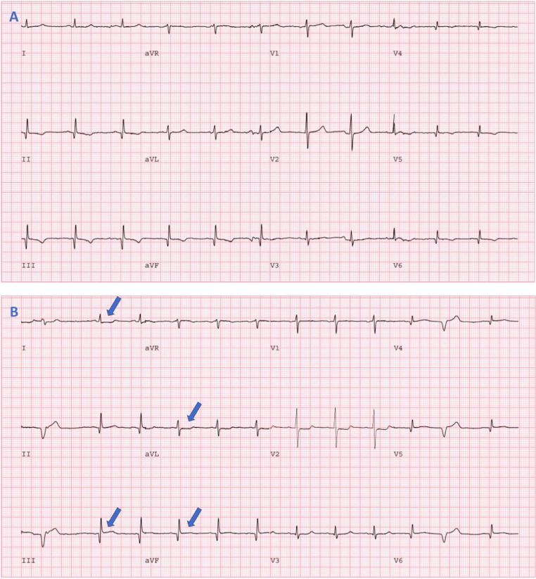
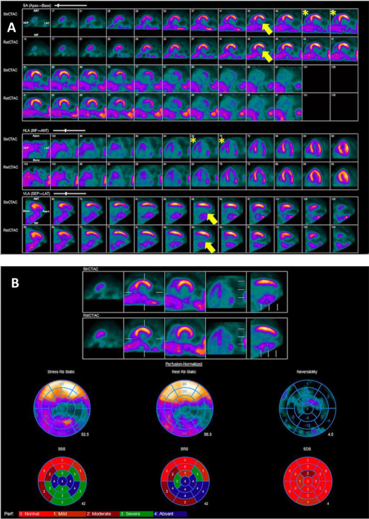
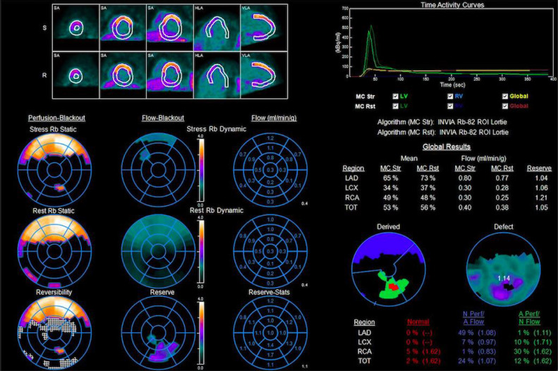
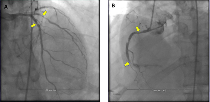
Myocardial perfusion imaging (MPI) with single photon emission computed tomography (SPECT) or positron emission tomography (PET) is a widely used technique for the evaluation of coronary artery disease (CAD). Interpreting physicians rely on regional variations in myocardial radiotracer uptake between rest and stress images to identify hemodynamically significant epicardial coronary artery stenosis. However, interpretation of MPI is very difficult in patients with large infarcts where there is no scintigraphically normal reference myocardium for comparison. In these patients, the stress and rest images appear similar due to balanced ischemia in the non-infarct territory. There are no clear guidelines on how to approach these cases. We present a case of MPI with a large right coronary artery territory (RCA) infarct where the left main (LM) coronary artery territory has no relative comparator and the images looked the same on stress and rest. However, the patient had multiple high-risk ancillary findings including electrocardiographic (ECG) changes with regadenoson, transient ischemic dilatation (TID), large severe inferior infarct, low myocardial blood flow (MBF) and myocardial flow reserve (MFR), but most notably increased right ventricular (RV) uptake on the stress images that was a subtle clue that we were dealing with LM equivalent in non-infarct zone. The coronary angiogram confirmed our findings. Through our case, we provide a comprehensive approach and review of literature on how to approach such challenging encounters.
Keywords: Multivessel coronary artery disease; Myocardial blood flow; Myocardial flow reserve; Rubidium-82 positron emission tomography; Transient ischemic dilatation.
© 2022 The Authors. Published by Elsevier Inc. on behalf of University of Washington.
Figures
References
-
- Ziadi M.C., et al. Does quantification of myocardial flow reserve using rubidium-82 positron emission tomography facilitate detection of multivessel coronary artery disease? J Nucl Cardiol. 2012;19(4):670–680. - PubMed
-
- Schindler T.H., et al. Cardiac PET imaging for the detection and monitoring of coronary artery disease and microvascular health. JACC Cardiovasc Imaging. 2010;3(6):623–640. - PubMed
-
- Lima R.S., et al. Incremental value of combined perfusion and function over perfusion alone by gated SPECT myocardial perfusion imaging for detection of severe three-vessel coronary artery disease. J Am Coll Cardiol. 2003;42(1):64–70. - PubMed
-
- Williams K.A., et al. Correct spatial normalization of myocardial perfusion SPECT improves detection of multivessel coronary artery disease. J Nucl Cardiol. 2003;10(4):353–360. - PubMed
-
- Berman D.S., et al. Underestimation of extent of ischemia by gated SPECT myocardial perfusion imaging in patients with left main coronary artery disease. J Nucl Cardiol. 2007;14(4):521–528. - PubMed
Publication types
LinkOut - more resources
Full Text Sources
Miscellaneous
