MALAT1/miR-320a in bone marrow mesenchymal stem cells function may shed light on mechanisms underlying osteoporosis
- PMID: 36457977
- PMCID: PMC9710279
- DOI: 10.5114/aoms/105838
MALAT1/miR-320a in bone marrow mesenchymal stem cells function may shed light on mechanisms underlying osteoporosis
Abstract
Introduction: Growing evidence supports the involvement of long noncoding RNAs (lncRNAs) in bone metabolism and diseases. This study aims to investigate the involvement of the lncRNA metastasis-associated lung adenocarcinoma transcript 1 (MALAT1) in the pathological process of osteoporosis and the effects of MALAT1 on regulation of BMSC differentiation through competitive endogenous RNA (ceRNA) mechanisms.
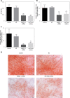
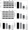
Material and methods: The expression of MALAT1 and miR-320a was determined using RT-PCR in bone tissue derived from female SD (Sprague Dawley) rats with osteoporosis. Immunohistochemical (IHC) staining was used to evaluate the expression of neuropilin-1 (NRP-1) and β-catenin. Bone marrow mesenchymal stem cells (BMSCs) were divided into 4 groups: control, NC (negative control), MALAT1 siRNA, and miR-320a mimics. Forty-eight hours later, the effect of MALAT1 on the miR-320a expression, proliferation and osteogenic differentiation of BMSCs was investigated. Two weeks later, the cell activity, alkaline phosphatase (ALP) activity, and mRNA expression of Osterix and Runx2 were evaluated. Three weeks later, alizarin red staining of calcified nodules and Western blot analysis of the expression of β-catenin, NRP-1, osteocalcin (OCN), and osteopontin (OPN) were performed.
Results: Downregulated MALAT1or upregulated miR-320a expression inhibited the activity and osteogenic differentiation of BMSCs, resulting in low ALP activity and NRP-1 expression, fewer calcified nodules, decreased mRNA levels of Osterix and Runx2, and inhibited expression of NRP-1, OCN, and OPN. MALAT1 silencing did not decrease the protein level of β-catenin in the cytoplasm but suppressed that in the nucleus.
Conclusions: Downregulated MALAT1 and upregulated miR-320a expression play an important role in the pathological process of osteoporosis, via inhibition of the osteogenic differentiation of BMSCs.
Keywords: bone marrow mesenchymal stem cells; competitive endogenous RNA; metastasis-associated lung adenocarcinoma transcript; miR-320a; osteoporosis.
Copyright: © 2021 Termedia & Banach.
Conflict of interest statement
The authors declare no conflict of interest.
Figures


Similar articles
-
LncRNA DANCR and miR-320a suppressed osteogenic differentiation in osteoporosis by directly inhibiting the Wnt/β-catenin signaling pathway.Exp Mol Med. 2020 Aug;52(8):1310-1325. doi: 10.1038/s12276-020-0475-0. Epub 2020 Aug 11. Exp Mol Med. 2020. PMID: 32778797 Free PMC article.
-
LncRNA metastasis-associated lung adenocarcinoma transcript-1 promotes osteogenic differentiation of bone marrow stem cells and inhibits osteoclastic differentiation of Mø in osteoporosis via the miR-124-3p/IGF2BP1/Wnt/β-catenin axis.J Tissue Eng Regen Med. 2022 Mar;16(3):311-329. doi: 10.1002/term.3279. Epub 2022 Jan 11. J Tissue Eng Regen Med. 2022. PMID: 34962086
-
Expression of lncRNA MALAT1 through miR-144-3p in Osteoporotic Tibial Fracture Rats and Its Effect on Osteogenic Differentiation of BMSC under Traction.Evid Based Complement Alternat Med. 2022 Jul 5;2022:2590055. doi: 10.1155/2022/2590055. eCollection 2022. Evid Based Complement Alternat Med. 2022. Retraction in: Evid Based Complement Alternat Med. 2023 Sep 27;2023:9783205. doi: 10.1155/2023/9783205. PMID: 35836824 Free PMC article. Retracted.
-
Long non-coding RNAs in bone formation: Key regulators and therapeutic prospects.Open Life Sci. 2024 Aug 16;19(1):20220908. doi: 10.1515/biol-2022-0908. eCollection 2024. Open Life Sci. 2024. PMID: 39156986 Free PMC article. Review.
-
The regulatory activities of MALAT1 in the development of bone and cartilage diseases.Front Endocrinol (Lausanne). 2022 Nov 14;13:1054827. doi: 10.3389/fendo.2022.1054827. eCollection 2022. Front Endocrinol (Lausanne). 2022. PMID: 36452326 Free PMC article. Review.
Cited by
-
Construction of dynamic ceRNA regulatory networks in osteogenesis during fracture healing based on transcriptomic analysis.Sci Rep. 2025 Jul 24;15(1):26946. doi: 10.1038/s41598-025-12505-6. Sci Rep. 2025. PMID: 40707643 Free PMC article.
-
Diagnostic value of circulating bone turnover markers osteocalcin, cathepsin K, and osteoprotegerin for osteoporosis in middle-aged and elderly postmenopausal women.Arch Med Sci. 2024 Oct 25;20(5):1727-1730. doi: 10.5114/aoms/193198. eCollection 2024. Arch Med Sci. 2024. PMID: 39649286 Free PMC article. No abstract available.
-
Long non-coding RNA small nucleolar RNA host gene 8 (SNHG8) sponges miR-34b-5p to prevent sepsis-induced cardiac dysfunction and inflammation and serves as a diagnostic biomarker.Arch Med Sci. 2024 Aug 22;20(4):1268-1280. doi: 10.5114/aoms/175468. eCollection 2024. Arch Med Sci. 2024. PMID: 39439678 Free PMC article.
-
MALAT1/miR-7-5p/TCF4 Axis Regulating Menstrual Blood Mesenchymal Stem Cells Improve Thin Endometrium Fertility by the Wnt Signaling Pathway.Cell Transplant. 2024 Jan-Dec;33:9636897241259552. doi: 10.1177/09636897241259552. Cell Transplant. 2024. PMID: 38847385 Free PMC article.
References
-
- Manolagas SC. Birth and death of bone cells: basic regulatory mechanisms and implications for the pathogenesis and treatment of osteoporosis. Endocr Rev 2000; 21: 115-37. - PubMed
-
- Che W, Dong Y, Quan HB. RANKL inhibits cell proliferation by regulating MALAT1 expression in a human osteoblastic cell line hFOB 1.19. Cell Mol Biol 2015; 61: 7-14. - PubMed
-
- Liang J, Liang L, Ouyang K, Li Z, Yi X. MALAT1 induces tongue cancer cells’ EMT and inhibits apoptosis through Wnt/beta-catenin signaling pathway. J Oral Pathol Med 2017; 46: 98-105. - PubMed
LinkOut - more resources
Full Text Sources
Research Materials
Miscellaneous
