

Antiviral response within different cell types of the CNS
- PMID: 36458002
- PMCID: PMC9706196
- DOI: 10.3389/fimmu.2022.1044721
Antiviral response within different cell types of the CNS
Abstract
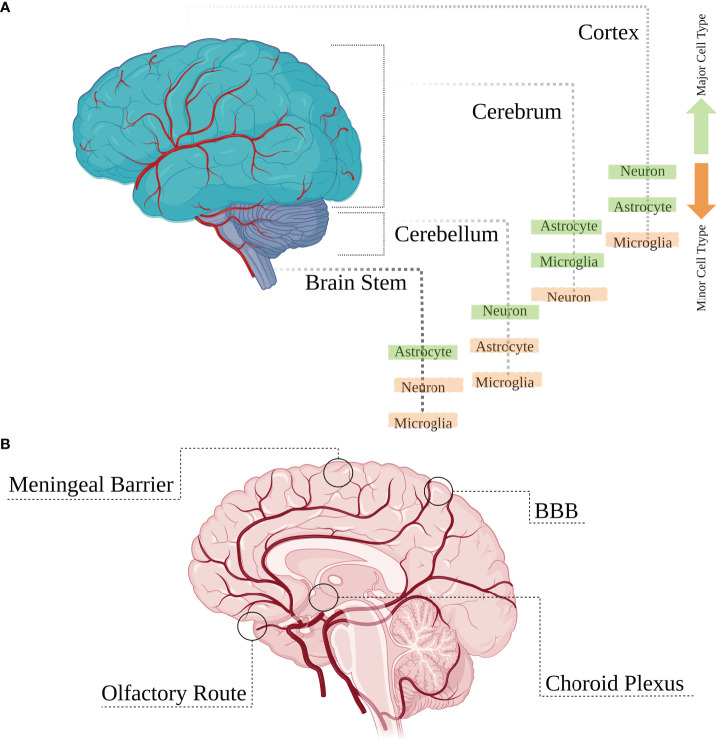
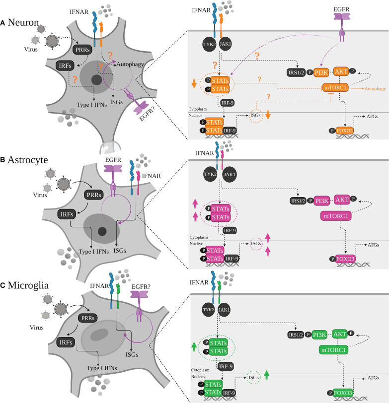
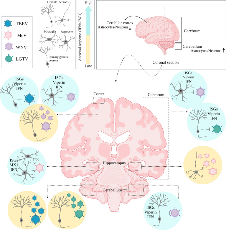
The central nervous system (CNS) is a constitutive structure of various cell types conserved by anatomical barriers. Many of the major CNS cell-type populations distributed across the different brain regions are targets for several neurotropic viruses. Numerous studies have demonstrated that viral susceptibility within the CNS is not absolute and initiates a cell-type specific antiviral defence response. Neurons, astrocytes, and microglial cells are among the major resident cell populations within the CNS and are all equipped to sense viral infection and induce a relative antiviral response mostly through type I IFN production, however, not all these cell types adopt a similar antiviral strategy. Rising evidence has suggested a diversity regarding IFN production and responsiveness based on the cell type/sub type, regional distinction and cell`s developmental state which could shape distinct antiviral signatures. Among CNS resident cell types, neurons are of the highest priority to defend against the invading virus due to their poor renewable nature. Therefore, infected and uninfected glial cells tend to play more dominant antiviral roles during a viral infection and have been found to be the major CNS IFN producers. Alternatively, neuronal cells do play an active part during antiviral responses but may adopt differential strategies in addition to induction of a typical type I IFN response, to minimize the chance of cellular damage. Heterogeneity observed in neuronal IFN responsiveness may be partially explained by their altered ISGs and/or lower STATS expression levels, however, further in vivo studies are required to fully elucidate the specificity of the acquired antiviral responses by distinct CNS cell types.
Keywords: brain; central nervous system; innate immunity; interferon; virus.
Copyright © 2022 Telikani, Monson, Hofer and Helbig.
Conflict of interest statement
The authors declare that the research was conducted in the absence of any commercial or financial relationships that could be construed as a potential conflict of interest.
Figures
Similar articles
-
Alpha/Beta Interferon (IFN-α/β) Signaling in Astrocytes Mediates Protection against Viral Encephalomyelitis and Regulates IFN-γ-Dependent Responses.J Virol. 2018 Apr 27;92(10):e01901-17. doi: 10.1128/JVI.01901-17. Print 2018 May 15. J Virol. 2018. PMID: 29491163 Free PMC article.
-
Neuronal Ablation of Alpha/Beta Interferon (IFN-α/β) Signaling Exacerbates Central Nervous System Viral Dissemination and Impairs IFN-γ Responsiveness in Microglia/Macrophages.J Virol. 2020 Sep 29;94(20):e00422-20. doi: 10.1128/JVI.00422-20. Print 2020 Sep 29. J Virol. 2020. PMID: 32796063 Free PMC article.
-
Microglia Activate Early Antiviral Responses upon Herpes Simplex Virus 1 Entry into the Brain to Counteract Development of Encephalitis-Like Disease in Mice.J Virol. 2022 Mar 23;96(6):e0131121. doi: 10.1128/JVI.01311-21. Epub 2022 Jan 19. J Virol. 2022. PMID: 35045263 Free PMC article.
-
Cytosolic DNA Sensors and CNS Responses to Viral Pathogens.Front Cell Infect Microbiol. 2020 Sep 16;10:576263. doi: 10.3389/fcimb.2020.576263. eCollection 2020. Front Cell Infect Microbiol. 2020. PMID: 33042875 Free PMC article. Review.
-
Control of glial immune function by neurons.Glia. 2001 Nov;36(2):191-9. doi: 10.1002/glia.1108. Glia. 2001. PMID: 11596127 Review.
Cited by
-
Viral immunological complications in neurological surgery: A comprehensive review of homeostatic disturbances and cognitive impairments.Surg Neurol Int. 2025 Jun 13;16:241. doi: 10.25259/SNI_337_2025. eCollection 2025. Surg Neurol Int. 2025. PMID: 40656505 Free PMC article. Review.
-
Antiviral immunity within neural stem cells distinguishes Enterovirus-D68 strain differences in forebrain organoids.J Neuroinflammation. 2024 Nov 5;21(1):288. doi: 10.1186/s12974-024-03275-5. J Neuroinflammation. 2024. PMID: 39501367 Free PMC article.
-
Expression Profiles of lncRNAs and mRNAs in the Mouse Brain Infected with Pseudorabies Virus: A Bioinformatic Analysis.Viruses. 2025 Apr 17;17(4):580. doi: 10.3390/v17040580. Viruses. 2025. PMID: 40285022 Free PMC article.
-
CNS disease associated with enhanced type I interferon signalling.Lancet Neurol. 2024 Nov;23(11):1158-1168. doi: 10.1016/S1474-4422(24)00263-1. Lancet Neurol. 2024. PMID: 39424561 Free PMC article. Review.
-
Ex vivo study of neuroinvasive and neurotropic viruses: what is current and what is next.FEMS Microbiol Rev. 2025 Jan 14;49:fuaf024. doi: 10.1093/femsre/fuaf024. FEMS Microbiol Rev. 2025. PMID: 40498321 Free PMC article. Review.
References
Publication types
MeSH terms
Substances
LinkOut - more resources
Full Text Sources