Review
. 2022 Nov 15:16:1042814.
doi: 10.3389/fnins.2022.1042814. eCollection 2022.

Neuroimaging in schizophrenia: A review article

Affiliations
Review

Neuroimaging in schizophrenia: A review article

Mona Dabiri et al. Front Neurosci. .

Abstract

In this review article we have consolidated the imaging literature of patients with schizophrenia across the full spectrum of modalities in radiology including computed tomography (CT), morphologic magnetic resonance imaging (MRI), functional magnetic resonance imaging (fMRI), magnetic resonance spectroscopy (MRS), positron emission tomography (PET), and magnetoencephalography (MEG). We look at the impact of various subtypes of schizophrenia on imaging findings and the changes that occur with medical and transcranial magnetic stimulation (TMS) therapy. Our goal was a comprehensive multimodality summary of the findings of state-of-the-art imaging in untreated and treated patients with schizophrenia. Clinical imaging in schizophrenia is used to exclude structural lesions which may produce symptoms that may mimic those of patients with schizophrenia. Nonetheless one finds global volume loss in the brains of patients with schizophrenia with associated increased cerebrospinal fluid (CSF) volume and decreased gray matter volume. These features may be influenced by the duration of disease and or medication use. For functional studies, be they fluorodeoxyglucose positron emission tomography (FDG PET), rs-fMRI, task-based fMRI, diffusion tensor imaging (DTI) or MEG there generally is hypoactivation and disconnection between brain regions. However, these findings may vary depending upon the negative or positive symptomatology manifested in the patients. MR spectroscopy generally shows low N-acetylaspartate from neuronal loss and low glutamine (a neuroexcitatory marker) but glutathione may be elevated, particularly in non-treatment responders. The literature in schizophrenia is difficult to evaluate because age, gender, symptomatology, comorbidities, therapy use, disease duration, substance abuse, and coexisting other psychiatric disorders have not been adequately controlled for, even in large studies and meta-analyses.

Keywords: DTI; MRI; PET; functional imaging; psychiatry; psychosis; schizophrenia.


Conflict of interest statement

Author DY reports royalties from Elsevier, personal fees as a Medicolegal consultant, he also received speaking and consulting fees from MRIOnline.com, outside the submitted work. The remaining authors declare that the research was conducted in the absence of any commercial or financial relationships that could be construed as a potential conflict of interest.

Figures

FIGURE 1
FIGURE 1
MPRAGE images of three patients with schizophrenia. These type of high resolution T1-weighted images allow quantitation of specific gray matter and cerebrospinal fluid (CSF) containing structures which has led to the conclusion that patients with schizophrenia have overall decreased gray matter volumes and enlarged CSF spaces. Specific cortical regions are best evaluated in group analyses and/or voxel-based morphometry analyses. Comparing these three subjects, one finds reduced right versus left hippocampal volume in the second and third subjects at group level, a finding that is not necessarily confirmed by visual analysis at individual level.
FIGURE 2
FIGURE 2
A low availability of the alpha7 nicotinic acetylcholine receptor in non-affective psychosis is supported by the binding (VT) values using 18F-ASEM PET. Mean parametric 18F-ASEM VT images derived from a study population of 15 controls (top), six patients with affective psychosis [Bipolar I disorder (middle)], and five patients with non-affective psychosis [Schizophrenia, Schizoaffective disorder (bottom)]. Images from left to right: axial, sagittal, and coronal views. VT is in reported in mL⋅cm–3 (Coughlin et al., 2018).
FIGURE 3
FIGURE 3
Regions with abnormal synchrony in rs-fMRI time-courses in early-schizophrenia patients (n = 58) compared to age- and sex-paired healthy controls. Blue codes decreased and red codes increased pairwise rs-fMRI correlations in schizophrenia patients. SP, superior parietal; Thal, thalamus; PrC, pre-central gyrus; PoC, post central gyrus; IO, inferior orbital; R, right; L, left (Faria et al., 2021).
FIGURE 4
FIGURE 4
Correlations between regional fractional anisotropy (FA) in diverse brain areas (Y axis) and performance in cognitive tests (X-axis). Red dots are early-stage schizophrenia patients (n = 58); blue is age- and sex-paired healthy controls. The shadow represents the 95% interval for the linear fitting (line). Significant correlations between FA and cognition were found in the schizophrenia group, but not in controls (Faria et al., 2019).
FIGURE 5
FIGURE 5
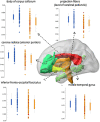
Significant differences in fractional anisotropy (FA) between a group of early-stage schizophrenia patients (n = 58; orange dots in the box-plots) and age- and sex-paired healthy controls (blue dots in the box-plots) (Faria et al., 2021).
FIGURE 6
FIGURE 6
Magnetic resonance (MR) spectra recorded at 7T in the centrum semiovale (CSO) white matter and anterior cingulate cortex (anterior cingulate cortex) using the STEAM pulse sequence and VAPOR water suppression in a xx-year-old subject with schizophrenia. Voxel locations and sizes are indicated overlaid on T1-weighted MR images. In the spectra, peaks are detected from lactate (Lac), lipid (Lip), N-acetyl aspartate (N-acetylaspartate), N-acetyl aspartylglutamate (N-acetylaspartateG, predominantly in white matter), glutamate (Glu), glutamine (Gln), glutathione (GSH), total creatine (tCr), total choline (tCho), myo-inositol (mI), and a combined glutamate and glutamine peak (Glx). While these spectra are virtually indistinguishable from those observed in healthy control subjects, quantitative analysis and studies in sufficient numbers of subjects reveal a number of statistically significant differences between patients and controls.
FIGURE 7
FIGURE 7
The cross-sectional study (left) observed reduced anterior cingulate cortex glutathione (GSH) levels and hippocampus volumes in non-responders; however, the longitudinal study data (right) found that anterior cingulate cortex GSH and hippocampus volume are stable over time in patients; these results imply that anterior cingulate cortex GSH and hippocampus volume may be trait markers for non-responders. Studying data from multimodal longitudinal cohorts may be a promising approach in the future to identify biomarkers for schizophrenia or a subset of schizophrenia patients. This figure shows anterior cingulate cortex GSH levels and the HP volume as potential biomarkers for TR to antipsychotics (Zhao et al., 2019; Picó-Pérez et al., 2022). Future studies are expected to identify additional biomarkers for TR and a combination of multimodal biomarkers that may be useful for the early diagnosis of TR in the clinic.

References

    1. Alamian G., Hincapié A. S., Pascarella A., Thiery T., Combrisson E., Saive A. L., et al. (2017). Measuring alterations in oscillatory brain networks in schizophrenia with resting-state MEG: State-of-the-art and methodological challenges. Clin. Neurophysiol. 128 1719–1736. 10.1016/j.clinph.2017.06.246 - DOI - PubMed
    1. Alsop D. C., Detre J. A., Golay X., Günther M., Hendrikse J., Hernandez-Garcia L., et al. (2015). Recommended implementation of arterial spin-labeled perfusion MRI for clinical applications: A consensus of the ISMRM perfusion study group and the European consortium for ASL in dementia. Magn. Reson. Med. 73 102–116. 10.1002/mrm.25197 - DOI - PMC - PubMed
    1. Andrade E. M., Geha L. M., Duran P., Suwwan R., Machado F., do Rosário M. C. (2016). Quality of life in caregivers of ADHD children and diabetes patients. Front. Psychiatry 7:127. 10.3389/fpsyt.2016.00127 - DOI - PMC - PubMed
    1. Andreasen N. C., Carpenter W. T., Jr., Kane J. M., Lasser R. A., Marder S. R., Weinberger D. R. (2005). Remission in schizophrenia: Proposed criteria and rationale for consensus. Am. J. Psychiatry 162 441–449. 10.1176/appi.ajp.162.3.441 - DOI - PubMed
    1. Andreasen N. C., Paradiso S., O’leary D. S. (1998). “Cognitive dysmetria” as an integrative theory of schizophrenia: A dysfunction in cortical-subcortical-cerebellar circuitry? Schizophr. Bull. 24 203–218. 10.1093/oxfordjournals.schbul.a033321 - DOI - PubMed

LinkOut - more resources