. 2022 Nov 15:27:491-501.
doi: 10.1016/j.omtm.2022.11.003. eCollection 2022 Dec 8.

Assessing production variability in empty and filled adeno-associated viruses by single molecule mass analyses

Affiliations

Assessing production variability in empty and filled adeno-associated viruses by single molecule mass analyses

Eduard H T M Ebberink et al. Mol Ther Methods Clin Dev. .

Abstract

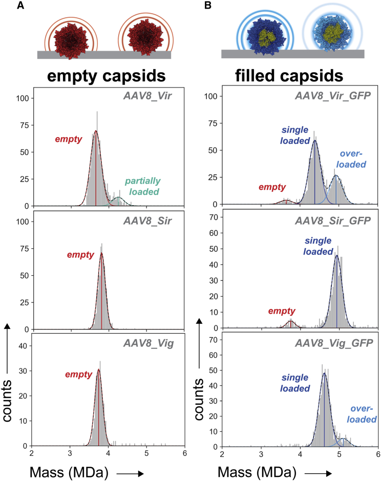
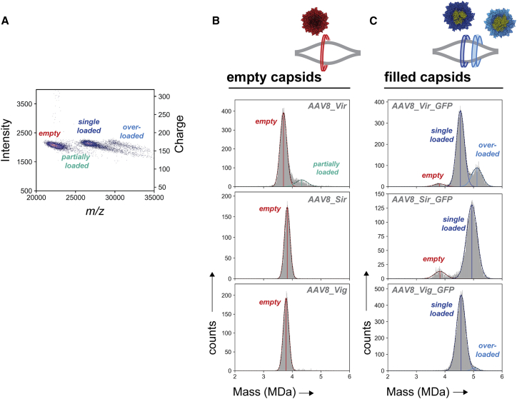
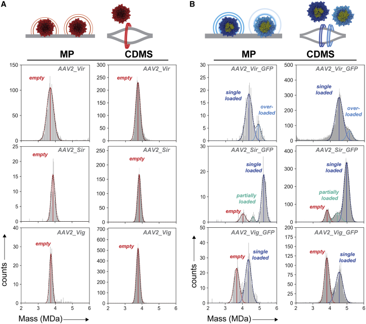
Adeno-associated viruses (AAVs) are useful vehicles for gene therapy because of their stability, low immunogenicity. and non-pathogenicity. However, disparity in AAV sample preparations (e.g., in capsid composition, DNA packaging, and impurities) gives rise to product heterogeneity, with possibly undesired effects on gene delivery. Ideally, AAV production should be with full control of AAV structure and genetic payload. Therefore, robust, efficient, and low material consuming methods are essential to characterize AAVs. Here, we use two emerging single-molecule techniques, mass photometry and Orbitrap-based charge-detection mass spectrometry, and show how they may efficiently and accurately characterize AAVs. We were able to resolve heterogeneous pools of particles, evaluating AAVs from two different serotypes (AAV8 and AAV2), produced by three independent production platforms, either lacking a genome or packed with a transgene. Together our data confirm that the different AAV production methods result in rather different and diverse AAV particle distributions. Especially for the packed AAVs, frequently additional subspecies were observed, next to the expected packed genome, mostly resulting from under- or overpackaging of genome material and/or residual empty particles. This work further establishes that both these single-particle techniques may become valuable tools in characterizing AAVs before they are used in gene therapy.

Keywords: AAV biomanufacturing; AAV2; AAV8; Adeno-associated virus; charge-detection mass spectrometry; empty-filled ratio; gene-delivery vector; mass photometry; native mass spectrometry; single molecule mass analyses.


Conflict of interest statement

A.R., M.N., and M.T. are employees of Roche Diagnostics GmbH, Penzberg, Germany, a company with interest in using AAV vectors for gene-delivery purposes.

Figures

None
Graphical abstract
Figure 1
Figure 1
Mass photometry analyses of empty and ssDNA packaged AAV8 capsids obtained from three different suppliers (A) Mass histograms of supposedly empty AAV8 capsids. The scattering event of each particle landing on the glass surface is translated into a particle mass and is classed in bins containing a bin width of 25 kDa. (B) Mass histograms of supposedly filled AAV8 capsids following production in the presence of a CMV-GFP transgene. Likewise, to the empty capsids, mass histograms were constructed with bin widths of 25 kDa. For each AAV8 sample, a single, representative mass histogram is displayed. For the most abundant species, Gaussian distributions were fitted and the mean masses are displayed as vertical lines. The average fitting over at least three mass distributions is given in Figure S3. Vir = Virovek; Sir = Sirion; Vig = Vigene; red = empty capsids; dark blue = single genome loaded capsids; light blue = overloaded capsids; turquoise = partially loaded capsids.
Figure 2
Figure 2
Orbitrap-based charge-detection mass spectrometry on empty and ssDNA packaged AAV8 capsids AAV8 capsids were electrosprayed into a UHMR Orbitrap analyzer to detect the charge and mass-over-charge ratio. (A) Displayed is an overlay of the 2D-histograms of m/z versus intensity and charge from AAV8_Vir and AAV8_Vir_GFP. Bin widths of 25 Th and 10 arbitrary units were used for, respectively, m/z and intensity. The color code represents the number of particles in each bin ranging from blue to red for respectively low and high counts. (B) The masses of empty capsids were calculated from the 2D-histogram and plotted in a 1D mass histogram with a bin width of 10 kDa. (C) Filled AAV8 capsids were processed the same way as empty capsids. Different species of AAVs can be distinguished containing different amounts of added mass in both the mass histograms as well as in the z-space 2D histograms. Vertical lines are drawn at the fitted mean. The average fitting over at least three independent measurements is given in Figure S3. Vir = Virovek; Sir = Sirion; Vig = Vigene; red = empty capsids; dark blue = single genome loaded capsids; light blue = overloaded capsids; turquoise = partially loaded capsids.
Figure 3
Figure 3
Mass photometry and charge-detection mass spectrometry on empty and ssDNA packaged AAV2 capsids (A) Constructed mass histograms of empty AAV2 capsids measured by MP and CDMS. (B) Filled AAV2 capsids contained a CMV-GFP transgene. Displayed are representative mass histograms as obtained by MP and CDMS. Mass histograms following MP and CDMS were plotted with, respectively, 25 kDa and 10 kDa bin widths. Vertical lines are drawn at the fitted mean. The average fitting over at least three measurements is given in Figure S3. Vir = Virovek; Sir = Sirion; Vig = Vigene; red = empty capsids; dark blue = single genome loaded capsids; light blue = overloaded capsids; turquoise = partially loaded capsids.
Figure 4
Figure 4
PTM profiling of viral protein subunits VP1, VP2, and VP3 derived from the different empty AAV8 and AAV2 samples Displayed are the deconvoluted masses of VP1, VP2, and VP3 following LC-MS on an Exploris 480 Orbitrap mass analyzer in intact protein mode of (A) AAV8 and (B) AAV2. Indicated with a black dashed line is the most abundant VP proteoform across the three sample sets (i.e., AAV_Vir, AAV_Sir, AAV_Vig). Major PTMs (i.e., phosphorylation, acetylation) are indicated with a red dashed line. Smaller PTMs are separately indicated in the individual plots. Indicated with an asterisk are VP2s that contain an unidentified modification of approximately +172 Da. The intensities of the deconvoluted masses are normalized to the most abundant peak in the plot. See Tables S3 and S4 for all measured masses and assignments.

Similar articles

Cited by

References

    1. Sha S., Maloney A.J., Katsikis G., Nguyen T.N.T., Neufeld C., Wolfrum J., Barone P.W., Springs S.L., Manalis S.R., Sinskey A.J., et al. Cellular pathways of recombinant adeno-associated virus production for gene therapy. Biotechnol. Adv. 2021;49:107764. - PubMed
    1. Li C., Samulski R.J. Engineering adeno-associated virus vectors for gene therapy. Nat. Rev. Genet. 2020;21:255–272. - PubMed
    1. Wu Z., Asokan A., Samulski R.J. Adeno-associated virus serotypes: vector toolkit for human gene therapy. Mol. Ther. 2006;14:316–327. - PubMed
    1. Keeler A.M., Flotte T.R. Recombinant adeno-associated virus gene therapy in light of luxturna (and zolgensma and glybera): where are we, and how did we get here? Annu. Rev. Virol. 2019;6:601–621. - PMC - PubMed
    1. Girod A., Wobus C.E., Zádori Z., Ried M., Leike K., Tijssen P., Kleinschmidt J.A., Hallek M. The VP1 capsid protein of adeno-associated virus type 2 is carrying a phospholipase A2 domain required for virus infectivity. J. Gen. Virol. 2002;83:973–978. - PubMed

LinkOut - more resources