

Histomorphometric Survey of Placentas of HIV-positive Mothers in Relation to their Clinical Stage in a Teaching Hospital in Uyo, South-South Nigeria
- PMID: 36458251
- PMCID: PMC9631121
- DOI: 10.5001/omj.2022.103
Histomorphometric Survey of Placentas of HIV-positive Mothers in Relation to their Clinical Stage in a Teaching Hospital in Uyo, South-South Nigeria
Abstract
Objectives: HIV infection in pregnancy affects the mother, her placenta, and fetus resulting in perinatal/maternal morbidity and mortality. Studies show that HIV-positive mothers have several placental morphological changes. This study aimed to describe the histomorphometric parameters/lesions of placentas of HIV-positive mothers in Uyo, Akwa Ibom State, Nigeria.
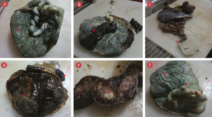
Methods: A prospective cross-sectional hospital-based analytical study was conducted at the departments of Obstetrics and Gynecology, and Histopathology, University of Uyo Teaching Hospital, Nigeria from December 2015 to May 2016. We studied 144 pregnant mothers (48 HIV-positive as the test group vs. 96 HIV-negative as controls). Their placentas (fetal membrane, umbilical cord, and placental disk) were collected post-delivery and evaluated (grossly/microscopically) to determine the range of histomorphometric placental parameters/lesions. Relevant obstetric data were obtained from their case notes.
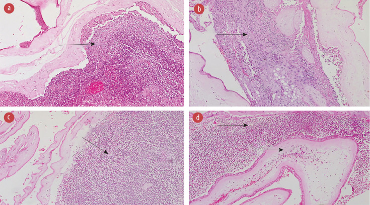
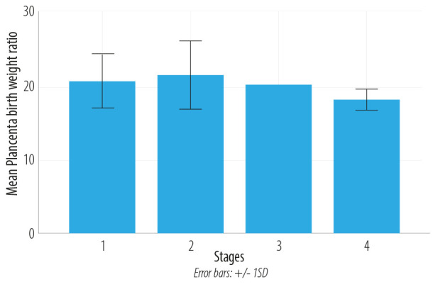
Results: The test group delivered more through cesarean section than the control group (52.1% vs. 31.3%), with mean birth weights of 2.8±0.7 and 3.1±0.6 kg (p = 0.004). The mean placental weights were 57±190.1 and 664.6±167.4 g (p = 0.003), with mean placenta-birth weight ratio of 20.1±4.8 and 20.5±4.57% (p = 0.33). The test groups placental fetal membranes, umbilical cords, and disks mainly displayed acute chorioamnionitis (47.9%), acute umbilical phlebitis (14.6%), and villous vasculopathy (33.3%). The test group had a higher stage/grade of placental inflammation than the control group. In the test group, two stage 4 HIV disease state cases presented with the most severe form of placental inflammatory lesions.
Conclusions: The commonest placental histomorphometric parameters/lesions were acute chorioamnionitis, acute umbilical phlebitis, and acute intervillositis. There was no significant association between HIV/AIDS disease stage with the most severe forms of placental inflammatory lesions.
Keywords: Acquired Immunodeficiency Syndrome; Chorioamnionitis; HIV Infections; Nigeria; Placenta; Pregnancy.
The OMJ is Published Bimonthly and Copyrighted 2022 by the OMSB.
Figures


Similar articles
-
Weights of Fetal Membranes and Umbilical Cords: Correlation With Placental Pathology.Pediatr Dev Pathol. 2020 Aug;23(4):249-252. doi: 10.1177/1093526619889460. Epub 2019 Nov 18. Pediatr Dev Pathol. 2020. PMID: 31739758
-
Pregnancy outcome and placental weights: their relationship to HIV-1 infection.East Afr Med J. 1993 Feb;70(2):85-9. East Afr Med J. 1993. PMID: 8513748
-
[Effect of rivanol-induced abortion on placental histology: pitfalls in pathological interpretations].Zhonghua Bing Li Xue Za Zhi. 2020 Aug 8;49(8):782-787. doi: 10.3760/cma.j.cn112151-20200331-00274. Zhonghua Bing Li Xue Za Zhi. 2020. PMID: 32746543 Chinese.
-
Pattern of placenta histopathology in low birth weight babies seen in a tertiary health centre in South-Western Nigeria.Niger J Med. 2014 Apr-Jun;23(2):149-52. Niger J Med. 2014. PMID: 24956688
-
HIV, Placental Pathology, and Birth Outcomes-a Brief Overview.J Infect Dis. 2021 Dec 8;224(12 Suppl 2):S683-S690. doi: 10.1093/infdis/jiab240. J Infect Dis. 2021. PMID: 33987644 Review.
References
-
- Gupta D, Bhatnagar S, Mishra S. Euthanasia: issues implied within. Internet J Pain Symptom Control Palliat Care 2006;4(2).
-
- Huettner PC. 2008 Short Course #06 - placental development, indications for and methods of examination. United States and Canadian Academy of Pathology. 2008 [cited 2015 Oct 23]. p. 1-80. Available from: http://www.uscapknowledgehub.org/newindex.htm?97th/shorth.htm.
LinkOut - more resources
Full Text Sources