. 2023 Mar;39(3):721-732.
doi: 10.1007/s00381-022-05776-1. Epub 2022 Dec 2.

Image quality and related outcomes of the ShuntScope for catheter implantation in pediatric hydrocephalus-experience of 65 procedures

Affiliations

Image quality and related outcomes of the ShuntScope for catheter implantation in pediatric hydrocephalus-experience of 65 procedures

Anna Prajsnar-Borak et al. Childs Nerv Syst. 2023 Mar.

Abstract

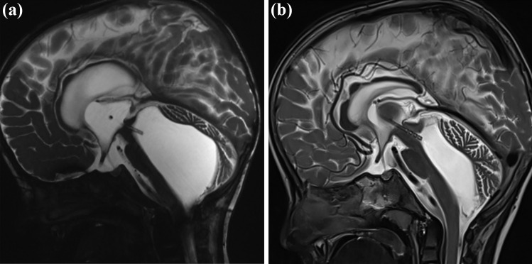
Purpose: Ventricular catheter implantation in pediatric hydrocephalus can become a highly challenging task due to abnormal anatomical configuration or the need for trans-aqueductal stent placement. Transluminal endoscopy with the ShuntScope has been invented to increase the rate of successful catheter placements. This study aims to evaluate ShuntScope's image qualities and related surgical outcomes in the pediatric population.

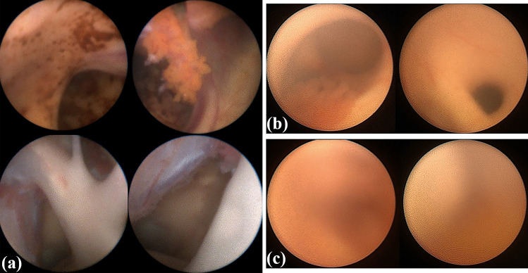
Methods: A retrospective analysis of all pediatric patients undergoing ventricular catheter placement using the ShuntScope from 01/2012 to 01/2022 in the author's department was performed. Demographic, clinical, and radiological data were evaluated. The visualization quality of the intraoperative endoscopy was stratified into the categories of excellent, medium, and poor and compared to the postoperative catheter tip placement. Follow-up evaluation included the surgical revision rate due to proximal catheter occlusion.

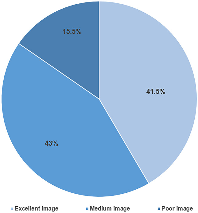
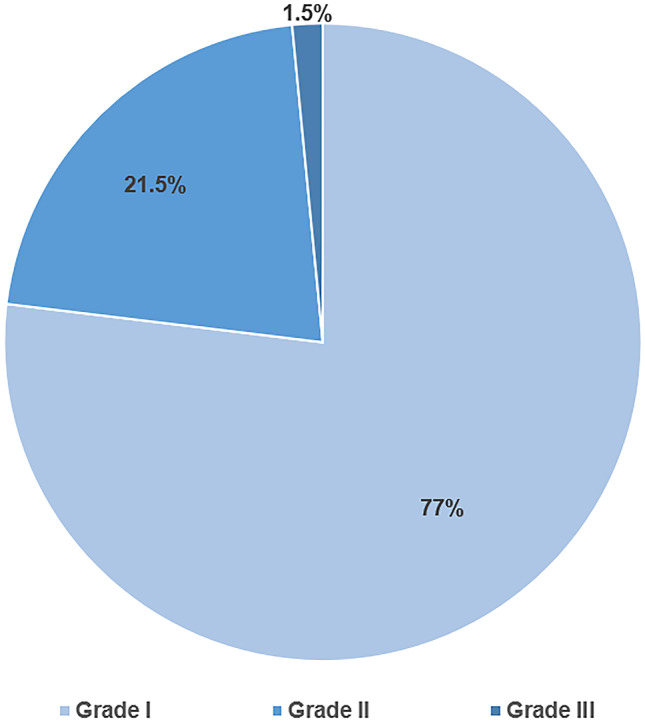
Results: A total of 65 ShuntScope-assisted surgeries have been performed on 51 children. The mean age was 5.1 years. The most common underlying pathology was a tumor- or cyst-related hydrocephalus in 51%. Achieved image quality was excellent in 41.5%, medium in 43%, and poor in 15.5%. Ideal catheter placement was achieved in 77%. There were no intraoperative complications and no technique-related morbidity associated with the ShuntScope. The revision rate due to proximal occlusion was 4.61% during a mean follow-up period of 39.7 years. No statistical correlation between image grade and accuracy of catheter position was observed (p-value was 0.290).

Conclusion: The ShuntScope can be considered a valuable addition to standard surgical tools in treating pediatric hydrocephalus. Even suboptimal visualization contributes to high rates of correct catheter placement and, thereby, to a favorable clinical outcome.

Keywords: Endoscopy; Pediatric hydrocephalus; ShuntScope; Shunting; Ventricular catheter.


Conflict of interest statement

There are no competing financial or non-financial interests. Joachim Oertel acted as a consultant for Karl Storz Company for spine surgery until 2020. No conflicting interests exist for the material and technique mentioned in the publication.

Figures

Fig. 1
Fig. 1
A semi-rigid ShuntScope, Karl Storz GmbH & Co.KG, Tuttlingen, Germany, with a length of 160 mm, an outer diameter of 1 mm, and an image resolution of 10,000 pixels
Fig. 2
Fig. 2
Visualization Grading of intraoperative ShuntScope achieved image quality. a Excellent image: The intraventricular landmarks are absolutely obvious and simply recognizable. The quality of the image is magnificent. Vascular structures such as choroid plexus, thalamostriate vein, septal vein, or basilar artery are definable. b Medium image: However, the suboptimal image quality is satisfactory for intraventricular orientation Foramen of Monro, choroid plexus, aqueduct entrance is adequately defined. c Poor image: an insufficient, blear, fuzzed image with highly restricted recognizability of intraventricular anatomy. The achieved image is not usable for intraoperative orientation during catheter placement
Fig. 3
Fig. 3
Distribution of achieved image quality during ShuntScope-assisted guidance based on visualization grading
Fig. 4
Fig. 4
Accuracy of VC placement in the postoperative image for 65 performed procedures
Fig. 5
Fig. 5
Distribution of image quality vs. Modified Hayhurst score
Fig. 6
Fig. 6
Distribution of image quality vs. Proximal shunt failure rate
Fig. 7
Fig. 7
Preoperative MR images. Axial T2—(a) and sagittal T2-(b) weighted MR images. Chronic hydrocephalus with an aspect of isolating fourth ventricle after shunting
Fig. 8
Fig. 8
Intraoperative ShuntScope-guided images. Sequential intraoperative photographs show the ShuntScope-controlled exploration and trans-aqueductal stent placement. Sufficient image obtained with recognition of the ventral and dorsal aspect of the third ventricle; right foramen of Monro, the floor of the third ventricle with the mamillary body (b, c), infundibular recess (d). Endoscopic view of pathologic cerebral aqueduct; narrow, obliterated aqueductal entrance, posterior commissure (e, f). Exploration of the fourth ventricle (g). The ShuntScope is stepwise withdrawn, transluminal image (h) by correctly positioning the trans-aqueductal stent
Fig. 9
Fig. 9
Postoperative control MR images. MR images were taken four days after surgery (a) and 14 months after surgery (b), confirming the trans-aqueductal position and functionality of the stent. The size of the fourth ventricle regressed revenant resulting in the resolving of previous brain compression. The prepontine cistern was again definable. The cerebellum was unfolded. All figures were created using Adobe Photoshop

Similar articles

Cited by

References

    1. Abdoh MG, Bekaert O, Hodel J, Diarra SM, Le Guerinel C, Nseir R, et al. Accuracy of external ventricular drainage catheter placement. Acta Neurochir (Wien) 2012;154:153–159. doi: 10.1007/s00701-011-1136-9. - DOI - PubMed
    1. Hsia CC, Chen YH, Wu HY, Liu MY. The misplacement of the external ventricular drain by the freehand method in emergent neurosurgery. Acta Neurol Belg. 2011;111:22–28. - PubMed
    1. Lind CR, Correia JA, Law AJ, Kejriwal E. A survey of surgical techniques for catheterizing the cerebral lateral ventricles. J Clin Neurosci. 2008;15:886–890. doi: 10.1016/j.jocn.2007.05.013. - DOI - PubMed
    1. Azeem SS, Origitano TC. Ventricular catheter placement with a frameless neuronavigational system: a 1- year experience. Neurosurgery. 2007;60(4):243–248. doi: 10.1227/01.NEU.0000255387.03088.53. - DOI - PubMed
    1. Clark S, Sangra M, Hayhurst C, et al. The use of noninvasive electromagnetic neuronavigation for slit ventricle syndrome and complex hydrocephalus in a pediatric population. J Neurosurg Pediatr. 2008;2(6):430–434. doi: 10.3171/PED.2008.2.12.430rg/. - DOI - PubMed