Review
. 2022 Dec 2;80(1):14.
doi: 10.1007/s00284-022-03103-0.

Insight into the Pathogenic Mechanism of Mycoplasma pneumoniae

Affiliations
Review

Insight into the Pathogenic Mechanism of Mycoplasma pneumoniae

Jie Hu et al. Curr Microbiol. .

Abstract

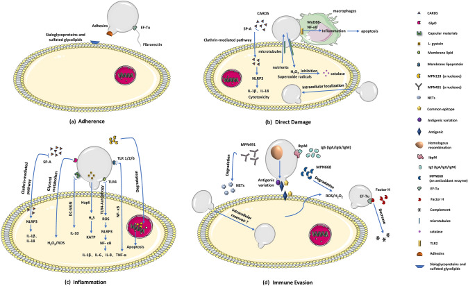
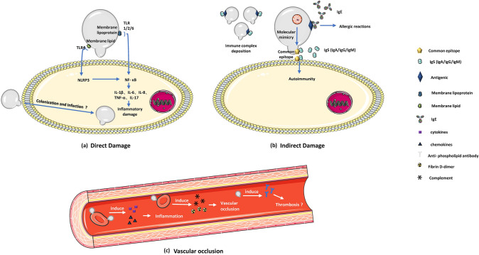
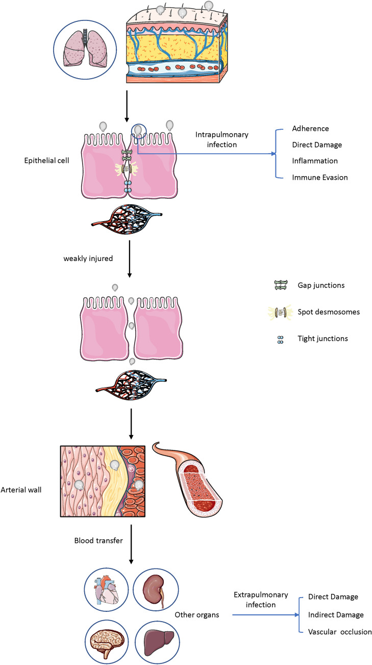
Mycoplasma pneumoniae, an obligate parasitic pathogen without cell wall, can cause severe upper and lower respiratory tract symptoms. It is the pathogen of human bronchitis and walking pneumonia, and named community-acquired pneumonia. In addition to severe respiratory symptoms, there are clinical extrapulmonary manifestations in the skin, brain, kidney, musculoskeletal, digestive system, and even blood system after M. pneumoniae infection. Hereby, we comprehensively summarized and reviewed the intrapulmonary and extrapulmonary pathogenesis of M. pneumoniae infection. The pathogenesis of related respiratory symptoms caused by M. pneumoniae is mainly adhesion damage, direct damage including nutrient predation, invasion and toxin, cytokine induced inflammation damage and immune evasion effect. The pathogenesis of extrapulmonary manifestations includes direct damage mediated by invasion and inflammatory factors, indirect damage caused by host immune response, and vascular occlusion. The intrapulmonary and extrapulmonary pathogenic mechanisms of M. pneumoniae infection are independent and interrelated, and have certain commonalities. In fact, the pathogenic mechanisms of M. pneumoniae are complicated, and the specific content is still not completely clear, further researches are necessary for determining the detailed pathogenesis of M. pneumoniae. This review can provide certain guidance for the effective prevention and treatment of M. pneumoniae infection.


Conflict of interest statement

The authors have no conflict of interest to declare.

Figures

Fig. 1
Fig. 1
Pathogenic mechanisms of M. pneumoniae intrapulmonary infection. a M. pneumoniae adhesion causes cell damage. Adhesins binds to sialoglycoproteins and sulfated glycolipids receptors on host cell surface to obtain nutrition for M. pneumoniae, which induces intracellular metabolism and ultrastructural changes in the infected cells. Additionally, EF-Tu can bind to a range of host molecules (such as fibronectin), increase mycoplasmas attach to tracheal epithelial cells. b M. pneumoniae releases CARD toxin, H2O2 and superoxide radicals into host cell to produce host cytotoxicity. c Inflammation-inducing factors (membrane lipid, lipoprotein, HapE enzyme, nuclease, oxidase GlpO, capsular materials) activate host inflammatory pathways to produce inflammatory damage. d M. pneumoniae produces a nuclease (encoded by MPN491) and an antioxidant enzyme (encoded by MPN668) to degrade NETs and peroxides, respectively. Homologous DNA recombination leads to antigenic variation. Furthermore, IbpM and EF-Tu enable M. pneumoniae evade the host's immune response
Fig. 2
Fig. 2
Component proteins of the internal structure and the surface adhesion complex of attachment organelle. The nap-like surface structure composed of the main adhesins (P1 and P30) and accessory proteins (P40 and P90) surrounding the cell membrane. The internal structure is made up of terminal button (HMW2, HMW3, P65), paired plates (HMW1, HMW2, CpsG, HMW3), and a bowl complex (Lon, P24, TopJ, P200, P41, MPN387, HMW2). HMW1, HMW2, HMW3 refer to three high molecular weight (HMW) proteins
Fig. 3
Fig. 3
Pathogenic mechanisms of M. pneumoniae extrapulmonary infection. a Direct invasion and inflammatory damage induced by cytokines lead to direct damage to host cells. b M. pneumoniae antigens mimic host cell components or cause changes in the structure of host cell membrane antigens to stimulate host autoimmunity. Immune complex deposition is considered responsible for M. pneumoniae extrapulmonary infection. Self-reactive IgEs promote the occurrence of allergic reactions and cause certain damage to tissue cells. c M. pneumoniae locally induces cytokines and chemokines to affect the vascular wall, causing vasculitis and thrombotic vascular occlusion through medical mediators (such as complement and fibrin D-dimer)
Fig. 4
Fig. 4
M. pneumoniae can be passively transferred into the circulation through weak gaps between injured epithelial cells in some patients with an immature or damaged immune barrier on the respiratory tract surface and transfer to other organs by adhering to erythrocytes to cause extrapulmonary infection

References

    1. Kumar S. Mycoplasma pneumoniae: a significant but underrated pathogen in paediatric community-acquired lower respiratory tract infections. Indian J Med Res. 2018;147(1):23–31. doi: 10.4103/ijmr.IJMR_1582_16. - DOI - PMC - PubMed
    1. Bajantri B, Venkatram S, Diaz-Fuentes G. Mycoplasma pneumoniae: a Potentially Severe Infection. J Clin Med Res. 2018;10(7):535–544. doi: 10.14740/jocmr3421w. - DOI - PMC - PubMed
    1. Kutty PK, Jain S, Taylor TH, Bramley AM, Diaz MH, Ampofo K, Arnold SR, Williams DJ, Edwards KM, McCullers JA, Pavia AT, Winchell JM, Schrag SJ, Hicks LA. Mycoplasma pneumoniae among children hospitalized with community-acquired pneumonia. Clin Infect Dis. 2019;68(1):5–12. - PMC - PubMed
    1. Waites KB, Xiao L, Liu Y, Balish MF, Atkinson TP. Mycoplasma pneumoniae from the respiratory tract and beyond. Clin Microbiol Rev. 2017;30(3):747–809. doi: 10.1128/CMR.00114-16. - DOI - PMC - PubMed
    1. Jiang Z, Li S, Zhu C, Zhou R, Leung P. Mycoplasma pneumoniae Infections: pathogenesis and vaccine development. Pathogens. 2021;10(2):119. doi: 10.3390/pathogens10020119. - DOI - PMC - PubMed

LinkOut - more resources