Review
. 2023 Jan;23(1):53-65.
doi: 10.1007/s11882-022-01055-w. Epub 2022 Dec 2.

Human Monoclonal IgE Antibodies-a Major Milestone in Allergy

Affiliations
Review

Human Monoclonal IgE Antibodies-a Major Milestone in Allergy

Scott A Smith et al. Curr Allergy Asthma Rep. 2023 Jan.

Abstract

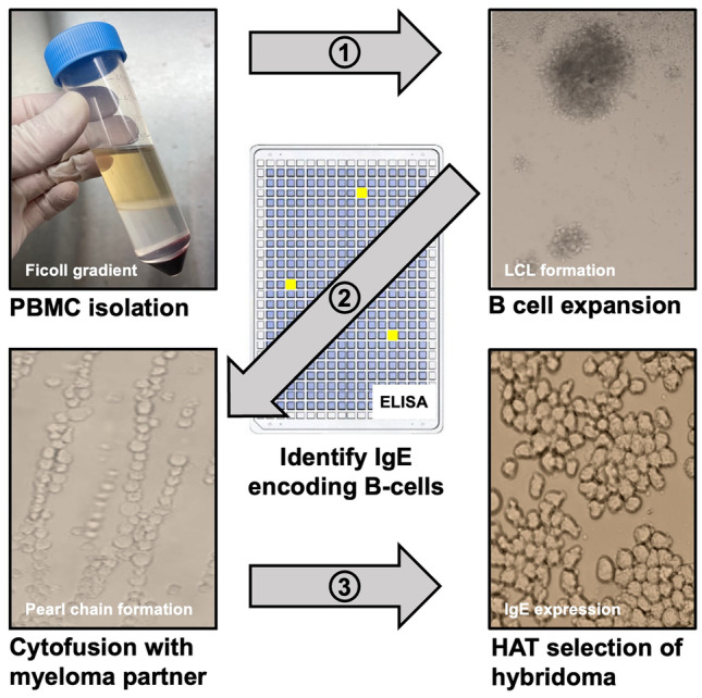
Purpose of review: Bound to its high affinity receptor on mast cells and basophils, the IgE antibody molecule plays an integral role in the allergic reaction. Through interactions with the allergen, it provides the sensitivity and specificity parameters for cell activation and mediator release that produce allergic symptoms. Advancements in human hybridoma technologies allow for the generation and molecular definition of naturally occurring allergen-specific human IgE monoclonal antibodies.

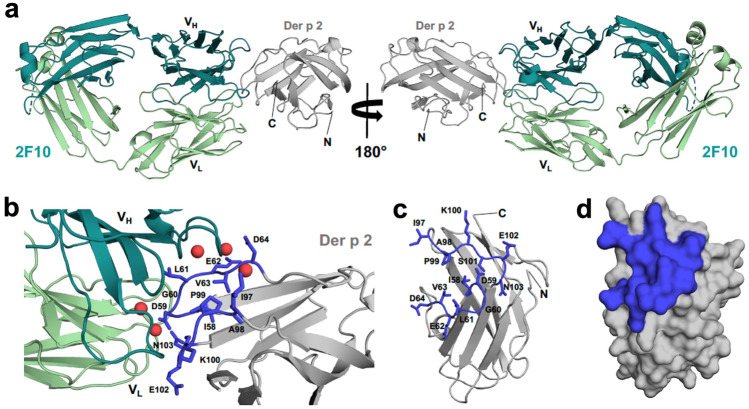
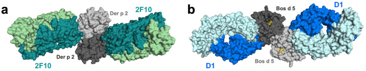
Recent findings: A high-resolution structure of dust mite allergen Der p 2 in complex with Fab of the human IgE mAb 2F10 was recently determined using X-ray crystallography. The structure reveals the fine molecular details of IgE 2F10 binding its 750 Å2 conformational epitope on Der p 2. This review provides an overview of this major milestone in allergy, the first atomic resolution structure of an authentic human IgE epitope. The molecular insights that IgE epitopes provide will allow for structure-based design approaches to the development of novel diagnostics, antibody therapeutics, and immunotherapies.

Keywords: Allergens; Allergy; Conformational epitope; Diagnosis; Human IgE monoclonal antibody; X-ray crystallography.


Conflict of interest statement

Scott A Smith reports grants from NIAID R21AI123307, and NIAID R01AI155668, during the conduct of the study; In addition, Dr. Smith has a patent 10,908,168 with royalties paid, a pending patents 63/159,764 and 63/125,099 with royalties paid. Maksymilian Chruszcz declares no conflict of interest. Martin D. Chapman reports grants from NIH—NIAID, during the conduct of the study; and License agreement with Vanderbilt University Medical Center for commercialization of human IgE monoclonal antibodies for research and diagnostic purposes. The hIgE mAb covered by this agreement are available from InBio (www.inbio.com). Anna Pomés reports a grant from NIH – NIAID 5R01AI077653-12 and is employed by InBio.

Figures

Fig. 1
Fig. 1
IgE secreting hybridoma generation overview. PBMCs are isolated from selected subject’s blood using a Ficoll density gradient method. In step 1, cells are grown in 96-well tissue culture plates for 7 days to expand all B cells, leading to the development of lymphoblastoid cell lines (LCLs). Cultures containing IgE are identified by ELISA and cells within wells containing IgE are fused with a myeloma partner by electrical cytofusion reactions in step 2. Human hybridomas are selected for in HAT medium in step 3 and biologically cloned using flow cytometric single cell sorting. Finally, IgE secreting hybridomas are grown in serum free culture medium and IgE monoclonal antibody purified by omalizumab chromatography
Fig. 2
Fig. 2
Complex between Der p 2.0103 and IgE 2F10 Fab. a Der p 2 and 2F10 Fab are shown in ribbon representation. Heavy chain of the antibody is shown in teal, while light chain is shown in pale green. b Interface between 2F10 and Der p 2. Residues forming the epitope are marked in blue and their side chains are shown in stick representation. 2F10 binding epitope (in blue) mapped on the structure of Der p 2 shown in ribbon c, and space filling d, representations. Water molecules are represented by red spheres. N- and C-terminal ends of Der p 2 are labeled in a and c
Fig. 3
Fig. 3
a 2F10 Fab binding to a putative Der p 2.0103 dimer. b D1 IgE Fab in complex with Bos d 5 dimer. Non-physiological Bos d 5 ligand (dodecyl-β-D-maltoside) is shown in stick representation

References

    1. Heeringa JJ, Rijvers L, Arends NJ, Driessen GJ, Pasmans SG, van Dongen JJM, de Jongste JC, van Zelm MC. IgE-expressing memory B cells and plasmablasts are increased in blood of children with asthma, food allergy, and atopic dermatitis. Allergy. 2018;73(6):1331–1336. doi: 10.1111/all.13421. - DOI - PubMed
    1. Achatz G, Nitschke L, Lamers MC. Effect of transmembrane and cytoplasmic domains of IgE on the IgE response. Science. 1997;276(5311):409–411. doi: 10.1126/science.276.5311.409. - DOI - PubMed
    1. Karnowski A, Achatz-Straussberger G, Klockenbusch C, Achatz G, Lamers MC. Inefficient processing of mRNA for the membrane form of IgE is a genetic mechanism to limit recruitment of IgE-secreting cells. Eur J Immunol. 2006;36(7):1917–1925. doi: 10.1002/eji.200535495. - DOI - PubMed
    1. Avery DT, Ma CS, Bryant VL, Santner-Nanan B, Nanan R, Wong M, Fulcher DA, Cook MC, Tangye SG. STAT3 is required for IL-21-induced secretion of IgE from human naive B cells. Blood. 2008;112(5):1784–1793. doi: 10.1182/blood-2008-02-142745. - DOI - PubMed
    1. Renz H, Mazer BD, Gelfand EW. Differential inhibition of T and B cell function in IL-4-dependent IgE production by cyclosporin A and methylprednisolone. J Immunol. 1990;145(11):3641–3646. doi: 10.4049/jimmunol.145.11.3641. - DOI - PubMed

Publication types