. 2022 Dec 6;119(49):e2211549119.
doi: 10.1073/pnas.2211549119. Epub 2022 Dec 2.

The molecular basis for pore pattern morphogenesis in diatom silica

Affiliations

The molecular basis for pore pattern morphogenesis in diatom silica

Christoph Heintze et al. Proc Natl Acad Sci U S A. .

Abstract

Biomineral-forming organisms produce inorganic materials with complex, genetically encoded morphologies that are unmatched by current synthetic chemistry. It is poorly understood which genes are involved in biomineral morphogenesis and how the encoded proteins guide this process. We addressed these questions using diatoms, which are paradigms for the self-assembly of hierarchically meso- and macroporous silica under mild reaction conditions. Proteomics analysis of the intracellular organelle for silica biosynthesis led to the identification of new biomineralization proteins. Three of these, coined dAnk1-3, contain a common protein-protein interaction domain (ankyrin repeats), indicating a role in coordinating assembly of the silica biomineralization machinery. Knocking out individual dank genes led to aberrations in silica biogenesis that are consistent with liquid-liquid phase separation as underlying mechanism for pore pattern morphogenesis. Our work provides an unprecedented path for the synthesis of tailored mesoporous silica materials using synthetic biology.

Keywords: ankyrin-repeat domain; biomineralization; mesoporous silica; phase separation; silica deposition vesicle.


Conflict of interest statement

The authors declare no competing interest.

Figures

Fig. 1.
Fig. 1.
Identification of valve SDV proteins. (A) Scheme of the cluster analysis workflow that established in a published transcriptomics dataset (22) 11 different gene expression profiles. The expression patterns in the sin1 cluster is shown: red line—sin1 profile, grey lines—profiles of the other 401 genes, blue area—cluster average within two standard deviations. (B) The Euler diagram shows the overlap between proteins identified in the valve SDV fraction (Dataset S1) and genes of the sin1 cluster (Dataset S2). (C) Heat map of mRNA expression of sin1 cluster genes that encode proteins identified in the valve SDV fraction. Gene IDs of sin1 (24710), Tp25735, and dank1-3 (Tp23225, Tp5147, Tp21058) are highlighted in red. The y-axis indicates time before (negative) and after addition of silicic acid, which triggers synchronous cell cycle progression. Valve SDV development occurs within hours 2.5–4.5 (23). (D) Scheme of a dividing diatom cell in cross section: black—cell wall, blue—plasma membrane, grey—protoplasm, green—valve SDV (other compartments not shown). (E-I) Confocal fluorescence microscopy images of individual dividing cells expressing the indicated proteins as GFP fusions (z-projection of optical slices through mid-cell region): green—GFP, red—chloroplast autofluorescence. (Scale bars: 2 µm.)
Fig. 2.
Fig. 2.
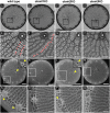
Valve morphologies in T. pseudonana wild type and dank knockout mutants. (A–D) TEM and (I–M) SEM images of entire valves. (EH, NQ) Details corresponding to the boxed areas in A–D and I–M, respectively. Red arrowheads point to cribrum pores and yellow arrowheads to the fultoportulae. The inset in L is a zoom-in of the region around a central fultoportula (yellow arrowhead). (Scale bars: 1 µm in A–D and I–M, 300 nm in E–H and N–Q, 200 nm in L, Inset.)
Fig. 3.
Fig. 3.
Analysis of the pore patterns in valve silica from wild type and dank knockout mutants. (A) Recognition of cribrum pores from a TEM image of an individual valve. The girdle band margin is cropped from the analysis and only pores in the area underneath the blue line were analyzed. The recognition algorithm enables measurement of the PD and the PDF. (B) Statistical analysis of the pore densities from wild type, dank knockout mutants, and dank rescue strains: central line—median value, box edges—25th and 75th percentiles, whiskers—minimum and maximum values, white circles—sample outliers (independent two-sample t test ***P < 0.001). Differences that were not statistically significant are denoted “ns.” The number of analyzed valves was 50 for the wild type and between 13 and 19 for each genetically modified strain. (C) Averaged PDF functions for the dank knockout strains within a domain of 300 nm × 300 nm. The central peak marks the position of the reference pore, while the two satellite peaks characterize the relative position of the closest neighbors. The color scale indicates normalized probabilities of finding a neighboring pore.
Fig. 4.
Fig. 4.
Morphogenesis of porous silica patterns in valves from T. pseudonana wild type, dank1KO, and dank3KO strains. TEM images show representative sub areas of nascent valve silica from (A–D) wild type, (E–H) dank1KO, and (I–M) dank3KO in different developmental stages proceeding from Left to Right. (Scale bars: 200 nm.) Based on these TEM data and assuming liquid-liquid phase separation to occur in the SDV (11, 12), we propose models for cribrum pore formation in valves of (A’–D’) wild type, (E’–H’) dank1KO, and (I’–M’) dank3KO strains. Black—silica ribs, orange—organic nanodroplets, grey—cribrum plate silica. Red arrowheads point to less electron dense spots assumed to originate from unstable cribrum pores that were filled in with silica. Yellow arrowheads indicate forming cribrum pores.
Fig. 5.
Fig. 5.
Putative model for dAnk-controlled liquid-liquid phase separation (LLPS) in the SDV (components are not drawn to scale). (A) Current model of an SDV. The lumen is assumed to contain an organic matrix composed of strongly charged, soluble biomacromolecules (e.g., zwitterionic silaffins, polyanionic silacidins, polycationic LCPA) that are prone to undergo phase separation (11, 30, 43). The SDV membrane contains transmembrane proteins Sin1, SAP1, SAP3 that might interact with LCPA and silaffins in the SDV lumen (14, 19). A V-type H+-ATPase establishes and acidic pH in the SDV lumen (24). The chemical structure of the silica precursor molecules and the Si-transporter that catalyzes their import into the SDV are yet unknown. dAnks associate with the cytoplasmic surface of the SDV by binding to yet unknown transmembrane proteins (coined dAnk receptors) resulting in a dAnk complex. (B) The binding of dAnk1 (light blue symbol) to its receptor (light green symbol) triggers liquid–liquid phase separation in the SDV lumen. Stability of the nanodroplets is controlled by an antagonism between dAnk2 (blue symbol) and dAnk3 (dark blue symbol) complexes with their specific receptors. While dAnk2 complexes promote the disassembly of nanodroplets, dAnk3 complexes have a stabilizing effect.

References

    1. Faivre D., Schüler D., Magnetotactic bacteria and magnetosomes. Chem. Rev. 108, 4875–4898 (2008). - PubMed
    1. Sun J., Bhushan B., Hierarchical structure and mechanical properties of nacre: a review. RSC Adv. 2, 7617–7632.
    1. Goessling J. W., Su Y., Kühl M., Ellegaard M., "Frustule Photonics and light harvesting strategies in diatoms" in Diatom Morphogenesis, Annenkov V., Seckbach J., Gordon R., Eds. (Scrivener Publishing, Beverly, 2022), pp. 269–300.
    1. Round F. E., Crawford R. M., Mann D. G., The Diatoms: Biology and Morphology of the Genera (Cambridge University Press, Cambridge, UK, 1990).
    1. Volcani B. E., "Cell wall formation in diatoms: Morphogenesis and biochemistry" in Silicon and Siliceous Structures in Biological Systems, Simpson T. L., Volcani B. E., Eds. (Springer, New York, NY, 1981), pp. 157–200.

Publication types

Substances

LinkOut - more resources