. 2022 Dec 1;29(12):1685-1702.e22.
doi: 10.1016/j.stem.2022.11.004.

A reference human induced pluripotent stem cell line for large-scale collaborative studies

Caroline B Pantazis  1 Andrian Yang  2 Erika Lara  1 Justin A McDonough  3 Cornelis Blauwendraat  4 Lirong Peng  5 Hideyuki Oguro  6 Jitendra Kanaujiya  6 Jizhong Zou  7 David Sebesta  8 Gretchen Pratt  8 Erin Cross  8 Jeffrey Blockwick  8 Philip Buxton  8 Lauren Kinner-Bibeau  8 Constance Medura  8 Christopher Tompkins  8 Stephen Hughes  8 Marianita Santiana  1 Faraz Faghri  9 Mike A Nalls  9 Daniel Vitale  9 Shannon Ballard  9 Yue A Qi  1 Daniel M Ramos  1 Kailyn M Anderson  1 Julia Stadler  1 Priyanka Narayan  10 Jason Papademetriou  1 Luke Reilly  1 Matthew P Nelson  1 Sanya Aggarwal  11 Leah U Rosen  12 Peter Kirwan  11 Venkat Pisupati  13 Steven L Coon  14 Sonja W Scholz  15 Theresa Priebe  16 Miriam Öttl  16 Jian Dong  16 Marieke Meijer  16 Lara J M Janssen  16 Vanessa S Lourenco  16 Rik van der Kant  17 Dennis Crusius  18 Dominik Paquet  19 Ana-Caroline Raulin  20 Guojun Bu  20 Aaron Held  21 Brian J Wainger  22 Rebecca M C Gabriele  23 Jackie M Casey  23 Selina Wray  23 Dad Abu-Bonsrah  24 Clare L Parish  25 Melinda S Beccari  26 Don W Cleveland  26 Emmy Li  27 Indigo V L Rose  27 Martin Kampmann  27 Carles Calatayud Aristoy  28 Patrik Verstreken  28 Laurin Heinrich  29 Max Y Chen  29 Birgitt Schüle  29 Dan Dou  30 Erika L F Holzbaur  30 Maria Clara Zanellati  31 Richa Basundra  31 Mohanish Deshmukh  31 Sarah Cohen  31 Richa Khanna  32 Malavika Raman  32 Zachary S Nevin  33 Madeline Matia  33 Jonas Van Lent  34 Vincent Timmerman  34 Bruce R Conklin  33 Katherine Johnson Chase  20 Ke Zhang  20 Salome Funes  35 Daryl A Bosco  35 Lena Erlebach  36 Marc Welzer  36 Deborah Kronenberg-Versteeg  36 Guochang Lyu  37 Ernest Arenas  37 Elena Coccia  38 Lily Sarrafha  38 Tim Ahfeldt  38 John C Marioni  39 William C Skarnes  40 Mark R Cookson  41 Michael E Ward  42 Florian T Merkle  43
Affiliations

A reference human induced pluripotent stem cell line for large-scale collaborative studies

Caroline B Pantazis et al. Cell Stem Cell. .

Abstract

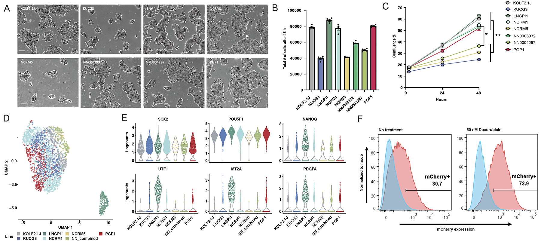
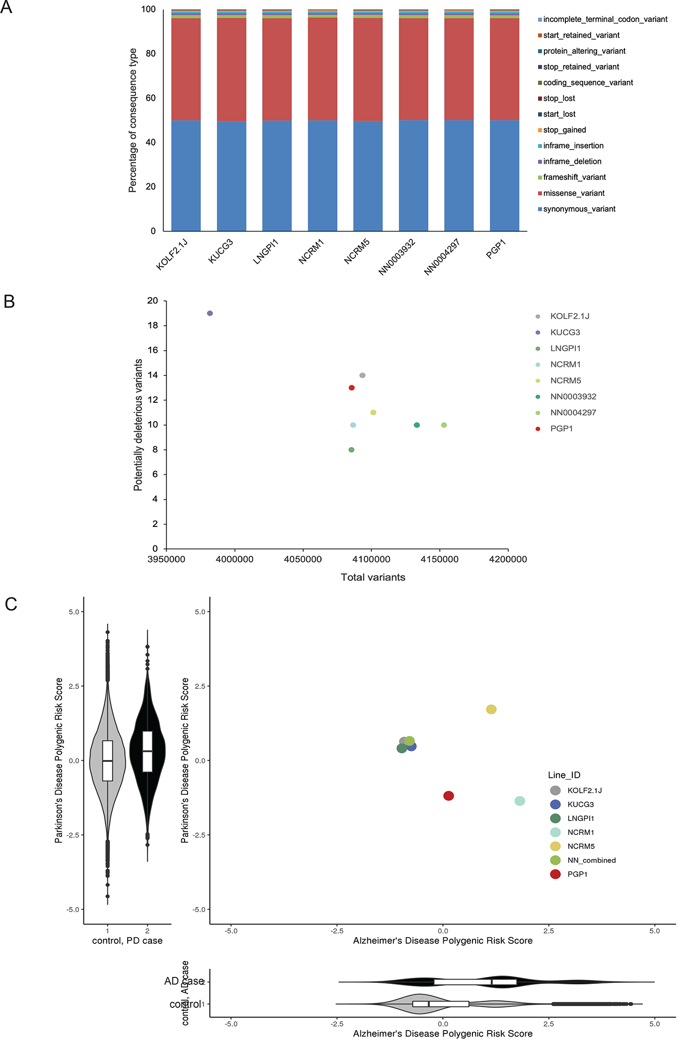
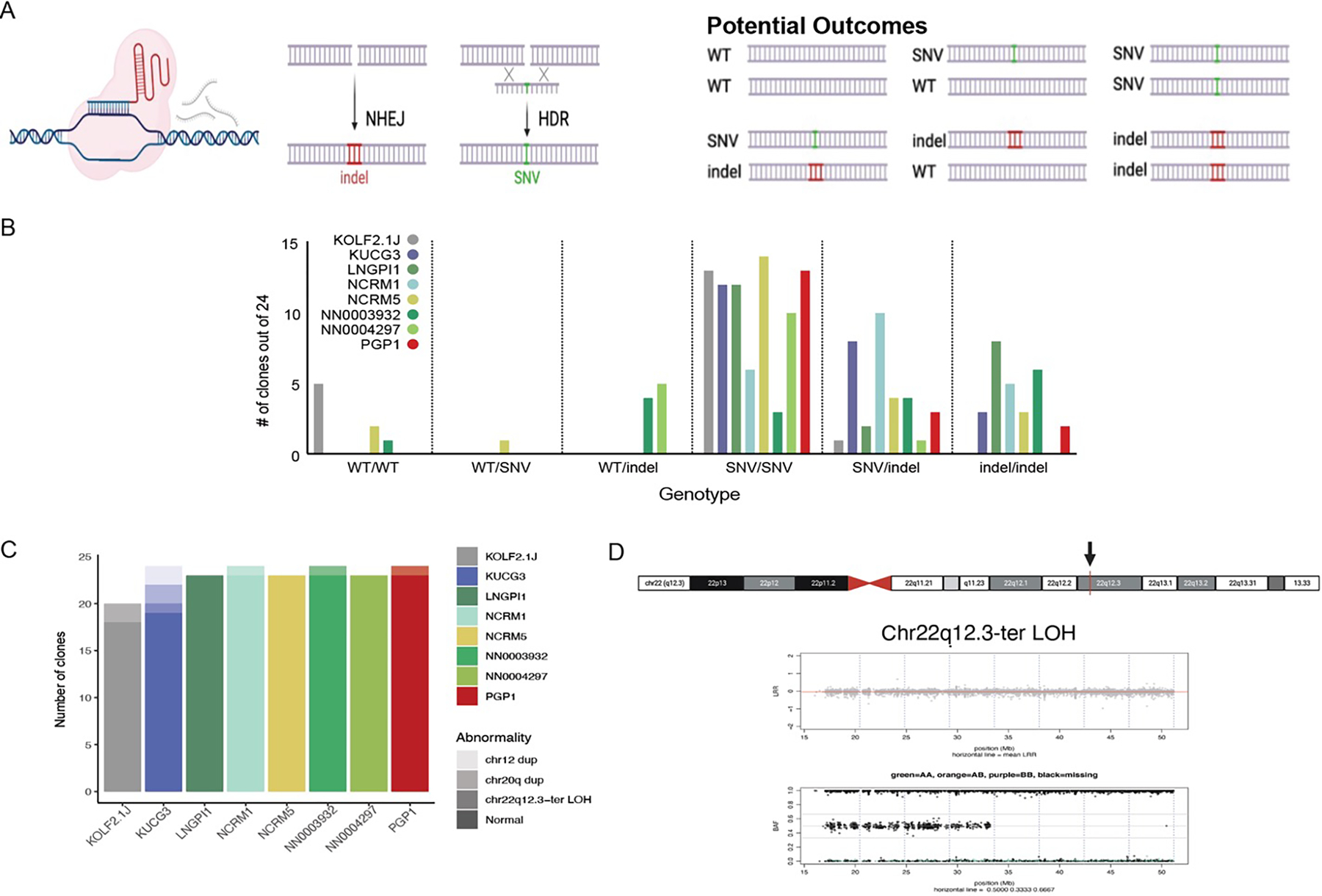
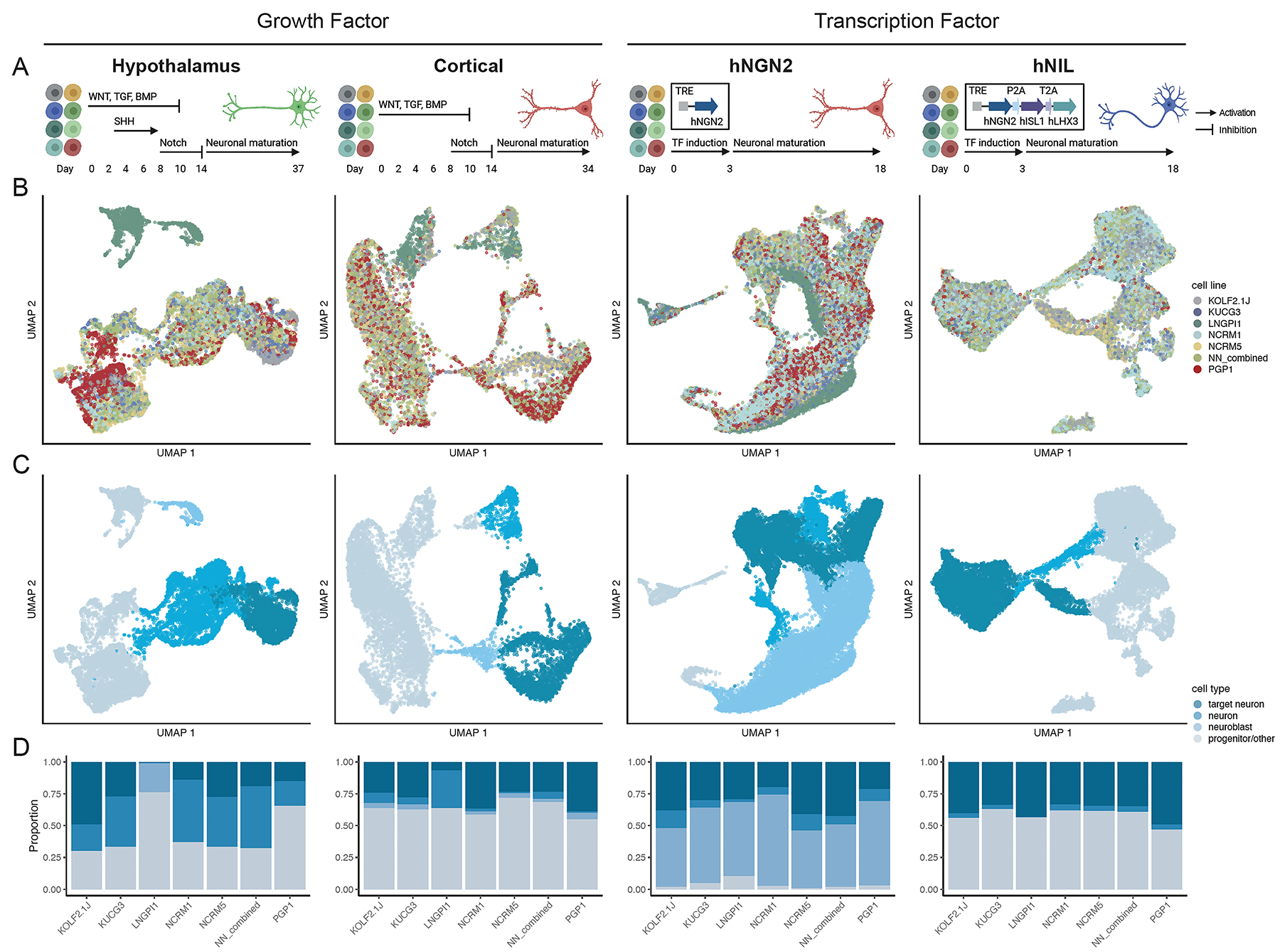
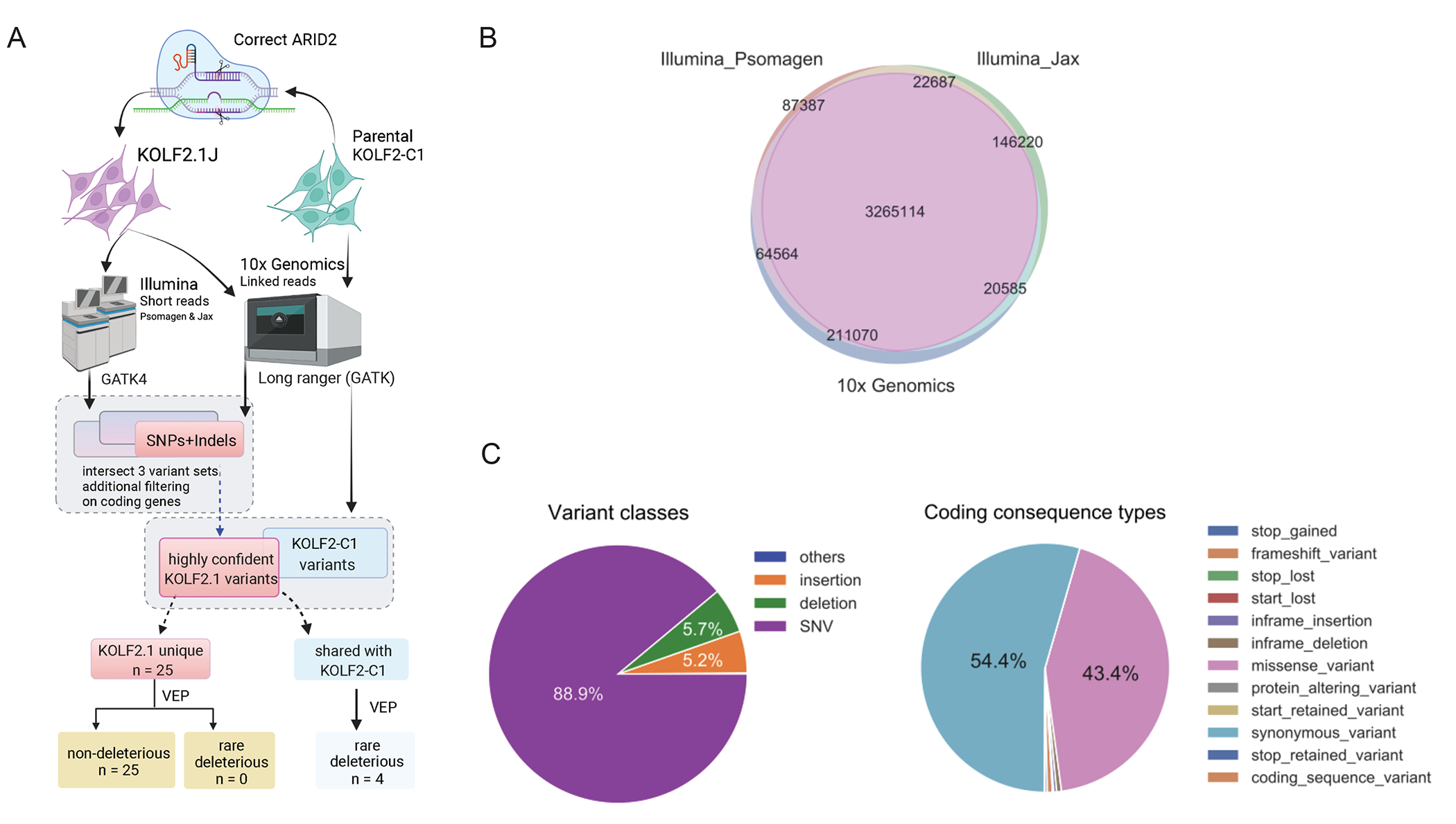
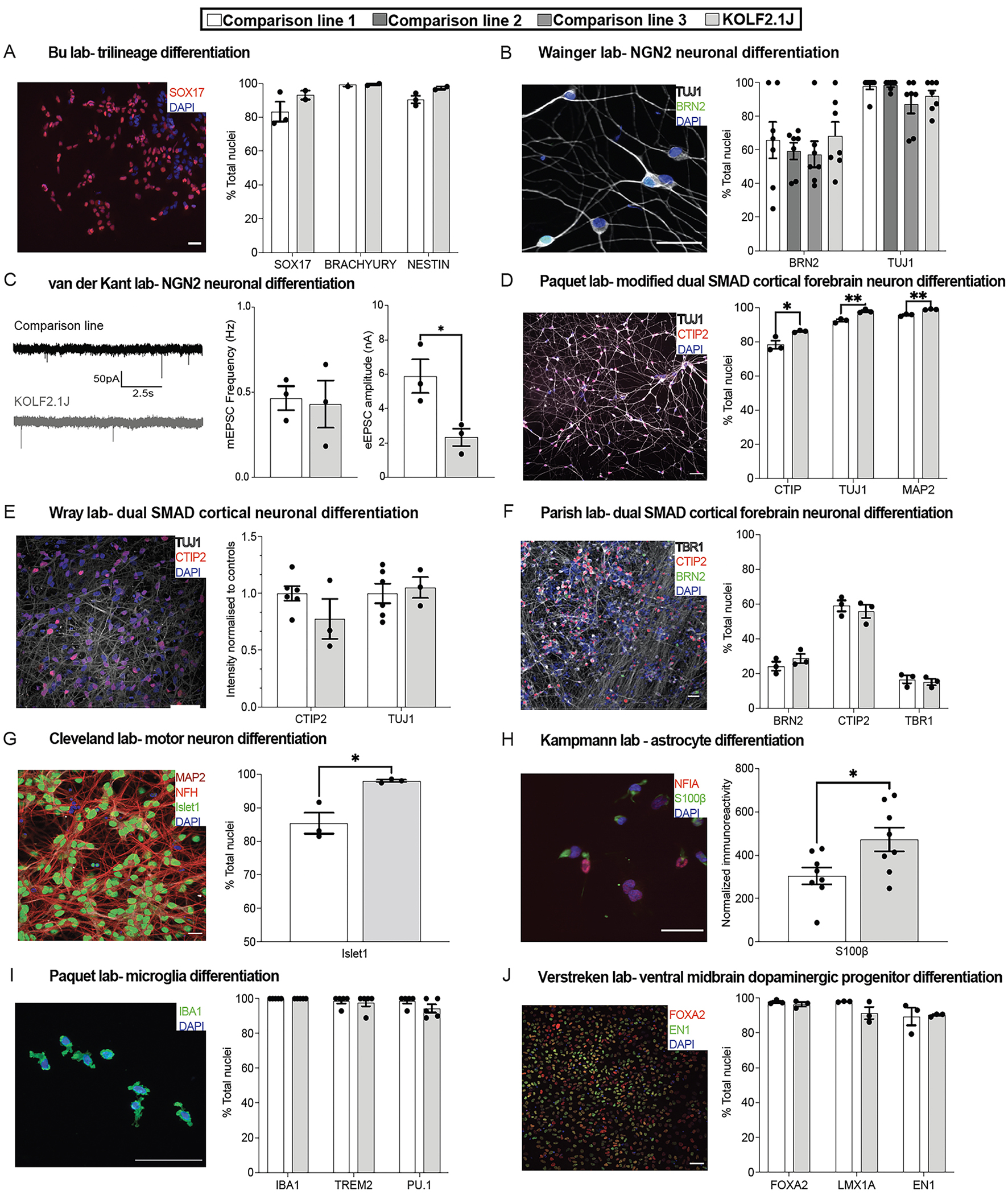
Human induced pluripotent stem cell (iPSC) lines are a powerful tool for studying development and disease, but the considerable phenotypic variation between lines makes it challenging to replicate key findings and integrate data across research groups. To address this issue, we sub-cloned candidate human iPSC lines and deeply characterized their genetic properties using whole genome sequencing, their genomic stability upon CRISPR-Cas9-based gene editing, and their phenotypic properties including differentiation to commonly used cell types. These studies identified KOLF2.1J as an all-around well-performing iPSC line. We then shared KOLF2.1J with groups around the world who tested its performance in head-to-head comparisons with their own preferred iPSC lines across a diverse range of differentiation protocols and functional assays. On the strength of these findings, we have made KOLF2.1J and its gene-edited derivative clones readily accessible to promote the standardization required for large-scale collaborative science in the stem cell field.

Keywords: CRISPR; differentiation; iPSC; karyotype; p53; pluripotent; reference; single-cell; stem cell; whole-genome.


Conflict of interest statement

Declaration of interests S.W.S. is on the scientific advisory council of the Lewy Body Dementia Association and the MSA Coalition. S.W.S. is an editorial board member for the Journal of Parkinson Disease and JAMA Neurology. S.W.S. received research support from Cerevel Therapeutics. M.K. serves on the scientific advisory boards of Engine Biosciences, Casma Therapeutics, Cajal Neuroscience, and Alector and is a consultant to Modulo Bio and Recursion Therapeutics. Participation by researchers from Data Tecnica International, LLC in this project was part of a competitive contract awarded to Data Tecnica International, LLC by the National Institutes of Health to support open science research. M.A.N. also currently serves on the scientific advisory board for Clover Therapeutics and is an advisor to Neuron23 Inc. E.A. is founder, shareholder, and scientific advisor of Cholestenix, Ltd.

Figures

Figure 1:
Figure 1:. Growth, gene expression, and P53 pathway integrity of candidate cell sub-lines.
(A) Representative phase contrast photomicrographs of colony morphology of the eight iPSC sub-lines on day 3 after replating. Scale bars indicate 100 μm. (B) Mean and SEM (n=4) of the total number of cells 48h after plating 30,000 cells/well. (C) Mean and SEM (n=6) of percent confluence at 0, 24 and 48 h after plating 30,000 cells/well. (D) UMAP projection of 2,270 iPSC cells color-coded by cell sub-line. There are two distinct groupings of cells, with the large group being composed of the 6 out of 7 sub-line and the small outlier group being composed of the LNGPI1 sub-line. (E) Beeswarm plots showing expression of selected genes associated with undifferentiated stem cells (top row) or poor neuronal differentiation potential (bottom row). (F) A mCherry PiggyBac reporter assay confirms the baseline activity (left plot) of the p53 pathway in KOLF2.1J (red) relative to TP53 knockout cells (blue), which is further inducible in response to the DNA damaging agent doxorubicin (right plot).
Figure 2.
Figure 2.. Genetic analyses of eight candidate iPSC sub-lines.
(A) The percentage of genetic variant types present in the 8 candidate iPSC sub-lines, grouped by their bioinformatically predicted consequences on coding sequences. (B) The number of rare (gnomAD AF<0.001) predicted deleterious (CADD phred >30) variants identified in the 8 iPSC sub-lines versus the total number of identified variants. (C) Polygenic risk scores (PRS) for Alzheimer’s disease (AD) and Parkinson’s disease (PD) are shown alongside the population-centered Z score distribution for PD PRS (y-axis) in 2995 PD cases (black) and 96,215 controls (grey) from the UK Biobank, and a similar PRS distribution for AD polygenic risk score (x-axis) in 2337 AD cases (grey) and the same controls.
Figure 3.
Figure 3.. Comparative gene editing efficiency.
(A) Schematic of the gene editing experiment, showing how a CRISPR/Cas9-induced double strand break can lead to the formation of insertion/deletion mutations (indels) via the non-homologous end joining (NHEJ) pathway or be repaired with a single-stranded oligodeoxynucleotide via the homology-directed repair (HDR) pathway to introduce a single nucleotide variant (SNV) of interest and resulting in 6 potential alleles. Figure created with BioRender.com (B) The number of clones out of 24 expressing each possible genotype at the targeted SNV for each analyzed sub-line. (C) Number of genomic abnormalities observed among 20–24 analyzed clones across the eight analyzed sub-lines. (D) Ideogram of chromosome 22 with the TIMP3 gene in 22q12.3 indicated by a red bar and black arrow, using the UCSC Genome Browser. NeuroArray genotyping revealed Chr22 CN-LOH from chr22q12.3-ter in clones derived from the KOLF2.1J sub-line. Upper plots show Log R ratio (LRR) where the mean LRR (red line) is 0 for the normal clone and >1 for the abnormal clones, and middle panels show the B allele frequency (BAF) for bi-allelic probes along the arrays with evidence of duplicated alleles across the chromosome.
Figure 4.
Figure 4.. Differentiation potential of candidate cell sub-lines.
(A) Schematic of experimental design for four differentiation protocols evaluated in this study: hypothalamus and cortical differentiation (growth factor-based protocols), and hNGN2 and hNIL differentiation (transcription factor-based protocols). See STAR methods for detailed descriptions of differentiation protocols. Figure created with BioRender.com (B and C) UMAP plot for each differentiation colored by cell sub-line (B) and cell type (C). Cell type classification is derived from grouping each cluster into target neuron, neuron, neuroblast and progenitor/other categories based on the cluster annotation (Figure S4). (D) Bar plot showing proportion of cells assigned to each cell type per cell sub-line.
Figure 5.
Figure 5.. Whole genome sequencing of KOLF2.1J and the KOLF2-C1 parental line.
(A) Flowchart of the discovery, filtering, annotations, and comparisons of SNP/indel variants in the ARID2-corrected KOLF2.1J and its parental line KOLF2-C1. Schematic created with BioRender.com (B) The consensus variant calls for KOLF2.1J from two Illumina short sequencing services (Psomagen and Jax) and one linked-read sequencing platform (10x Genomics). (C) The genetic compositions of the variant classes (left) and their effect on coding genes (right) for the 3.28 million high-confidence SNPs and indels in KOLF2.1J.
Figure 6.
Figure 6.. Performance of KOLF2.1J and established hPSC lines in head-to-head comparisons
In all figure panels, the performance of KOLF2.1J is shown relative to a comparison line, with the providing laboratory and differentiation method indicated. Photomicrographs of immunostaining (or recording traces) are shown to the left of each panel, where scale bars represent 50 μm and nuclear markers are in blue. Quantification of phenotypic outcomes are given on the right, where N=3 differentiations were analyzed for each cell line and unpaired t-tests were used to calculate significance unless otherwise stated. *p<0.05, **p<0.01. (A) Multi-lineage differentiation to endoderm (SOX17), mesoderm (brachyury), or ectoderm (nestin) reveals no significant differences between KOLF2.1J and APOE3 ALSTEM. All data not shown. (B) NGN2-induced neuronal differentiation quantified by the expression of the neuronal marker TUJ1 (grey) and the cortical layer 2/3 marker BRN2 (green) show similar expression across KOLF2.1J and comparison lines ND50003, ND50004, and 11a. n=7 wells/line, N=1 differentiation. Kruskal-Wallis test for each marker. (C) Patch clamp analysis of NGN2-induced neurons derived from BioniC13 and KOLF2.1J lines show spontaneous miniature excitatory postsynaptic currents (mEPSCs, left) in similar frequencies between the lines (center), though KOLF2.1J-derived cells appeared to have a lower excitatory EPSC amplitude (right). (D) A greater proportion of KOLF2.1J-derived cortical neurons generated by directed differentiation expressed the deep-layer cortical marker CTIP2 and TUJ1 than the Thermo Fisher A18944 comparison line (D). (E) In another group, directed cortical neuron differentiation yielded similar expression of Tuj1 (grey) CTIP2 (red) as the comparison lines RBi001 and Ctrl1. Comparison lines averaged together, N=3–6 differentiations/line. (F) KOLF2.1J and H9 neurons had similar expression of BRN2 (green), CTIP2 (red), and TBR1 (grey) following directed differentiation. (G) Greater efficiency of Islet1 expression was observed following differentiation of KOLF2.1J into motor neurons, compared to the CVB cell line. (H) KOLF2.1J and WTC11 cell lines were differentiated into astrocytes and immunostained for S100β (green) and NFIA (red, left), revealing a higher intensity of S100β immunoreactivity in KOLF2.1J. N=8 differentiations. (I) Microglial differentiation of KOLF2.1J and A18944 revealed no significant differences in the expression of the IBA1, TREM2, or PU.1 marker genes. Mann-Whitney test for each marker. (J) Differentiation of KOLF2.1J and SFC065 towards ventral midbrain dopaminergic neurons showed similar percentages of cells expressing the marker genes EN1 (green) and FOXA2 (red). Mann-Whitney test for EN1 expression.

Similar articles

Cited by

References

    1. Kwart D, Gregg A, Scheckel C, Murphy EA, Paquet D, Duffield M, Fak J, Olsen O, Darnell RB, and Tessier-Lavigne M (2019). A Large Panel of Isogenic APP and PSEN1 Mutant Human iPSC Neurons Reveals Shared Endosomal Abnormalities Mediated by APP beta-CTFs, Not Abeta. Neuron 104, 256–270 e255. 10.1016/j.neuron.2019.07.010. - DOI - PubMed
    1. Konttinen H, Cabral-da-Silva MEC, Ohtonen S, Wojciechowski S, Shakirzyanova A, Caligola S, Giugno R, Ishchenko Y, Hernandez D, Fazaludeen MF, et al. (2019). PSEN1DeltaE9, APPswe, and APOE4 Confer Disparate Phenotypes in Human iPSC-Derived Microglia. Stem Cell Reports 13, 669–683. 10.1016/j.stemcr.2019.08.004. - DOI - PMC - PubMed
    1. Guttikonda SR, Sikkema L, Tchieu J, Saurat N, Walsh RM, Harschnitz O, Ciceri G, Sneeboer M, Mazutis L, Setty M, et al. (2021). Fully defined human pluripotent stem cell-derived microglia and tri-culture system model C3 production in Alzheimer’s disease. Nat Neurosci 24, 343–354. 10.1038/s41593-020-00796-z. - DOI - PMC - PubMed
    1. Doetschman T (2009). Influence of genetic background on genetically engineered mouse phenotypes. Methods Mol Biol 530, 423–433. 10.1007/978-1-59745-471-1_23. - DOI - PMC - PubMed
    1. Bonyadi M, Rusholme SA, Cousins FM, Su HC, Biron CA, Farrall M, and Akhurst RJ (1997). Mapping of a major genetic modifier of embryonic lethality in TGF beta 1 knockout mice. Nat Genet 15, 207–211. 10.1038/ng0297-207. - DOI - PubMed

Publication types