. 2022 Dec 2;13(1):7451.
doi: 10.1038/s41467-022-35156-x.

Nascent peptide-induced translation discontinuation in eukaryotes impacts biased amino acid usage in proteomes

Affiliations

Nascent peptide-induced translation discontinuation in eukaryotes impacts biased amino acid usage in proteomes

Yosuke Ito et al. Nat Commun. .

Abstract

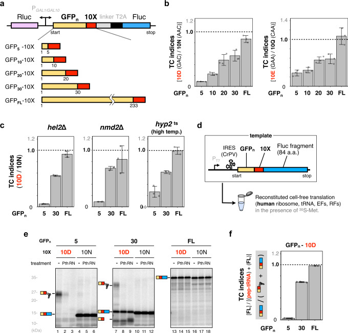
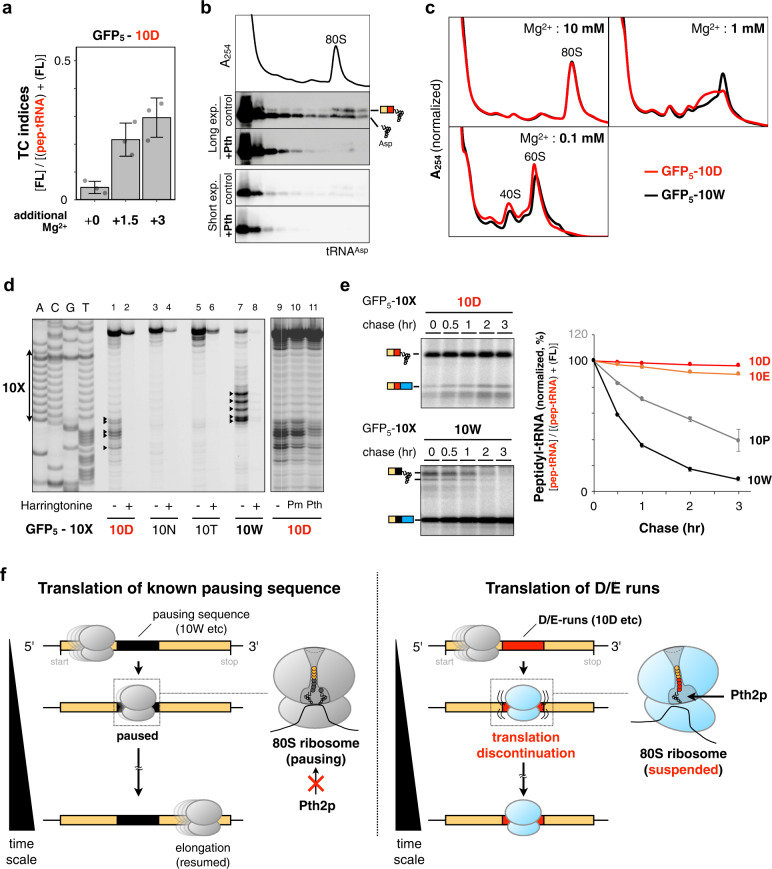
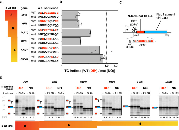
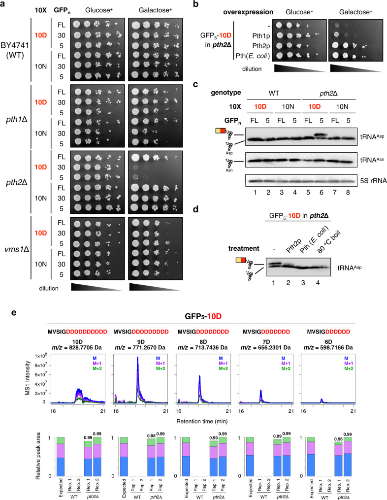
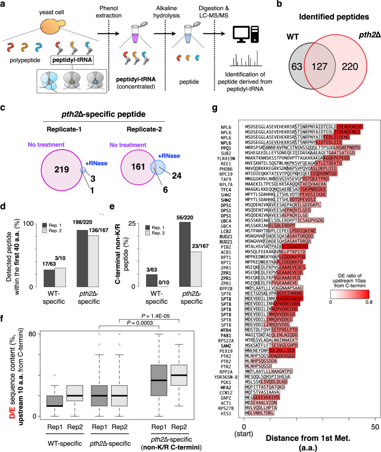
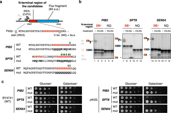
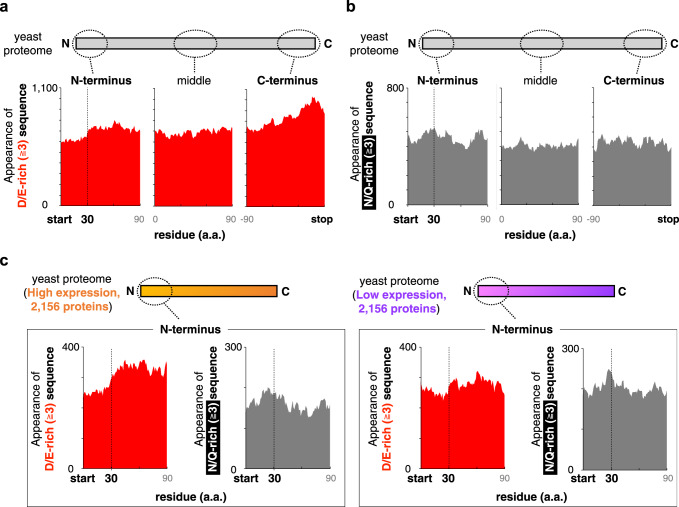
Robust translation elongation of any given amino acid sequence is required to shape proteomes. Nevertheless, nascent peptides occasionally destabilize ribosomes, since consecutive negatively charged residues in bacterial nascent chains can stochastically induce discontinuation of translation, in a phenomenon termed intrinsic ribosome destabilization (IRD). Here, using budding yeast and a human factor-based reconstituted translation system, we show that IRD also occurs in eukaryotic translation. Nascent chains enriched in aspartic acid (D) or glutamic acid (E) in their N-terminal regions alter canonical ribosome dynamics, stochastically aborting translation. Although eukaryotic ribosomes are more robust to ensure uninterrupted translation, we find many endogenous D/E-rich peptidyl-tRNAs in the N-terminal regions in cells lacking a peptidyl-tRNA hydrolase, indicating that the translation of the N-terminal D/E-rich sequences poses an inherent risk of failure. Indeed, a bioinformatics analysis reveals that the N-terminal regions of ORFs lack D/E enrichment, implying that the translation defect partly restricts the overall amino acid usage in proteomes.


Conflict of interest statement

The authors declare no competing interests.

Figures

Fig. 1
Fig. 1. Consecutive negatively charged amino acids in the N-terminal regions prematurely terminate translation.
a Schematic representation of the dual-luciferase assay. Homo-decamers of amino acids (10X) were inserted between varying lengths of the N-terminal region of GFP (GFPn; n = 5, 10, 20, 30, and 233 as full-length: FL) and a linker. Firefly (Fluc) and Renilla (Rluc) luciferases were used as reporter enzymes to investigate the translation continuation and an internal translation control, respectively. The GAL1-GAL10 promoter is a galactose-inducible bidirectional promoter. T2A is the self-cleaving peptide from Thosea asigna virus 2A. b The translation efficiencies of ORFs harboring 10X in S. cerevisiae were evaluated by the reporter system shown in a. After induction by galactose, the translation efficiencies were measured as the relative activity of Fluc to that of Rluc. Translation continuation (TC) indices were calculated by the following formula: {Fluc activity/Rluc activity (10D)} / {Fluc activity/Rluc activity (10 N)}. Error bars indicate standard deviations (SD) from three biological replicates. The codons encoding 10X are indicated in parentheses. c TC indices evaluated using the reporter system in the hel2Δ, nmd2Δ, and hyp2 temperature-sensitive (ts) strains are shown. The hyp2ts cells were grown at 33 °C instead of 30 °C, the normal cultivation temperature. Error bars indicate standard deviations (SD) from three biological replicates. d Schematic representation of the fusion gene, GFPn-Methionine-10X-truncated Fluc, for the cell-free assay. A methionine codon was inserted between GFP and 10X to label the translation product with 35S-methionine. e The fusion genes were translated using the human factor-based reconstituted cell-free translation system (HsPURE system) including 35S-methionine, followed by puromycin treatment. The products were further treated with a peptidyl-tRNA hydrolase (yeast Pth2p: Pth) as indicated and separated by neutral pH SDS-PAGE with optional RNase A (RN) treatment. Radioactive bands were developed by using an imaging plate. Peptidyl-tRNAs and tRNA-cleaved polypeptides are indicated by schematic labels. f TC indices calculated from the band intensities in e. The TC index was defined by the following formula: {full-length polypeptide (FL: 10D)} / {full-length polypeptide (FL: 10D) + peptidyl-tRNA (10D)}. Error bars indicate standard deviations (SD) from three technical replicates.
Fig. 2
Fig. 2. Characterization of the 80S ribosome translating the DE-runs.
a Effect of Mg2+ on the 10D sequence-dependent translation attenuation. The GFP5−10D sequences were translated by the HsPURE system supplemented with additional Mg2+ as indicated, and individual TC indices were calculated. Error bars indicate standard deviations (SD) from three technical replicates. b The HsPURE system mixture translating the GFP5−10D peptidyl-tRNA was fractionated by sucrose density gradient ultracentrifugation, separated by SDS-PAGE, and detected by northern blotting. Distributions of the ribosomes were monitored by A254 measurement. The chemiluminescence images with long- or short-time exposure were shown. Translation mixture was treated with yeast Pth2p (Pth) if indicated. A representative of three independent experiments is shown. c The HsPURE system mixture translating the GFP5−10D or GFP5−10W sequence was fractionated as shown in b except that the Mg2+ concentration in the fractionation buffer was arranged (10, 1, or 0.1 mM). A representative of two independent experiments is shown. d Toeprint analysis. The HsPURE system mixture was directed by the GFP5−10D, 10 N, 10 T, or 10 W template (lanes 1–8), in the presence or absence of 10 mM harringtonine, respectively. Reaction mixtures were then subjected to reverse transcription using a downstream, fluorescent primer. Dideoxy sequencing reactions were also primed by the same primer (lanes A, C, G, and T). The reaction mixture of GFP5−10D was treated with puromycin (lane 10) or Pth2p (lane 11) before the reverse transcription. A representative of three independent experiments is shown. e Inactivation of the 80 S ribosome by the translation of D/E-runs. The GFP5−10D, 10E, 10 P, or 10 W mRNA was translated by the HsPURE system including 35S-methionine for 1 h. Then excessive unlabeled methionine (2 mg/mL) was added to stop the incorporation of 35S-methionine and further incubated for the indicated time. The 35S-labeled translational products at each time point were withdrawn and monitored by gel electrophoresis (left). The amount of peptidyl-tRNA was calculated as following formula {peptidyl-tRNA (10X)} / {full-length polypeptide (10X) + peptidyl-tRNA (10X)}, and their normalized values (0 min: 100) were plotted with standard deviations (SD) from three technical replicates. f Schematic representation for a plausible model based on biochemical experiments. Left, translation of known pausing sequences such as 10 W pauses but resumes the translation elongation. Pth2p cannot cleave the corresponding peptidyl-tRNA in the 80 S ribosome. Right, translation of polyacidic sequences alters the conformation of the 80 S ribosome in an altered state, and then causes translation discontinuation. Pth2p can cleave the peptidyl-tRNA associated with the 80 S ribosome.
Fig. 3
Fig. 3. Translation of endogenous genes that have multiple Asp/Glu (D/E) residues in the N-terminal regions.
a The list of representative genes with 8, 6, and 4 D/Es in the N-terminal 11 amino acids. The N-terminal sequences (wt) are shown with the mutant sequences (mut), in which D/Es are substituted by N/Qs. b The N-terminal 10 amino acid sequences except for the first methionine were inserted instead of the 10X in the dual-luciferase reporter construct shown in Fig. 1a. TC indices were calculated by the following formula: {Fluc activity (wt) / Rluc activity (wt)} / {Fluc activity (mut)/Rluc activity (mut)}. Error bars indicate standard deviations (SD) from three biological replicates. c, d The N-terminal 11 amino acids of the genes with multiple D/Es were fused with truncated Fluc (c). d The genes were translated using the HsPURE system, as described in Fig. 1e.
Fig. 4
Fig. 4. Pth2p, one of the peptidyl-tRNA hydrolases, is responsible for hydrolyzing the peptidyl-tRNAs derived from the D/E-rich sequence-induced premature termination.
a Wild-type (WT), pth1Δ, pth2Δ, and vms1Δ strains harboring the plasmids for the expression of the 10X-containing ORFs shown in Fig. 1a were spotted on SD (Glucose+) or SG (Galactose+) plates lacking uracil and cultivated at 30 °C for 2 days. Cultures were 10-fold serially diluted before spotting. b Complementation assay of Pth variants. The pth2Δ mutant harboring the GFP5−10D construct showing the lethal phenotype, the transformed empty vector and plasmids carrying pth1, pth2, and pth from E. coli, were spotted on SD or SG plates lacking uracil and leucine and incubated at 30 °C for 2 days. c Northern blot analysis with anti-tRNAAsp and tRNAAsn probes showing tRNAAsp with or without a short peptide (peptidyl-tRNA or tRNA, respectively) depending on strains and constructs. The results of 5S rRNA are also shown as a loading control. The result represents one of the two similarly conducted experiments. d Northern blot analysis with the anti-tRNAAsp probe, showing tRNAAsp with or without a short peptide depending on the hydrolysis treatment. RNA extract was incubated with 1 μM of purified Pth2p (Pth2p, at 30 °C for 20 min), 1 μM of purified E. coli Pth {Pth (E. coli), at 37 °C for 20 min}, or was heated under alkaline conditions (80 °C boil, at 80 °C for 20 min). The result represents one of the two similarly conducted experiments. e (Upper) Extracted ion chromatograms of the peptide fragment expressed from the GFP5-10D sequence in the phenol-extracted RNA fraction of the pth2∆ strain. Each panel represents extracted ion chromatograms of MS1 intensity derived from a GFP5-(X)D peptide. A chromatogram of monoisotopic ion is depicted in blue, and its +1 and +2 isotopes are depicted in purple and green, respectively. The specific m/z value of the monoisotopic ion is shown at the top. (Lower) Relative peak areas of MS1 chromatogram in the phenol-extracted RNA fraction of the wild-type (WT) and pth2∆ strain (two technical replicates). Areas of monoisotopic ions, +1 isotopic ions, and +2 isotopic ions are depicted in blue, purple, and green, respectively.
Fig. 5
Fig. 5. Identification of the abortive peptidyl-tRNAs accumulated in the pth2Δ strain by LC-MS/MS analysis.
a Flowchart of the analysis of the abortive peptidyl-tRNAs in yeast cells. Peptidyl-tRNAs, which are enriched in the phenol-extracted RNA fraction from wild-type (WT) or pth2Δ strains, are chemically hydrolyzed and then digested by trypsin and LysC peptidase. The resultant peptides are identified by LC-MS/MS. b The numbers of peptides identified in each strain prepared from mid-log culture and their overlap are represented in the Venn diagram. c The numbers of pth2∆-specific peptides prepared from mid-log cultured cells. The results of the two biological replicates are shown. To confirm that most of these peptides were derived from peptidyl-tRNAs, the numbers of peptides from the RNA fraction treated by RNase during the sample preparation and their overlap are represented in the Venn diagram. d The ratio of WT-specific and pth2∆-specific peptides whose C-terminal position was within 40 amino acids from the first methionine. The results of the two biological replicates from mid-log cultured cells are shown. The numbers above the bar represent the number of all detected peptides and the peptides within the first 40 amino acids. e The ratio of WT-specific and pth2∆-specific peptides whose C-terminal amino acid was not Lys/Arg (C-terminal non-K/R). The results of the two biological replicates from mid-log cultured cells are shown. The numbers above the bar represent the number of all detected peptides and the C-terminal non-K/R peptides. f The distribution of the relative content of D/E residues in the upstream ten amino acids from the C-terminus of the detected peptides. The results of the two biological replicates from mid-log cultured cells are shown. The box portions and the central bands are described according to the 25th percentile and the median, respectively. The whiskers indicate the maximum and minimum values excluding outliers defined by 1.5 times the length of the box. P-value was obtained by Wilcoxon’s rank sum test (two-sided). g Mapping of the identified C-terminal non-K/R peptides specific to the pth2Δ strain prepared from mid-log cultured cells. Box represents the identified peptides by LC-MS/MS. The relative contents of D/E residues in the upstream ten amino acids from the C-terminus of the detected peptides are shown as a red gradient. A bold letter in the gene name indicates that the corresponding peptide was identified from both two biological replicates.
Fig. 6
Fig. 6. IRD during the translation of LC-MS/MS-identified endogenous genes.
a Schematic representation of the sequences from endogenous genes for the HsPURE in vitro translation assay and the spot assay. N-terminal amino acid motifs detected by MS in pth2Δ cells (with 3 a.a. extension) were substituted for GFPn−10X of the constructs in Fig. 1a and Fig. 1d. b IRD during the translation of endogenous genes depending on the D/E-rich sequences in the vicinity of the N-terminal region. The reporter genes shown in Fig. 6a were translated using the HsPURE system, as described above. Representatives of two technical replicates are shown. c Toxicity upon the expression of endogenous IRD-inducing motifs. Wild-type (WT) and pth2Δ strains harboring plasmids expressing Fluc fused with the N-terminal sequence of the candidates were spotted on SD and SG plates lacking uracil and grown at 30 °C for 2–3 days.
Fig. 7
Fig. 7. Yeast proteome avoids positioning D/E-rich sequences in the N-terminal regions.
a, b Amino acid composition of the yeast proteome. The vertical axis represents the number of proteins harboring ≥3 D/E (a) or N/Q (b) in a 10 residue-moving window. N-terminus, C-terminus, and middle region are defined as N-terminal 100 amino acids excluding the first methionine, C-terminal 100 amino acids, and middle 100 amino acids of the 5,540 yeast proteins that have lengths greater than 130 amino acids. c Amino acid composition of the yeast proteome, depending on the protein abundance. The abundance data were obtained from the literature.

Similar articles

Cited by

References

    1. Wilson DN, Beckmann R. The ribosomal tunnel as a functional environment for nascent polypeptide folding and translational stalling. Curr. Opin. Struct. Biol. 2011;21:274–282. doi: 10.1016/j.sbi.2011.01.007. - DOI - PubMed
    1. Duc KD, Batra SS, Bhattacharya N, Cate JHD, Song YS. Differences in the path to exit the ribosome across the three domains of life. Nucleic Acids Res. 2019;47:4198–4210. doi: 10.1093/nar/gkz106. - DOI - PMC - PubMed
    1. Ito K, Chiba S. Arrest peptides: cis-acting modulators of translation. Annu. Rev. Biochem. 2013;82:171–202. doi: 10.1146/annurev-biochem-080211-105026. - DOI - PubMed
    1. Yanofsky C. Attenuation in the control of expression of bacterial operons. Nature. 1981;289:751–758. doi: 10.1038/289751a0. - DOI - PubMed
    1. Wolin SL, Walter P. Ribosome pausing and stacking during translation of a eukaryotic mRNA. EMBO J. 1988;7:3559–3569. doi: 10.1002/j.1460-2075.1988.tb03233.x. - DOI - PMC - PubMed

Publication types